Special Article- Multiple Myeloma
Ann Hematol Oncol. 2014;1(3): 1014.
Melanoma Metastasis Mimicking Plasmacytoma in a Patient with Multiple Myeloma
David Azoulay1*, Vadim Sonkin2, Celia Suriu1, Luiza Akria1 and Andrei Braester1
1Hematology & Blood Transfusion Unit, Galilee Medical Center, Israel
2Department of Pathology, Galilee Medical Center, Israel
*Corresponding author: David Azoulay, Laboratory for Clinical Cell Analysis & Translational Research, Hematology & Blood Transfusion Unit, Galilee Medical Center, P.O.B. 21, Nahariya 22100, Israel
Received: November 28, 2014; Accepted: December 01, 2014; Published: December 03, 2014
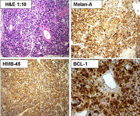
Analysis of a core biopsy from an inguinal lymph node of a 74-year-old male with multiple myeloma referred to our medical center due to the appearance of a new suspected Plasmacytoma in the right side of his penis. Flow cytometry analysis showing pathological cells with near hexaploid DNA content (DI=2.94), weaker than usual plasma cell CD38/CD138 intensity, negative cytoplasmic CD79a immunoreactivity, and lack of Kappa or Lambda light chain restriction (Figure 1). Immunohistochemical analysis showing immunoreactivity for melan-A, HMB-45, and BCL-1, suggesting the diagnosis of melanoma metastasis (Figure 2). Syndecan-1 (CD138) is a well-known marker for plasma cells. However, its expression by other non-hematopoietic tumor cells [1] could be misleading. This unusual case, which shows the development of secondary melanoma in a patient with multiple myeloma, emphasizes the need to use multiple markers for the Immunophenotyping of plasma cells and being aware of non-hematopoietic cells mimicking plasma cells.
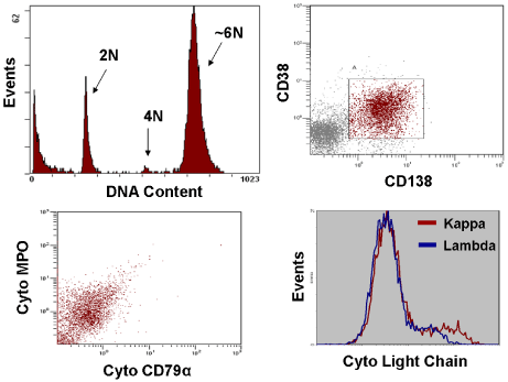
    Figure 1:  Flow cytometry analysis showing pathological cells with near
hexaploid DNA content (DI=2.94), weaker than usual plasma cell CD38/
CD138 intensity, negative cytoplasmic CD79a immunoreactivity, and lack of
Kappa or Lambda light chain restriction.