Case Report
Ann Hematol Onco. 2024; 11(2): 1453.
Unveiling The Rarity: A Case Report of TdT Negative Gamma Delta Acute Lymphoblastic Leukemia
Gupta Leena¹; Chopra Anita¹*; Meena Jagdish Prasad²
¹Laboratory Oncology, Dr. B.R.A.I.R.C.H, All India Institute of Medical Sciences, New Delhi, India
²Department of Pediatric Oncology Dr. B.R.A.I.R.C.H, All India Institute of Medical Sciences, New Delhi, India
*Corresponding author: Chopra Anita Laboratory Oncology, Dr. BRAIRCH, AIIMS Ansari Nagar, Room No. 423, 4th floor, New Delhi-110029, India. Tel: 91-11-29575415; Fax: 91-11-26588663 Email: chopraanita2005@gmail.com
Received: May 29, 2024 Accepted: June 24, 2024 Published: July 01, 2024
Abstract
Acute leukemias are frequently diagnosed malignancies in children, with T-Acute Lymphoblastic Leukemia (T-ALL) representing approximately 15% of childhood cases, of which Gamma Delta (γδ) T-ALL comprises 9-12%. This report details the case of a 7-year-old boy presenting with an acute onset of fever, cough, shortness of breath along with a neck swelling. γδ T-ALL was diagnosed through peripheral blood morphology examination and flow cytometric immunophenotyping that revealed 90% immature T-cell population positive for cytoplasmic CD3 (cCD3), surface CD3 (sCD3), CD5, T-cell receptor (TCR) gamma delta (γδ), CD99, CD7, CD2, CD4 (heterogenous), CD38 and negative for cytoplasmic terminal deoxynucleotidyl transferase (cyto-TdT). Here, we present a case of T-ALL exhibiting the absence of typical flow cytometry immaturity marker TdT and CD34 alongside positive gamma/delta receptor expression, which can be frequently misdiagnosed as mature γδ T cell neoplasm.
Keywords: T-cell leukemia; Gamma-Delta; TdT; T-cell acute lymphoblastic leukemia; Flow cytometry; Rare case
Abbreviations: T-ALL: T-Acute Lymphoblastic Leukemia; γδ: Gamma Delta; TCR: T-Cell Receptor; Cyto-Tdt: Cytoplasmic Terminal Deoxynucleotidyl Transferase; ALL: Acute Lymphoblastic Leukemia; Hb: Haemoglobin; TLC: Total Leucocyte Count; MPO: Myeloperoxidase; T-LGL Leukemia: T-cell Large Granular Lymphocytic Leukemia;
Introduction
Acute Lymphoblastic Leukemia (ALL) is a haematological malignancy characterized by abnormal proliferation of clonal progenitor lymphoid cells in the peripheral blood, bone marrow or extramedullary sites [1]. It is the most common malignancy in children, occurring mostly below 6 years of age [2]. T-ALL accounts for nearly 15% childhood ALL cases and up to 25% of adult ALL cases, the rest and majority being B-ALL [3]. In the vast majority of T-ALL cases, bone marrow is invariably affected, often accompanied by mediastinal or thymic involvement [4]. The γδ -ALL is a rare and aggressive type of T-ALL, constitutes 9-12% cases of T-ALL [5]. In contrast with aβ T-ALL, γδ T-ALL typically shows lower Hemoglobin (Hb) levels in children, increased incidence of splenomegaly, and higher Total Leucocyte Count (TLC) in adults [6]. We present a case of γδ-T-ALL distinguished by the lack of the typically observed immaturity marker i.e TdT and CD34 and the presence of gamma-delta T-cell receptor.
Case
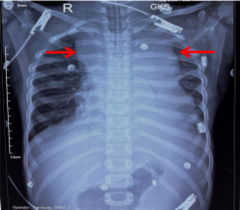
A 7-year-old boy, without any prior medical or surgical history, presented to emergency room with a 15-day history of high-grade fever, dry cough and shortness of breath associated with a rapidly increasing neck swelling. On examination, he had multiple cervical and axillary lymphadenopathy, as well as hepatosplenomegaly. In view of severe respiratory distress and stridor, the child was intubated and mechanically ventilated following which a chest X-ray was done that revealed a large mediastinal mass obscuring the left heart border. However, the lung parenchyma appeared normal (Figure 1). In view of superior mediastinal syndrome, the child was immediately started on intravenous dexamethasone, allopurinol and hydration while a definitive diagnosis was awaited.

Figure 1: Chest X-ray AP view showing a large mediastinal mass causing mediastinal widening (red arrows) with an endotracheal tube and nasogastric tube in situ.
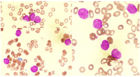
A complete blood count revealed leucocytosis with a TLC of 30× 109/L and bicytopenia with a Hb of 10.1 g/dL, and platelet count of 49× 109/L. The peripheral blood smear showed 2% neutrophils, 7% lymphocytes, and 91% blasts. The blasts were small to medium in size, with high nuclear-cytoplasmic ratio, slightly condensed chromatin, inconspicuous nucleoli and scant cytoplasm (Figure 2). These were negative for Myeloperoxidase (MPO) cytochemistry. Hence a provisional diagnosis of MPO negative acute leukemia was made. Multiparametric flow cytometry detected 90% blasts that were positive for cCD3, sCD3, CD5, TCR-γδ, CD99, CD7(bright), CD2, CD4 (heterogenous), CD38, and negative for CD34, TdT, CD8, CD56, CD16, cMPO, TCR-aβ, and CD1a (Figure 3A-I). Based on the morphology and immunophenotyping, a diagnosis of γδ+ acute lymphoblastic leukemia was made. Unfortunately, the child succumbed to his illness even before the definitive therapy could be administered.

Figure 2: Peripheral blood smear showing small to medium sized blasts (red arrow) with scant cytoplasm. (Jenner-Giemsa staining, original magnification X1000).

Figure 3: Flow cytometry analysis of the peripheral blood sample showed that 90% of the T-cells (highlighted in red) were abnormal. These cells were positive for (A) CD45(dim); (B) CD7 (bright); (C) cCD3, sCD3; (D) CD4 (heterogenous); (E) CD5, CD38; (F) TCR-γδ, CD99; (I) CD2 and TdT(dim), while negative for (B) CD16, CD56; (D) CD8; (F)TCR-aβ; (G)TdT ; (H) CD1a (I) CD34 and other markers tested.
Discussion
T-cells have two kinds of receptors: alpha-beta TCR, which are the main type in most peripheral T lymphocytes, and gamma-delta TCR, which are present in a smaller proportion (about 1-5%) of T lymphocytes [7]. γδ T-cell neoplasms are characterized by the expression of γδ -TCRs. Various neoplasms including γδ T-cell Large Granular Lymphocytic (T-LGL) Leukemia, Hepatosplenic T-cell lymphoma, skin and mucosal γδ T-cell lymphoma and γδ T-ALL are characterised by γδ –TCR expression [8].
The majority of T-ALL neither show expression of aβ nor γδ TCR. In less than 35% of cases, there is expression of either aβ or γδ TCR, with γδ expression occurring in only 9-12% of T-ALLs and around 2% of all ALL [6]. γδ T-ALL, a rare and aggressive subtype of T-ALL, constitutes approximately 9-12% of all T-ALL cases. This subtype is linked to an unfavorable prognosis [5].
The diagnosis of T-ALL relies on crucial elements such as lineage determination, assessment of clonality, and the presence of immaturity markers [9]. For assigning T-lineage either cytoplasmic CD3 (cCD3) or/and surface CD3 (sCD3) is required [10]. Demonstrating clonality usually entails examining the Vβ repertoire via flow cytometry or evaluating TCR rearrangements using molecular methods. However, these approaches can be costly and may not be universally available [11]. Most common marker associated with T-cell immaturity is TdT [10]. TdT, a DNA polymerase found within the nucleus, is typically present only in the initial stages of lymphocyte development and is frequently utilized to indicate the immaturity of T lineage cells [5]. Amongst all the immaturity marker,s TdT is detected in 90-95% of lymphoblastic lymphomas and helps in differentiating T-ALL from other leukemias and mature lymphomas [7]. Its absence can present a challenge in diagnosis. Additionally, its absence with concurrent expression of gamma/delta TCR, can erroneously diagnose the condition as mature gamma-delta T-cell leukemia/ lymphoma. Hence, it is advisable to evaluate other immaturity markers such as CD99, CD34, CD1a, HLA-DR, and CD117 in such instances [10]. Among these, CD99 emerges as particularly significant as an immaturity marker following TdT [7].
Several T-cell abnormalities were observed, such as bright CD7 expression and dim CD2 and heterogeneous CD4 expression, indicating the presence of T-cell clonality. While the normal γδ T cells lack both CD4 and CD8 markers, certain γδ T-ALLs may show expression of either CD4, CD8, or both [6]. Crucially, the absence of TdT does not definitely rule out the diagnosis of T-ALL, as demonstrated by documented cases of TdT-negative instances in existing literature. Faber et al. reported three cases of T-ALL with negative TdT expression out of total 200 cases in their publication [12] Richaet.al studied 38 patients and reported TdT negativity in 60.5% cases [9] In the study by Zhou et al, 7 out of 59 (12%) exhibited a near complete absence of TdT expression [13] Faraz et al. reported 2 cases of TdT negative γδ T-ALL [14].
Conclusion
Achieving an accurate diagnosis depends greatly on correlating clinical presentation with both morphological and immunophenotypic findings. The absence of frequently observed immaturity indicators such as TdT and CD34 does not definitively exclude the possibility of T-ALL; rather, additional immaturity markers like CD99, CD1a, HLA-DR, and CD117 should be considered for evaluation. In our case, an accurate diagnosis was achieved through the detection of CD99 expression along with CD3. Given the aggressive nature of the disease, early diagnosis and prompt treatment play pivotal roles in improving prognosis for these patients.
References
- Angsubhakorn N, Suvannasankha A. Acute lymphoblastic leukaemia with osteolytic bone lesions: diagnostic dilemma. BMJ Case Rep. 2018; 2018: bcr-2018-225008.
- Terwilliger T, Abdul-Hay M. Acute lymphoblastic leukemia: a comprehensive review and 2017 update. Blood Cancer J. 2017; 7: e577–e577.
- Arora RS, Arora B. Acute leukemia in children: A review of the current Indian data. South Asian J Cancer. 2016; 5: 155–60.
- Wei EX, Leventaki V, Choi JK, Raimondi SC, Azzato EM, Shurtleff SA, et al. γδ T-Cell Acute Lymphoblastic Leukemia/Lymphoma: Discussion of Two Pediatric Cases and Its Distinction from Other Mature γδ T-Cell Malignancies. Case Rep Hematol. 2017; 2017: 5873015.
- Pui CH, Pei D, Cheng C, Tomchuck SL, Evans SN, Inaba H, et al. Treatment response and outcome of children with T-cell acute lymphoblastic leukemia expressing the gamma-delta T-cell receptor. Oncoimmunology. 2019; 8: 1599637.
- Matos DM, Rizzatti EG, Fernandes M, Buccheri V, Falcao RP. Gammadelta and alphabeta T-cell acute lymphoblastic leukemia: comparison of their clinical and immunophenotypic features. Haematologica. 2005; 90: 264–6.
- Hassan M, Abdullah HMA, Wahid A, Qamar MA. Terminal deoxynucleotidyl transferase (TdT)-negative T-cell lymphoblastic lymphoma with loss of the T-cell lineage-specific marker CD3 at relapse: a rare entity with an aggressive outcome. BMJ Case Rep. 2018; 2018: bcr2018224570.
- Wang W, Li Y, Ou-Yang M, Zhang M, Zhao L, Liu J, et al. Gamma-Delta T-Cell Acute Lymphoblastic Leukemia/Lymphoma: Immunophenotype of Three Adult Cases. J Hematol. 2019; 8: 137-140.
- Gupta R, Garg N, Kotru M, Kumar D, Pathak R. Immunophenotypic characteristics of T lineage acute lymphoblastic leukemia: absence of immaturity markers-TdT, CD34 and HLADR is not uncommon. Am J Blood Res. 2022; 12: 1–10.
- Jaffe ES. Diagnosis and Classification of Lymphoma: Impact of Technical Advances. Semin Hematol. 2019; 56: 30–6.
- Gorczyca W, Weisberger J, Liu Z, Tsang P, Hossein M, Wu CD, et al. An approach to diagnosis of T-cell lymphoproliferative disorders by flow cytometry. Cytometry. 2002; 50: 177–90.
- Faber J, Kantarjian H, Roberts MW, Keating M, Freireich E, Albitar M. Terminal deoxynucleotidyl transferase-negative acute lymphoblastic leukemia. Arch Pathol Lab Med. 2000; 124: 92–7.
- Zhou Y, Fan X, Routbort M, Cameron Yin C, Singh R, Bueso-Ramos C, et al. Absence of terminal deoxynucleotidyl transferase expression identifies a subset of high-risk adult T-lymphoblastic leukemia/lymphoma. Mod Pathol. 2013; 26: 1338–45.
- Faraz M, Parmigiani A, Monkash N, Chen A. T-Cell Acute Lymphoblastic Leukemia/Lymphoma (T-ALL) With Negative Screening Immaturity Markers and Gamma-Delta Receptor Expression. Cureus. 2024; 16: e57399.