Case Report
Ann Hematol Onco. 2024; 11(4): 1463.
Isolated Unilateral Optic Nerve Involvement as the Presenting Feature of Early Relapse in Philadelphia Chromosome Positive Acute Lymphoblastic Leukemia
Salman Alharbi¹*; Anne Tierens¹; Jonathan Micieli²; Dawn Maze¹
¹University of Toronto, Princess Margaret Cancer Center, Ontario, Canada
²University of Toronto, Toronto Western Hospital, Ontario, Canada
*Corresponding author: Salman Alharbi University of Toronto, Princess Margaret Cancer Center, Ontario, Canada. Email: salmanjh@hotmail.com
Received: September 16, 2024 Accepted: October 04, 2024 Published: October 11, 2024
Abstract
Central Mervous System involvement (CNS) in Acute Lymphoblastic Leukemia (ALL) can be present at initial diagnosis or at disease relapse and early detection is crucial for prompt intervention. Optic nerve infiltration by leukemic cells is an oncologic emergency that requires urgent therapy to spare vision along with systemic therapy, with limited data about the optimal therapeutic strategy. Isolated optic nerve Involvement at relapse is rare; described in Philadelphia chromosome negative (Ph-) ALL in 2.2% of relapses in children and exceeding rare in Philadelphia chromosome positive (Ph+) cases, particularly in the absence of systemic disease. Here we report isolated unilateral optic nerve involvement as the sole feature of early disease relapse in Ph+ ALL in the absence of systemic disease, leading to early detection of molecular relapse.
Keywords: Optic nerve; CNS; Acute lymphoblastic Leukemia; Dasatinib
Case Presentation
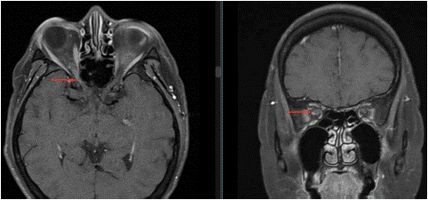
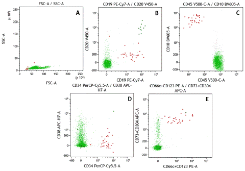
A 49-year-old female, initially presented to emergency with a 2 week history of bruising, epistaxis, and fever. Peripheral blood showed WBC 59 with 98% blasts, Platelet: 6, Hb 57. Her bone marrow was hypercellular and almost entirely replaced by blasts. Flow cytometry detected 86% CD34+ blasts which showed strong: CD10, CD19, CD20, CD22, CD34, CD38, CD58, CD66c, CD79a, HLA-DR, TdT, while Negative for: sCD3, cyCD3, CD7, MPO, TSLPR. Cytogenetics showed t(9;22) (q34.1;q11.2) while molecular testing confirmed BCR-ABL1 at p210 breakpoint. Diagnostic Lumbar puncture was negative for CNS involvement. She was induced with Dana Farber Cancer Institute (DFCI) chemotherapy protocol for age <60y (Ref), plus Imatinib at 400 mg. She achieved complete remission with negative MRD by flow cytometry, and her molecular testing showed 2.4 log reduction in BCR-ABL1 transcripts with lower limit of detection of 5.0 to 5.5 log reduction (0.0008 to 0.0003% IS). She completed maintenance phase of therapy while kept on Imatinib. Total of 12 intrathecal chemotherapy were received. She remained in remission over the whole treatment period with no evidence of disease relapse and her molecular MRD remained undetectable in her bone marrow. During follow up period, she was on concomitant treatment with ophthalmologist for myopia and glaucoma in addition to intraocular lens implantation for cataract with residual degree of vision impairment which remains stable. 3 years from initial diagnosis, she presented with gradually worsening loss of vision in her right eye with sensitivity to light over a period of 4 weeks, which was started after 10 days of undergoing LASER eye procedure for symptomatic high intraocular pressure following cataract surgery. CT brain was done and was unremarkable, followed by MRI which showed Enhancement and thickening of the right optic nerve, with optic nerve head protrusion, giving the differential of optic neuritis, which can be of infectious, inflammatory or Ischemic (Figure 1). She was evaluated by ophthalmology team and examination revealed impaired visual acuity with afferent pupillary defect of the right eye, with preservation of extraocular movements, while fundoscopic examination showed right optic disc edema with normal retina and macula. These findings supported the high suspicion of leukemic infiltration of the right optic nerve. The full neurological examination didn’t reveal any other significant findings, maintaining intact sensory function, motor power and normal reflexes. Her blood counts at that time showed WBC 3.6 with no peripheral blasts, Hb: 115, PLT: 169. A diagnostic lumbar puncture showed a clear CSF with no WBC by morphology. However, flow cytometry of the sample detects a very small B-cell precursor population consisting of approximately 0.9% of total cells with aberrant immunophenotype: Positive: CD10, CD19, CD38(dim), CD66c+123, CD73+304. While negative for CD20, (Figure 2). Such findings were supportive of involvement by the patient's previously diagnosed B-ALL. Bone Marrow Aspiration and biopsy showed morphological remission with <5% blasts with a very tiny population of CD19+ B-cell precursors with the aberrant immunophenotype are detected at 0.009% of total cells by flowcytometry which are CD10/19/20 heterogeneous/CD34/38(dim)/45(dim/66c+123). Molecular testing showed 2.4 log reduction of BCR/ABL1 transcripts indicating an early molecular relapse of the disease. ABL kinase domain mutation testing showed no clinically relevant variants.

Figure 1: MRI of the brain showing Enhancement and thickening of the right optic nerve, with optic nerve head protrusion.

Figure 2: The following antibodies were used: CD10BV605, CD19PeCy7, CD20V450, CD34PerCpCy5.5, CD38APC-H7, CD45V500-c, CD66cPe, CD73APC, CD81Fitc, CD123Pe, CD304APC. Data analysis was performed using Kaluza 2.0 (Beckman Coulter, Miami, FL, USA). The two-dimensional dot plots A, B, C, D and E show, respectively, the light scatter, CD19 vs CD20, CD10 vs CD45, CD34 vs CD38 and CD (66c+123) vs CD (CD73 vs CD304). The red and green dots represent B-lymphoblasts and mature lymphocytes. The B-lymphoblasts are positive for CD19, CD34, CD (66c and/or123) and (CD73 and/or CD123) and are negative for CD20, CD45 and CD38.
She was started on intravenous methylprednisolone at 1g/day for 5 days to spare the vision and referred to radiation oncology. She completed 10 fractions of whole brain radiation as the entire area is at risk at 180 cGy/fraction. She was also started on twice weekly intrathecal chemotherapy consists of Methotrexate 12 mg, Cytarabine 40 mg and hydrocortisone 15 mg until clearance of CSF, which was achieved on subsequent 2 samples by morphology and flowcytometry.
Due to evidence of early systemic relapse, Imatinib was discontinued, and she was started on Dasatinib at 70 mg bid given its established potency (1) and ability to cross blood brain barrier (2). She was started on HyperCVAD-arm B. Bone marrow post re-induction showed morphologic remission with negative MRD by flowcytometry and PCR showed 4.9 log reduction of BCR-ABL1 transcripts.
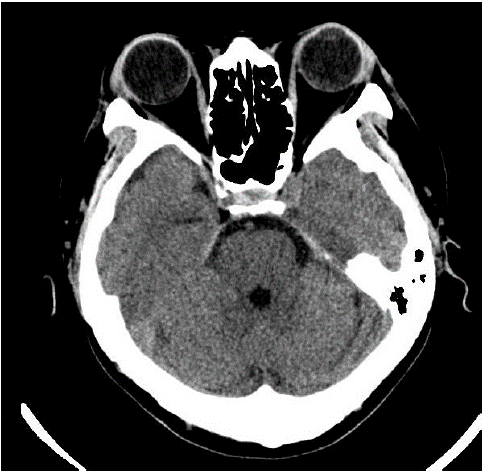
Our patient had no further visual deterioration but with an established optic atrophy. A repeated CT orbits showed no mass with decrease in size of the right intraorbital optic nerve sheath complex confirming no evidence of active ocular malignancy (Figure 3).

Figure 3: CT Orbit post treatment showing no mass with decrease in size of the right intraorbital optic nerve sheath complex.
Discussion
CNS involvement in ALL is well established, occurring in 5-7% at initial presentation [3,4], and considered the most common site of extramedullary relapse in 30% of pediatric cases [5]. Isolated unilateral optic nerve involvement as initial presentation of CNS relapse of the disease extremely rare, described in 2.2% of relapses [6] and was reported in few cases of Philadelphia chromosome negative (Ph-) pediatric patients [7,8].
A single case was reported by Satake et. al. on a relapsed Ph+ B-ALL with CNS and optic nerve involvement with concomitant hematological relapse while on Imatinib maintenance, which was successfully treated with Dasatinib plus one dose of triple intrathecal therapy [9].
To our knowledge, this is the first case report of isolated optic nerve involvement as initial presentation of CNS relapse of Ph+ B-cell ALL in adults, which prompted the early detection of molecular relapse in the absence of systemic features or morphologic evidence of disease in the blood or bone marrow. Given the negative morphologic evidence of relapse in CSF for our patient, the high clinical suspicion indicated further testing by flow cytometry which was able to detect the small leukemic clone and might be considered as the follow up marker of clearance in subsequent samples. Measures should be urgently addressed to spare vision in optic nerve involvement including radiotherapy, steroid and the use of highly CNS-penetrating TKI, like Dasatinib, and treat systemic disease followed by transplant. In our case, urgent treatment did not reverse the visual changes, but may have prevented further deterioration. While AlloSCT is among considered therapeutic options in our patient, conditioning regimen is to be discussed and decided given that she already received whole brain radiation as part of CNS directed salvage therapy to optic nerve involvement.
Conclusion
An evidence of progressive unilateral optic nerve malfunction in Ph+ ALL patients who are in remission represents a high clinical suspicion of disease relapse, which needs a thorough assessment followed by prompt treatment including radiation, steroids and intrathecal chemotherapy in addition to a high CNS penetrant TKI to spare vision and improve outcome as soon as the diagnosis is suspected.
Author Statements
Consent
The patient was consented to obtain and share her medical case with corresponding teams and publication.
References
- O’Hare T, Walters DK, Stoffregen EP, Jia T, Manley PW, Mestan J, et al. In vitro activity of Bcr-Abl inhibitors AMN107 and BMS-354825 against clinically relevant imatinib-resistant Abl kinase domain mutants. Cancer Res. 2005; 65: 4500-5.
- Porkka K, Koskenvesa P, Lundán T, Rimpiläinen J, Mustjoki S, Smykla R, et al. Dasatinib crosses the blood-brain barrier and is an efficient therapy for central nervous system Philadelphia chromosome-positive leukemia. Blood. 2008; 112: 1005-12.
- Lazarus HM, Richards SM, Chopra R, Litzow MR, Burnett AK, Wiernik PH, et al. Central nervous system involvement in adult acute lymphoblastic leukemia at diagnosis: results from the international ALL trial MRC UKALL XII/ECOG E2993. Blood. 2006; 108: 465-72.
- Kantarjian HM, O’Brien S, Smith TL, Cortes J, Giles FJ, Beran M, et al. Results of treatment with hyper-CVAD, a dose-intensive regimen, in adult acute lymphocytic leukemia. J Clin Oncol. 2000; 18: 547-61.
- Baytan B, Evim MS, Güler S, Gunes AM, Okan M. Acute Central Nervous System Complications in Pediatric Acute Lymphoblastic Leukemia. Pediatr Neurol. 2015; 53: 312-8.
- Somervaille TC, Hann IM, Harrison G, Eden TOB, Gibson BE, Hill FG, et al. Intraocular relapse of childhood acute lymphoblastic leukaemia. Br J Haematol. 2003; 121: 280-288.
- MacDonell-Yilmaz RE, Statler B, Channa P, Janigian RH Jr, Welch JG. Leukemic Infiltration of the Optic Nerve. JCO Oncol Pract. 2020; 16: 139-141.
- Kuan HC, Mustapha M, Oli Mohamed S, Abdul Aziz RA, Loh CK, Mohammed F, et al. Isolated Infiltrative Optic Neuropathy in an Acute Lymphoblastic Leukemia Relapse. Cureus. 2022; 14: e25625.
- Satake A, Okada M, Asada T, Fujita K, Ikegame K, Tamaki H, et al. Dasatinib is effective against optic nerve infiltration of Philadelphia chromosome-positive acute lymphoblastic leukemia. Leuk Lymphoma. 2010; 51: 1920-2.