Clinical Image
Ann Hematol Oncol. 2015;2(2): 1021.
Involvement of Bone Marrow by Reed - Stenberg Cells
Derdabi S1*, Rozen L1, Drowart A2, Efira A2and Demulder A1
1Laboratoy of Hematology and Hemostasis, Belgium
2Department of Hematology/Oncology, CHU Brugmann, Free University of Brussels, Belgium
*Corresponding author: Derdabi S, Laboratoy of Hematology and Hemostasis, Free University of Brussels, Belgium
Received: December 05, 2014; Accepted: January 05, 2015; Published: January 07, 2015
Clinical Image
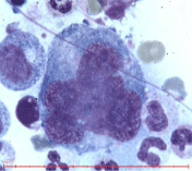
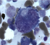
A 33 year Armenian man consulted at the emergency department for pain in the lower legs, lower back and left flank. One year ago, he suffered from Hodgkin lymphoma Stage II A, with involvement of lymph nodes in the maxillary region. He was treated in Armenia with an intensive treatment of six consecutive cures of chemotherapy BEACOPP and radiotherapy. Clinical examination upon admission was unremarkable but MRI showed pathological infiltration of the bone marrow of all vertebral bodies and pelvis, with a lytic and heterogeneous image of L3, and swelling of the left poses muscle. Imprints of bone biopsy revealed Reed- Sternberg cells (Figures 1and 2) and macrophage activation. Reed-Sternberg cells are large and are either multinucleated, ("Popcorn" appearance) or have a bilobed nucleus (thus resembling an "owl's eye" appearance) with prominent eosinophilic inclusion-like nucleoli. They are located mostly in the lymph nodes but are rarely found in bone marrow aspiration or biopsy.

Figure 1: Reed-Sternberg cell resembling popcorn (magnification x1000).
