Case Report
Ann Hematol Oncol. 2016; 3(3): 1082.
Primary Cutaneous Cryptococcosis in a Patient with Chronic Lymphocytic Leukemia: A Case Report
Ajam T¹, Hyun G², Blue BJ³ and Rajeh N³*
¹Department of Medicine, Saint Louis University School of Medicine, USA
²Medical Student, Saint Louis University School of Medicine, USA
³Division of Hematology, Saint Louis University School of Medicine, USA
*Corresponding author: Nabeel Rajeh, Division of Hematology, Saint Louis University School of Medicine, 3655 Vista Ave, West Pavilion, 3rd Floor, Saint Louis, MO 63110, USA
Received: March 11, 2016; Accepted: May 03, 2016; Published: June 06, 2016
Abstract
Cryptococcus neoformans is a fungal infection, which usually infects the lungs, however, certain subtypes can cause cutaneous infections. Primary Cutaneous Cryptococcosis is a rare presentation of cryptococcal infection and usually reserved for immunocompetent hosts. We present a case of a patient who develops a worsening skin lesion on her right forearm after planting vegetables and flowers in her garden. She suffers from relapsed chronic lymphocytic leukemia and is on ibrutinib. Culture of aspirate from the wound site revealed growth of Cryptococcus neoformans. She was started on fluconazole, and the patient achieved resolution of lesion on two month follow-up. The clinical and microbiological characteristics of primary cutaneous cryptococcosis are discussed.
Keywords: Cutaneous Cryptococcus; CLL; Ibrutinib; Cryptococcus
Abbreviations
PCC: Primary Cutaneous Cryptococcosis; CLL: Chronic Lymphocytic Leukemia
Introduction
Cryptococcus neoformans is yeast present in the environment and the main portal of entry for infecting particles is the respiratory tract [1]. C. neoformans has been recovered in soil contaminated with avian excreta, decaying wood, dust, vegetables, and fruit [2]. Primary cutaneous cryptococcosis (PCC) is a rare and clinically distinct mycosis caused by C. neoformans or C. gattii that usually presents as a unique skin lesion in the absence of disseminated disease [3].
This is the first reported case of PCC in a patient with chronic lymphocytic leukemia or a patient on ibrutinib. We report a 76-year old Caucasian woman with relapsed chronic lymphocytic leukemia on ibrutinib who presented with an ulcerated skin lesion on her right forearm. Subsequent skin biopsy and culture were positive for C. neoformans. Ibrutinib is an irreversible inhibitor of Bruton tyrosine kinase that blocks downstream B-cell receptor activation [4], and the most common adverse events include diarrhea, fatigue, and upper respiratory tract infections [5]. The case represents an uncommon presentation of cutaneous cryptococcosis in an immune compromised patient. Though clinicians are familiar with the presentation and management of systemic cryptococcal infection in patients with impaired immunity, primary cutaneous cryptococcosis is a rare presentation that should not be overlooked.
Case Presentation
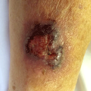
A 76-year-old woman with relapsed chronic lymphocytic leukemia diagnosed in 2000 and an avid gardener on ibrutinib presented with a progressive worsening skin lesion. Significant past medical history includes a splenectomy in 2012. She had no history of recent travel, and she lived in the Mississippi river valley. In her garden she is exposed to soil, compost, rose thorns, and vegetables. Two weeks prior to presentation, she developed an erythematous papule without any conceivable cause that appeared on her right dorsal forearm, and was progressively increasing in size. At home she developed a fever of 101°F with associated fatigue. At first she used over-the-counter antibiotic ointments, and was later prescribed amoxicillin/clavulanic acidby her haematologist without clinical improvement. Upon general examination, she was a febrile and no involvement of other skin sites or organs. The skin manifestation was an erythematous 3 by 4 cm plaque with central irregular ulceration with pink fibrinous base (Figure 1) on the right dorsal forearm. There was no regional lymphadenopathy or any other cutaneous lesion. A sporotrichosis was suspected, itraconazole 100 mg/12 h was started, and a skin biopsy was performed for histopathological and microbiological studies.

Figure 1:
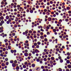
Microscopic examination of the skin lesion biopsy revealed vascular proliferation, fibroblasts, and an oedematous stroma. There was dermal haemorrhage with a collection of neutrophils in the dermis. A Grocott’s methenamine silver stain showed fungal yeast cells with broad-based budding (Figure 2). Gram stain was negative. Periodic Acid Schiff staining showed an encapsulated organism with surrounding clear space. A mucicarmine stain highlighted the capsule and a colloidal iron stain weakly highlighted a mucinous capsule.

Figure 2:
Preliminary fungal culture was reported to be positive for cryptoccoccus, and tissue culture confirmed the presence of C. neoformans. Itraconazole was discontinued for further evaluation by lumbar puncture of systemic cryptococcal infection. Antibodies to hepatitis B and C and HIV were negative.
Chest X-ray and cerebral spinal fluid analysis did not show any systemic involvement. She tested negative for HIV. CSF Cryptococcal antigen, India ink stain, and culture were all negative.
A six month course of fluconazole 400 mg daily was started. Two week follow-up with improvement of skin lesion and complete resolution of the lesion at eight week follow-up. There was no sign any new lesions in other parts of the body.
Discussion
C. neoformans is an opportunistic yeast with four different serotypes and two varieties. A and D serotypes correspond to neoformans variety and B and C serotypes correspond to gattii variety [6]. C. neoformans classically affects patients of compromised immunity secondary to HIV, malignancy, or other causes by inhalation of non-encapsulated spores. Spores from pigeon droppings, vegetables and fruits are typically the source for this infection [1]. Encapsulation in the alveolar spaces protects the fungus from phagocytosis, and it can later spread by the bloodstream. The skin is involved in 10-20% of cases [7].
Cryptococcus infections have a high mortality among immunocompromised patients. Skin manifestations are often secondary to disseminated systemic infection. Innate and T-cell mediated immunity mediate host defences against Cryptococcus. There is decreased production of pro-inflammatory cytokines and a resultant decreased development of T helper cells [8], which is thought to increase the risk of early cryptococcal infection.
PCC is a rare skin disorder caused by the encapsulated yeasts C. neoformans or C. gattii. It was first reported in the 1950’s (reviewed in [9], and the majority cases were reported from Europe and Japan [10]).
Skin lesions found in PCC may differ from those seen in disseminated cryptococcosis [11]. In secondary cutaneous cryptococcosis, the lesions are typically multiple and scattered most commonly found on the head and neck whereas PCC is solitary to a limited area located on exposed areas of the skin [12]. However disseminated cryptococcosis may include every type of lesion including extensive cellulitis, polymorphic lesions [13], and pyoderma gangrenosum-like lesions [14].
The most common presentations of cryptococcal infection in the community are meningeal encephalopathy and pneumonia. Cutaneous manifestations are rare, and are usually secondary to haematogenous dissemination. Unlike secondary cutaneous cryptococcosis, PCC is diagnosed by a solitary skin lesion in the absence of systemic disease with culture positive for Cryptococcus. In addition to culture, further identification can be made by virulence factors such as polysaccharide capsule, melanin production, or enzyme secretions [15].
Our patient was treated with oral fluconazole. Although there is no well-established protocol for treatment exclusively for PCC, fluconazole appears to be the most appropriate therapy. Duration of therapy vary between reports.
In conclusion, although rare, PCC should be included in the differential diagnosis for an isolated skin lesion in the setting of an immunocompromised patient. Clinicians should be aware of the appropriate risk factors such as age and history of skin injury, outdoor activities, or environmental exposures, and that C. neoformans can be responsible for unexplained skin lesions [16].
Acknowledgements
We would like to thank the patient for volunteering her information. Also we would like to thank the faculty of Saint Louis University School of Medicine and SLU Care Cancer Center.
References
- Powell KE, Dahl BA, Weeks RJ, Tosh FE. Airborne Cryptococcus neoformans: particles from pigeon excreta compatible with alveolar deposition. J Infect Dis. 1972; 125: 412-415.
- Misra VC, Randhawa HS. Occurrence and significance of Cryptococcus neoformans in vegetables and fruits. Indian J Chest Dis Allied Sci. 2000; 42: 317-321.
- Revenga F, Paricio JF, Merino FJ, Nebreda T, Ramirez T, Martinez AM. Primary cutaneous cryptococcosis in an immunocompetent host: case report and review of the literature. Dermatology. 2002; 204: 145-149.
- Herman SE, Gordon AL, Hertlein E, Ramanunni A, Zhang X, Jaglowski S, et al. Bruton tyrosine kinase represents a promising therapeutic target for treatment of chronic lymphocytic leukemia and is effectively targeted by PCI-32765. Blood. 2011; 117: 6287-6296.
- Byrd JC, Furman RR, Coutre SE, Flinn IW, Burger JA, Blum KA, et al. Targeting BTK with ibrutinib in relapsed chronic lymphocytic leukemia. N Engl J Med. 2013; 369: 32-42.
- Wadhwa PD, Morrison VA. Infectious complications of chronic lymphocytic leukemia. Semin Oncol. 2006; 33: 240-249.
- Hernandez AD. Cutaneous cryptococcosis. Dermatol Clin. 1989; 7: 269-274.
- Schupbach CW, Wheeler CE Jr, Briggaman RA, Warner NA, Kanof EP. Cutaneous manifestations of disseminated cryptococcosis. Arch Dermatol. 1976; 112: 1734-1740.
- Kaiko GE, Horvat JC, Beagley KW, Hansbro PM. Immunological decision-making: how does the immune system decide to mount a helper T-cell response? Immunology. 2008; 123: 326-338.
- Baes H, Van Cutsem J. Primary cutaneous cryptococcosis. Dermatologica. 1985; 171: 357-361.
- Miura T, Akiba H, Saito N, Seiji M. Primary cutaneous cryptococcosis. Dermatologica. 1971; 142: 374-379.
- Murakawa GJ, Kerschmann R, Berger T. Cutaneous Cryptococcus infection and AIDS. Report of 12 cases and review of the literature. Arch Dermatol. 1996; 132: 545-548.
- Neuville S, Dromer F, Morin O, Dupont B, Ronin O, Lortholary O, et al. Primary cutaneous cryptococcosis: a distinct clinical entity. Clin Infect Dis. 2003; 36: 337-347.
- Crounse RG, Lerner AB. Cryptococcosis; case with unusual skin lesions and favorable response to amphotericin B therapy. AMA Arch Derm. 1958; 77: 210-215.
- Massa MC, Doyle JA. Cutaneous cryptococcosis simulating pyoderma gangrenosum. J Am Acad Dermatol. 1981; 5: 32-36.
- Casadevall A, Rosas AL, Nosanchuk JD. Melanin and virulence in Cryptococcus neoformans. Curr opin Microbiol. 2000; 4: 354-358.