Case Report
Ann Hematol Oncol. 2016; 3(6): 1098.
Chronic Lymphocytic Leukemia Presenting with Concurrent Isolated CNS Parenchymal and Leptomeningeal Large B-Cell Lymphoma
Dunbar M
Dilli E
Slack GW
Villa D
1Department of Pediatrics, University of British Columbia, Canada
2Division of Neurology, University of British Columbia, Canada
3Department of Pathology and Laboratory Medicine, British Columbia Cancer Agency, Canada
4Division of Medical Oncology and Centre for Lymphoid Cancer, British Columbia Cancer Agency, Canada
*Corresponding author: Diego Villa, Division of Medical Oncology and Centre for Lymphoid Cancer, British Columbia Cancer Agency, 600 West 10th Avenue, Vancouver, BC V5Z 4E6, Canada
Received: June 20, 2016; Accepted: August 06, 2016; Published: August 09, 2016
Abstract
A previously well 59-year-old man presented with two weeks of progressive vertigo, ataxia, vomiting and diplopia. Computed tomography (CT) revealed multifocal areas of cerebral and cerebellar edema with contrast enhancement and magnetic resonance imaging with gadolinium also showed diffuse leptomeningeal enhancement with evidence of parenchymal invasion. Cerebrospinal fluid analysis identified malignant lymphocytes consistent with a large B-cell lymphoma. CT of the neck, chest and abdomen did not demonstrate hepatosplenomegaly or lymphadenopathy and peripheral blood counts showed a mild lymphocytosis. Bone marrow biopsy was consistent with chronic lymphocytic leukemia (CLL). Symptoms initially improved with corticosteroids, but the patient declined active therapy and died one month later of progressive central nervous system (CNS) disease. Patients with CLL may rarely develop CNS involvement. Even more rarely, some of these patients can present with aggressive histology lymphoma in the CNS. There is no standard treatment for CNS CLL or large B-cell lymphoma in these patients; although agents that cross the blood-brain barrier and intrathecal chemotherapy are most commonly used. The presence of large B-cell lymphoma involving the CNS appears to double six-month mortality compared to secondary CNS involvement by CLL.
Keywords: Chronic lymphocytic leukemia; CNS lymphoma; Richter syndrome; Transformation
Abbreviations
CT: Computed Tomography; CLL: Chronic Lymphocytic Leukemia; CNS: Central Nervous System; RS: Richter Syndrome; DBLCL: Diffuse Large B-cell Lymphoma; MRI: Magnetic Resonance Imaging; CSF: Cerebrospinal Fluid
Introduction
The natural history of chronic lymphocytic leukemia (CLL) is variable: approximately one third of patients experience longterm survival without need for treatment, one third are initially asymptomatic but eventually experience disease progression requiring therapy and one third have symptomatic disease requiring immediate treatment [1]. Approximately 2-10% patients may experience transformation to an aggressive lymphoma, commonly known as Richter’s syndrome (RS) [2]. RS is a rare and fatal complication of CLL with an average survival of 8-12 months [3]. The most common aggressive histology is diffuse large B cell lymphoma (DLBCL) [3].
Symptomatic central nervous system (CNS) involvement in CLL is known to be rare, with only 92 cases described in a recent review of the literature [4]. Asymptomatic CNS involvement identified on autopsy is much more common, between 6-50% [5-7]. In most cases, CNS involvement in CLL occurs within five years of diagnosis, although in a very small subset CNS symptoms are the first presentation of CLL [8-16]. RS can rarely present in the CNS with parenchymal invasion causing encephalopathy, seizures and hemiparesis, or with lymphomatous meningitis [17-32]. Regardless of histology, leptomeningeal infiltration is associated with a median overall survival of 4-6 weeks if untreated [33].
It is exceptionally uncommon for patients to present with newlydiagnosed CLL together with DLBCL involving exclusively the CNS [34-36]. Here we present a case of CLL presenting concurrently with large B-cell parenchymal and leptomeningeal involvement and review the clinical characteristics, treatment and prognosis of this uncommon disease presentation.
Case Presentation
A 59 year-old right-handed man of Chinese ancestry presented with a two-week history of worsening vertigo, ataxia, vomiting and diplopia. His past medical history was only significant for mild hypertension and remote smoking.
Temperature was 37 degrees Celsius, blood pressure estimated by an automatic blood pressure monitor was 153/90 mmHg, heart rate was 72 beats per minute and respiratory rate was 16 breaths per minute. There was no peripheral lymphadenopathy or hepatosplenomegaly. Mental status and language were normal. The pupils were equal and reactive to light and funduscopic examination was normal. There was right beating nystagmus upon right, upward and downward gaze. Saccades were hypometric to the right. There was decreased auditory perception in the left ear. The remainder of the cranial nerve exam was normal. In all four limbs, power was intact (grade 5/5), with decreased tone and reduced reflexes. There was bilateral dysmetria, left greater than right, as well as slow rapid alternating movements in the upper extremities. There was marked ataxia in both legs and gait was ataxic and right-veering.
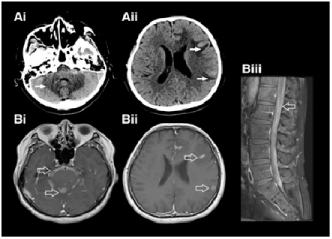
Peripheral blood cell counts showed white blood cells 13.5x10⁹/L (absolute lymphocyte count 4.7x10⁹/L), hemoglobin 134 g/L, platelets 215x109/L. Serum sodium was 140 mmol/L, potassium 3.9 mmol/L, urea 6.7 mmol/L, creatinine 99 mmol/L, alanine aminotransferase 21 U/L, aspartate aminotransferase 10 U/L, lactate dehydrogenase 125 U/L. Hepatitis B, Hepatitis C and Human Immunodeficiency Virus serologies were negative. Computed tomography (CT) of the neck, chest, abdomen and pelvis did not reveal hepatosplenomegaly or lymphadenopathy. CT head demonstrated bilateral multifocal cerebral and cerebellar areas of vasogenic edema with nodular cortical and cerebellar enhancement and increased vascularity on the CT angiogram (Figure 1A). Caudal images of the head/neck CT identified possible paralysis of the left vocal cord. Magnetic resonance imaging (MRI) with gadolinium demonstrated diffuse contrast enhancement throughout the entire subarachnoid space of the brain and spinal canal. There were multiple leptomeningeal foci along the left cerebral hemisphere and superior cerebellum with marked intraparenchymal edema and restricted diffusion suspicious for parenchymal invasion (Figure 1B).

    Figure 1: A: Computed Tomography (CT) head (axial) demonstrating
vasogenic edema (filled arrows) in the cerebellum (i) and cortex (ii). B:
Magnetic resonance imaging with gadolinium (axial) demonstrating diffuse
leptomeningeal enhancement (open arrows) in the posterior fossa (i) cortex
(ii) and sagital lumbar spine (iii).
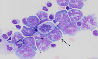
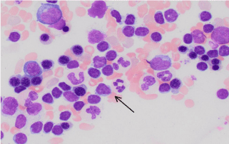
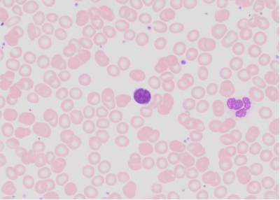
Cerebrospinal fluid (CSF) analysis via lumbar puncture demonstrated white blood cells 341x106/L, red blood cells <500 x106/L, glucose 6.3 mmol/L, protein 12,202 mg/L. CSF was thick and straw coloured and flow cytometry identified an abnormal B-cell population positive for CD19, CD20 (bright), CD10, FMC-7 with monotypic expression of kappa surface immunoglobulin light chain. CD5 and CD23 were negative. CSF cytology revealed the presence of malignant lymphocytes with large multilobulated nuclei consistent with large B-cell lymphoma (Figure 2). Bone marrow biopsy showed 30-40% cellularity with an infiltrate of small atypical lymphocytes (Figure 3). They exhibited a different immunophenotype by flow cytometry: positive for CD19, CD20 (dim), CD5 and CD23 with excess surface kappa immunoglobulin light chain expression; but negative for CD10, FMC-7 and CD38. Immunoglobulin VH mutation status, ZAP-70 and cytogenetics (including TP53) were not performed. Peripheral blood smear showed small, atypical lymphocytes similar in appearance to those in the bone marrow (Figure 4), although peripheral blood flow cytometry was not performed. The findings were consistent with discordant involvement of the bone marrow with chronic lymphocytic leukemia. Polymerase chain reaction for B-cell receptor clonality was not performed on either specimen.

    Figure 2: Cerebrospinal fluid cytospin preparation showing large atypical
lymphocytes with multilobulated nuclei, prominent nucleoli, and moderate
amounts of basophilic cytoplasm (arrow). A mitotic figure is present in the
center of the field. (Wright stain, 40x).

    Figure 3: Bone marrow aspirates showing increased lymphocytes, which are
small and mature in appearance (arrow). (Wright stain, 40x).

    Figure 4: Peripheral blood smears showing small and mature lymphocytes
similar in appearance to those in the bone marrow aspirate. (Wright stain,
40x).
He was initially treated with oral dexamethasone 8 mg twice daily, with significant improvement of the ataxia and diplopia. Given the aggressive presentation of the leptomeningeal disease, systemic and intrathecal chemotherapy was recommended, including methotrexate, cytarabine, fludarabine and rituximab. Craniospinal radiotherapy was also presented as an alternative. The patient understood the treatment options and given the poor longterm prognosis afforded by the extent of CNS involvement, after a comprehensive informed consent discussion, he opted not to undergo active therapy for CLL or CNS lymphoma. He experienced progressive worsening of neurologic deficits despite dexamethasone and had a tonic-clonic seizure 38 days after his initial presentation. He did not regain consciousness and died a few days later.
Discussion
This case describes a previously healthy man with a sub acute history of cerebellar and cranial nerve deficits secondary to lymphomatous meningitis by aggressive histology B-cell lymphoma. In secondary CNS malignancies, leptomeningeal involvement is the third most common complication in the CNS, after parenchymal masses and epidural spinal cord compression [33]. The hematologic malignancies most likely to present with neoplastic meningitis are lymphoblastic leukemia/lymphoma, Burkitt lymphoma, acute myelomonocytic leukemia and primary CNS lymphoma [37,38].
Because immunoglobulin heavy chain rearrangement studies were not performed on the CSF or bone marrow specimens, we cannot determine whether the CNS disease was a true Richter transformation of systemic CLL or whether it was coincidental, de novo primary CNS lymphoma. The disparity between CD10, FMC7 and kappa light chain expression between CSF and bone marrow samples suggests there was no clonal connection between both lymphomas. In a separate report of a case with a very similar presentation, authors clearly demonstrated clonality between the CNS tumor and CLL, indicating that in some instances both histologies are indeed related [39]. Ultimately, this question did not turn out to be significant clinically in our patient, as it did not alter treatment or prognosis.
A MEDLINE search was performed to identify case reports, case series, or cohort studies in English or French of patients with CLL presenting with neurologic involvement. A total of 23 patients with CLL involving the CNS between 1982-2013 were identified from 22 reports [8-17,39-50]. In a separate search to identify cases of CNS DLBCL, there were 15 cases of CNS DLBCL in known CLL from 12 reports [17,19,21,25,26,28-32,51] and two review papers each briefly described 5 additional cases [22,24]. In addition, 4 cases in whom CLL was diagnosed simultaneously with CNS DLBCL were identified from 3 reports [18,23,52]. These findings suggest that involvement of the CNS in patients with CLL is exceedingly uncommon and is more likely to occur in patients with a pre-existing history of CLL. Our patient’s presentation with CNS large B-cell lymphoma concurrently with CLL is therefore the least frequent and constitutes the fifth reported case in the literature.
There is no standard of care for the treatment of CNS CLL or RS. Primary and secondary CNS lymphomas are known to be poorly responsive to a variety of agents alone or in combination, presumably due to the genetic abnormalities acquired during its evolution [3,53- 56]. The most frequently used therapy across groups in this review was intrathecal methotrexate (47% of those with RS and pre-existing CLL, 40% of those with RS without pre-existing CLL and 26% of those with CNS CLL). Treatment of CNS involvement in CLL has been most successful with IT methotrexate and cranial radiation, which in one retrospective series resulted in symptom improvement in 83% of cases [17]. Due to the enormous variability of treatments, small number of cases and the high mortality, no strong conclusions can be made from this review regarding the success of any particular treatment type or combination.
Despite the apparently favorable prognosis based on Rai staging for a majority of patients, the overall survival of patients with CNS involvement is poor. Of the 43 patients reviewed, 19 (44%) were reported to die in the year after CNS diagnosis, 17 of whom died within the first six months. There was a trend towards better survival in those with CNS CLL compared to CNS DLBCL. In those with untransformed CLL presenting in the CNS, 14 of 23 (58%) were alive at the time of follow up, median 12 months, compared to only 6 of 15 (33%) with RS and prior CLL alive at the time of follow up, median 3 months. In the 5 patients with CNS DLBCL and simultaneous CLL diagnosis, two died within 6 weeks of presentation, two were alive at one year and one was known to be alive at 3 month follow up, though with recurrent lymphoma. The two who survived to one year presented with parenchymal tumors and one received radiation, the other systemic and IT chemotherapy.
This case report and literature review demonstrates that patients with CLL may rarely present with CNS involvement in the form of CNS CLL or aggressive histology B-cell lymphoma. Prognosis appears most influenced by the presence of DLBCL, with a high mortality rate at 6 months. There is no gold standard therapy, though intrathecal methotrexate was most commonly used in the reported cases. Treatment can be effective for symptom relief, but is rarely curative. In the very rare case of CLL presenting in the CNS with DLBCL, death occurs within six weeks if untreated, but survival may be improved by several months with treatment.
References
- Dighiero G. Unsolved issues in CLL biology and management. Leukemia. 2003; 17: 2385-2391.
- Tsimberidou AM, Keating MJ. Richter syndrome: biology, incidence, and therapeutic strategies. Cancer. 2005; 103: 216-228.
- Jamroziak K, Tadmor T, Robak T, Polliack A. Richter syndrome in chronic lymphocytic leukemia: updates on biology, clinical features and therapy. Leuk Lymphoma. 2015; 56: 1949-1958.
- de Souza SL, Santiago F, Ribeiro-Carvalho Mde M, Arnóbio A, Soares AR, Ornellas MH. Leptomeningeal involvement in B-cell chronic lymphocytic leukemia: a case report and review of the literature. BMC Res Notes. 2014; 7: 645.
- Cramer SC, Glaspy JA, Efird JT, Louis DN. Chronic lymphocytic leukemia and the central nervous system: a clinical and pathological study. Neurology. 1996; 46: 19-25.
- Bojsen-Møller M, Nielsen JL. CNS involvement in leukaemia. An autopsy study of 100 consecutive patients. Acta Pathol Microbiol Immunol Scand A. 1983; 91: 209-216.
- Barcos M, Lane W, Gomez GA, Han T, Freeman A, Preisler H, Henderson E. An autopsy study of 1206 acute and chronic leukemias (1958 to 1982). Cancer. 1987; 60: 827-837.
- Korsager S, Laursen B, Munck Mortensen T. Dementia and central nervous system involvement in chronic lymphocytic leukaemia. Scand J Haematol. 1982; 29: 283-286.
- Boogerd W, Vroom TM. Meningeal involvement as the initial symptom of B cell chronic lymphocytic leukemia. Eur Neurol. 1986; 25: 461-464.
- Pérez Fernández I, Salgado Ordóez F, Rueda Dominguez A, Sevilla García I, Siles Rodriguez A. Infiltration of the central nervous system as a presentation form of early stage chronic lymphocyte leukemia. Am J Hematol. 1998; 58: 339-340.
- Poplawska-Szczyglowska L, Walewski J, Pienkowska-Grela B, Rymkiewicz G, Mioduszewska O. Chronic lymphocytic leukaemia presenting with central nervous system involvement. Med Oncol. 1999; 16: 65-68.
- Akintola-Ogunremi O, Whitney C, Mathur SC, Finch CN. Chronic lymphocytic leukemia presenting with symptomatic central nervous system involvement. Ann Hematol. 2002; 81: 402-404.
- Kaiser U. Cerebral involvement as the initial manifestation of chronic lymphocytic leukemia. Acta Haematol. 2003; 109: 193-195.
- Kalac M, Suvic-Krizanic V, Ostojic S, Kardum-Skelin I, Barsic B, Jaksica B, et al. Central nervous system involvement of previously undiagnosed chronic lymphocytic leukemia in a patient with neuroborreliosis. Int J Hematol. 2007; 85: 323-325.
- Tonino SH, Rijssenbeek AL, Oud ME, Pals ST, van Oers MH, Kater AP, et al. Intracerebral infiltration as the unique cause of the clinical presentation of chronic lymphocytic leukemia/small lymphocytic leukemia. J Clin Oncol. 2011; 29: e837-839.
- Chan K-L, McKelvie P, Firkin F, Bazargan A, Tam CS. Chronic lymphocytic leukemia presenting as an intracranial epidural mass in a patient with myeloproliferative neoplasm associated with JAK2 V617F mutation. Leuk Lymphoma. 2013; 54: 1110-1112.
- Moazzam AA, Drappatz J, Kim RY, Kesari S. Chronic lymphocytic leukemia with central nervous system involvement: report of two cases with a comprehensive literature review. J Neurooncol. 2012; 106: 185-200.
- Lane PK, Townsend RM, Beckstead JH, Corash L. Central nervous system involvement in a patient with chronic lymphocytic leukemia and non-Hodgkin“s lymphoma (Richter”s syndrome), with concordant cell surface immunoglobulin isotypic and immunophenotypic markers. Am J Clin Pathol. 1998; 89: 254-259.
- O'neill BP, Habermann TM, Banks PM, O'Fallon JR, Earle JD. Primary central nervous system lymphoma as a variant of Richter's syndrome in two patients with chronic lymphocytic leukemia. Cancer. 1989; 64: 1296-1300.
- Bayliss KM, Kueck BD, Hanson CA, Matthaeus WG, Almagro UA. Richter's syndrome presenting as primary central nervous system lymphoma. Transformation of an identical clone. Am J Clin Pathol. 1990; 93: 117-123.
- Ng K, Nash J, Woodcock BE. High grade lymphoma of the cerebellum: a rare complication of chronic lymphatic leukaemia. Clin Lab Haematol. 1991; 13: 93-97.
- Robertson LE, Pugh W, O'Brien S, Kantarjian H, Hirsch-Ginsberg C, Cork A, et al. Richter's syndrome: a report on 39 patients. J Clin Oncol. 1993; 11: 1985-1989.
- Mahé B, Moreau P, Bonnemain B, Letortorec S, Menegali D, Bourdin S, et al. Isolated Richter's syndrome of the brain: two recent cases. Nouv Rev Fr Hematol. 1994; 36: 383-385.
- Giles FJ, O'Brien SM, Keating MJ. Chronic lymphocytic leukemia in (Richter's) transformation. Semin Oncol. 1998; 25: 117-125.
- Agard G, Hamidou M, Leautez S, Garand R, Grolleau JY. [Neuro-meningeal location of Richter syndrome]. Rev Med Interne. 1999; 20: 64-67.
- Robak T, Góra-Tybor J, Tybor K, Jamroziak K, Robak P, Kordek R, et al. Richter's syndrome in the brain first manifested as an ischaemic stroke. Leuk Lymphoma. 2004; 45: 1261-1267.
- Yamamoto Y, Tsujimoto M, Konoike Y, Nakamine H, Morii T, Kimura H. Richter's syndrome presenting as a nasal lymphoma. Leuk Lymphoma. 2004; 45: 1919-1923.
- Schmid C, Diem H, Herrmann K, Voltz R, Ott G, Hallek M. Unusual sites of malignancies: CASE 2. Central neurogenic hyperventilation as a complication of Richter's syndrome. J Clin Oncol. 2005; 23: 2096-2098.
- Bagic A, Lupu VD, Kessler CM, Tornatore C. Isolated Richter's transformation of the brain. J Neurooncol. 2007; 83: 325-328.
- Calvo-Villas J-M, Fernández JA, la Fuente de I, Godoy AC, Mateos MC, Poderós C. Intrathecal liposomal cytarabine for treatment of leptomeningeal involvement in transformed (Richter's syndrome) and non-transformed B-cell chronic lymphocytic leukaemia in Spain: a report of seven cases. Br J Haematol. 2010; 150: 618-620.
- Fløisand Y, Delabie J, Fosså A, Helseth E, Jacobsen EA, Rolke J, et al. Richter syndrome presenting as a solitary cerebellar tumor during first-line treatment for chronic lymphocytic leukemia. Leuk Lymphoma. 2011; 52: 2007-2009.
- Jain P, Benjamini O, Pei L, Caraway NP, Landon G, Kim S, Shivaprasad S. Central nervous system Richter's transformation and parvovirus B19 infection. Leuk Lymphoma. 2013; 54: 2070-2072.
- Chamberlain MC1. Leptomeningeal metastasis. Curr Opin Oncol. 2010; 22: 627-635.
- Tadmor T, Shvidel L, Bairey O, Goldschmidt N, Ruchlemer R, Fineman R, et al. Richter's transformation to diffuse large B-cell lymphoma: A retrospective study reporting clinical data, outcome, and the benefit of adding rituximab to chemotherapy, from the Israeli CLL Study Group. Am J Hematol. 2014; 89: E218-E222.
- Parikh S, Bernard G, Leventer RJ, van der Knaap MS, van Hove J, Pizzino A, et al. A clinical approach to the diagnosis of patients with leukodystrophies and genetic leukoencephelopathies. Mol Genet Metab. 2015; 114: 501-515.
- Parikh SA, Rabe KG, Call TG, Zent CS, Habermann TM, Ding W, et al. Diffuse large B-cell lymphoma (Richter syndrome) in patients with chronic lymphocytic leukaemia (CLL): a cohort study of newly diagnosed patients. Br J Haematol. 2013; 162: 774-782.
- Chamberlain MC, Nolan C, Abrey LE. Leukemic and lymphomatous meningitis: incidence, prognosis and treatment. J Neurooncol. 2005; 75: 71-83.
- Scott BJ, Douglas VC, Tihan T, Rubenstein JL, Josephson SA. A systematic approach to the diagnosis of suspected central nervous system lymphoma. JAMA Neurol. 2013; 70: 311-319.
- Oshimi K, Akahoshi M, Hagiwara N, Tanaka M, Mizoguchi H. A case of T-cell chronic lymphocytic leukemia with an unusual phenotype and central nervous system involvement. Cancer. 1985; 55: 1937-1942.
- Cash J, Fehir KM, Pollack MS. Meningeal involvement in early stage chronic lymphocytic leukemia. Cancer. 1987; 59: 798-800.
- Hagberg H, Lannemyr O, Acosta S, Birgegård G, Hagberg H. Successful treatment of syndrome of inappropriate antidiuretic secretion (SIADH) in 2 patients with CNS involvement of chronic lymphocytic leukaemia. Eur J Haematol. 1997; 58: 207-208.
- Elliott MA, Letendre L, Li CY, Hoyer JD, Hammack JE. Chronic lymphocytic leukaemia with symptomatic diffuse central nervous system infiltration responding to therapy with systemic fludarabine. Br J Haematol. 1999; 104: 689-694.
- Remková A, Bezayová T, Vyskocil M. B cell chronic lymphocytic leukemia with meningeal infiltration by T lymphocytes. Eur J Intern Med. 2003; 14: 49-52.
- Nimubona S, Bernard M, Morice P, Brassier G, Caulet-Maugendre S, Carsin B, et al. Complications of malignancy: case 3. Chronic lymphocytic leukemia presenting with panhypopituitarism. J Clin Oncol. 2004; 22: 374-376.
- Knop S, Herrlinger U, Ernemann U, Kanz L, Hebart H. Fludarabine may induce durable remission in patients with leptomeningeal involvement of chronic lymphocytic leukemia. Leuk Lymphoma. 2005; 46: 1593-1598.
- Kiewe P, Dallenbach FE, Fischer L, Hoecht S, Kombos T, Thiel E, et al. Isolated B-Cell Lymphoproliferative Disorder at the Dura Mater with B-Cell Chronic Lymphocytic Leukemia Immunophenotype. Clin Lymphoma Myeloma. 2007; 7: 594-596.
- Kelly K, Lynch K, Farrell M, Rawluk D, Kaliaperumal C, Murphy P. Intracranial small lymphocytic lymphoma. Br J Haematol. 2008; 141: 411.
- Kakimoto T, Nakazato T, Hayashi R, Hayashi H, Hayashi N, Ishiyama T, et al. Bilateral occipital lobe invasion in chronic lymphocytic leukemia. J Clin Oncol. 2010; 28: e30-32.
- Cohen JB, Cavaliere R, Byrd JC, Andritsos LA. Hearing Loss due to Infiltration of the Tympanic Membrane by Chronic Lymphocytic Leukemia. Case Rep Hematol. 2012.
- PÃ3Å,torak M, CzÅ,onkowska A, Nowicka K. Chronic lymphocytic leukemia: study of cell subsets in cerebrospinal fluid and peripheral blood. Eur Neurol. 1983; 22: 289-292.
- Resende LS, Bacchi CE, Resende LA, Gabarra RC, Niéro-Melo L. [Isolated Richter's syndrome in central nervous system: case report]. Arq Neuropsiquiatr. 2005; 63: 530-531.
- Bayliss EA, Steiner JF, Fernald DH, Crane LA, Main DS. Descriptions of barriers to self-care by persons with comorbid chronic diseases. Ann Fam Med. 2003; 1: 15-21.
- Tsimberidou AM, Kantarjian HM, Cortes J, Thomas DA, Faderl S, Garcia-Manero G, et al. Fractionated cyclophosphamide, vincristine, liposomal daunorubicin, and dexamethasone plus rituximab and granulocyte-macrophage-colony stimulating factor (GM-CSF) alternating with methotrexate and cytarabine plus rituximab and GM-CSF in patients with Richter syndrome or fludarabine-refractory chronic lymphocytic leukemia. Cancer. 2003; 97: 1711-1720.
- Tsimberidou AM, Wierda WG, Wen S, Plunkett W, O'Brien S, Kipps TJ, et al. Phase I-II clinical trial of oxaliplatin, fludarabine, cytarabine, and rituximab therapy in aggressive relapsed/refractory chronic lymphocytic leukemia or Richter syndrome. Clin Lymphoma Myeloma Leuk. 2013; 13: 568-574.
- Tsimberidou AM, Wierda WG, Plunkett W, Kurzrock R, O'Brien S, Wen S, et al. Phase I-II study of oxaliplatin, fludarabine, cytarabine, and rituximab combination therapy in patients with Richter's syndrome or fludarabine-refractory chronic lymphocytic leukemia. J Clin Oncol. 2008; 26: 196-203.
- Tsimberidou AM, Murray JL, O'Brien S, Wierda WG, Keating MJ. Yttrium-90 ibritumomab tiuxetan radioimmunotherapy in Richter syndrome. Cancer. 2004; 100: 2195-2200.