Case Report
Ann Hematol Oncol. 2016; 3(9): 1110.
Diagnosis and Treatment of Sacrococcygeal Myeloid Sarcoma Involving Multiple Positions without Leukemia: A Case Report
Hou XW¹, Yu HT¹, Zhang XJ¹*, Wang L², Xian CJ²*
1Department of Radiology, Aerospace Central Hospital, China
2Sansom Institute for Health Research, University of South Australia, Australia
*Corresponding author: Xiaojin Zhang, Department of Radiology, Aerospace Central Hospital, 15 Yu’quan Road, Haidian District, Beijing 100049, China
Cory Xian, School of Pharmacy and Medical Sciences, University of South Australia, GPO Box 2471, Adelaide 5001, Australia
Received: August 22, 2016; Accepted: September 24, 2016; Published: September 26, 2016
Abstract
Myeloid sarcoma is a rare malignant haematologic tumour, and cases of sacral and tail presentation involving multiple organs without leukemia are particularly rare. Herein we report a case of a previously healthy man who presented with progressive backache, hip pain, leg numbness, walking difficulty, constipation, dysuria, and urinary incontinence. An ultrasound scan revealed a large sacrococcygeal mass. Spine MRI revealed an epidural irregular mass in the sacral vertebral canal, expansion of the intervertebral foramen, nerve root involvement, infiltrating paraspinal musculature in the lumbosacral area with propagation into pelvis, and formation of a large soft tissue mass in the sacral space. His baseline laboratory data were normal. Two times ultrasound-guided needle biopsies and immunohistochemistry confirmed the myeloid sarcoma diagnosis. A bone marrow biopsy revealed grade-III bone marrow morphology. Immediate induction chemotherapy significantly improved his symptoms. A 6-month following-up did not show leukemia development. Thus, when a large sacrococcygeal mass is seen by imaging, instead of an immediate decision of surgical removal, a needle biopsy should be taken for pathological tests. Immunohistochemistry, in conjunction with the clinical, imaging and laboratory investigations, played an essential role in achieving the correct diagnosis. Immediate and adequate chemotherapy is crucial for achieving the optimal outcomes.
Keywords: Myeloid sarcoma; Ultrasound; MRI; Pathology; Immunohistochemistry; Chemotherapy
Introduction
Myeloid sarcoma (MS), is a focal tumor composed of primitive myeloid cells at an extramedullary site, which has often been described in association with leukemia or myeloid proliferative disorders. It was previously known as granulocytic sarcoma, chloroma, or extramedullary myeloid tumor; and the 2008 World Health Organization classification adopted the term “myeloid sarcomas” as a subgroup of “acute myeloid leukemias, not otherwise categorized” [1]. Myeloid sarcoma most commonly occurs in the soft tissues, bone, and skin, while rarely in the central nervous system (CNS) [2]. This condition can precede, follow, or occur simultaneously with chemotherapy in patients with acute myeloid leukemia (AML), bone marrow hyperplasia syndrome, bone marrow proliferation diseases and other diseases of the blood system. Because of the poor specificity of the morphology and clinical manifestations of MS, this condition (particularly with a single tumour) is easily misdiagnosed as malignant lymphoma or other tumours. Here, we present a case study of an MS located in the sacrum and tail with propagation into the pelvis and adjacent soft tissues but without evidence of systemic myeloid disease.
Case Presentation
Patient presentation
A 45-year-old male farmer, who had been healthy previously, presented with buttock and lower extremity numbness, with mild pain but without an apparent inducing cause. Subsequently, these symptoms progressed to involve severe pain accompanied by difficulty in walking, constipation, dysuria, and occasional urinary incontinence. No fever, cough, sputum, chest tightness, and chest pain were present. The patient had no history of medical or psychological illnesses, including AML and genetic disorders. Because of persistent and worsening symptoms, his physician suggested that he undergo supplementary examinations. The current study adhered to ethical guidelines on informed consent and the authors affirm that written informed consent was obtained from the patient for the study and for publication of this case report and any accompanying images.
Radiological examinations and tumour diagnosis
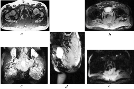
An ultrasound scan revealed a large tail mass with a large echo and unclear boundaries (not shown). A lumbar spine magnetic resonance image (MRI) revealed an irregular epidural spinal mass. The lesion involved multiple organs including the sacral and tail vertebrae, the intervertebral foramen, the nerve root, the surrounding soft tissues, the left iliac bone, and the rectum. The mass exhibited intermediate signal intensity on the T1-weighted images, was slightly hyper-intense on the T2-weighted images, and was hyper-intense on diffusion-weighted images. Expansion into the intervertebral foramen and the nerve root involvement were observed, and the boundary was unclear (Figure 1). The irregular dorsal soft tissue mass was approximately 11x5 cm, and its boundaries were unclear. A large presacral gap irregular mass of approximately 12x6 cm was present. No visible peripheral capsule, central necrosis or calcification was observed. Bone involvement of the lumbar and sacral vertebrae and the left iliac bone (in which the signal was increased) was observed, but no obvious bone destruction was present. The rectum was pressed forward, and the bladder was slightly compressed. Therefore, the radiologist diagnosed a malignant tumour.

    Figure 1:  Magnetic resonance imaging findings of sacrococcygeal myeloid
sarcoma. The sarcoma mass involved multiple organs/tissues including
the sacral and tail vertebrae, the intervertebral foramen, the nerve root,
the surrounding soft tissues, the left iliac bone, and the rectum, with some
boundaries being unclear. The mass exhibited intermediate signal intensity
on the T1-weighted images (a), was slightly hyper-intense on the T2-weighted
images (b, c, d), and was hyper-intense on diffusion-weighted images (e).
Biopsy histopathological and immunohistochemical examinations and myeloid sarcoma diagnosis
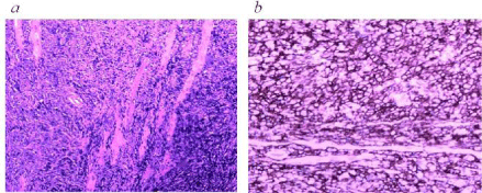
Following the radiological diagnosis of a malignant tumour, the patient did not undergo surgery, and instead an ultrasound-guided needle biopsy was taken for pathological and immunohistochemical examinations and diagnosis. Pathological examination of the needle biopsy revealed diffuse infiltration of tumour cells (Figure 2A). An immunohistochemical study of the tumour cells revealed positivities for myeloperoxidase (MPO), cluster of differentiation (CD) 34, CD43 (Figure 2B), and CD117; a Ki67 index of 60%; and a positivity for leukocyte common antigen. Negative staining results were obtained for the following molecules: CD3, CD20, CD5, CD79, CD19, CD21, CD99, Syn, and C-myc. Based on these pathological and immunohistochemical findings, this rare condition was diagnosed as MS. Other conditions that were considered but ruled out at the end included aggressive lymphoma, chordoma, and neurogenic tumors.

    Figure 2:  Representative images for the pathological examination and
immunohistochemical studies. (a) Diffuse infiltration of tumour cells in
muscle tissue (H&E image, original magnification 4x); and (b) positive
immunostaining for CD43 (DAB peroxidase immunostaining reaction image,
original magnification 10x).
Chemotherapeutic treatment and following up
Upon diagnosis of myeloid sarcoma, the patient underwent 6 therapeutic courses of induction chemotherapy (idarubicin and arabinocytidine) with good responses, then received several months of consolidation chemotherapy. The patient showed good treatment adherence/tolerability without significant adverse/unanticipated events. He was followed up by an ultrasound scan which found lesion reduction, and he showed substantial improvements to his clinical symptoms. He was followed up for 6 months, with results of his peripheral white blood cell and platelet counts being normal. He did not develop leukemia.
Discussion
The adult incidence of MS is 1/100 million [3]. MS affects people of all ages, and the incidence significantly differs between men and women. The occurrence of MS is related to bone marrow diseases, and it is present in approximately 2-8% of patients with AML and approximately 4-4.5% of those with chronic myeloid leukemia. The common sites of tumour occurrence are the lymph nodes, skin, bone and muscles. Other rarely occurring sites include the brain, testis, epididymis, vagina, lungs and bladder. An MS located in the sacral vertebral canal with multiple infiltrations has not previously been reported.
MSs are divided into isolated MSs and leukemic MSs based on whether the patient has myeloid leukemia. Isolated MS is also defined as non-leukemic MS, and this type of MS is found within 30 days after a diagnosis based on a bone marrow biopsy without exceptions [4]. MS is more common among patients younger than 10 years old and those between 20 and 44 years old. The rates in men and women do not differ. The major manifestations include different parts of the body mass. The present patient was followed up for six months, and he did not develop leukemia. Therefore, this case was classified as a solitary MS because of the lack of specific clinical manifestations and other cancers (i.e., lymphoma, anaplastic carcinoma, Ewing’s sarcoma and phase identification). If a mass is not obvious, then a variety of other conditions should be considered, including lumbar disc herniation and other symptoms of nerve root involvement. The appearances of different types of MS include those that are fish-shaped with delicate textures and grey or green sections. Tumour-cell-specific expression of MPO, CD43 and CD68 is essential to diagnose MS.
On MRI, MS exhibits specific characteristics [5]. MS presents as a soft-tissue mass with homogeneous signal intensities featuring a lack of cystic change, calcification or necrosis and no obvious bone destruction. These features can aid its differentiation from other cancers, including neurofibroma, schwannoma, and malignant fibrous histiocytoma. When the MRI features above are present in the sacrococcygeal region in patients without histories of leukemia, MS should be considered.
The average survival period for patients with MS is 2.5-22 months [6]. Tsimberidou, et al. [7] reported that patients who receive chemotherapy live longer than those who do not. The current recommended treatment regimen in patients with isolated MS or with MS presenting concomitantly with AML is the conventional AMLFigure type chemotherapy protocols, and the survival time for isolated MS in non-leukemic patients is significantly longer than that associated with leukaemia [8]. In the current case, after 6 courses of induction adjuvant chemotherapy, the symptoms were greatly improved; the patient could walk, and his defecation was not compromised. His condition has not developed into AML and at 6-month follow up he was not suffering from leukaemia.
Immediate adjuvant chemotherapy is important for reducing neurological sequelae and improving the survival rate. If necessary, this treatment should be combined with radiotherapy, surgery, and bone marrow transplantation. While the best treatment for an isolated MS is unclear, it has been recommended that the treatment of an isolated MS should be similar to that of AML, and induction chemotherapy is now the standard of care. Isolated MS is generally thought to precede AML, although cases without progression even upon long-term following up have been reported [9].
In conclusion, to improve prognosis, clinical tests should be performed as soon as possible to diagnose MS, especially for primary MS with the acute myeloid disease. MRI imaging of MSs has certain characteristics. If a radiologist does not consider a sacrococcygeal tumour as a common type, then an isolated MS should be considered. We believe that immediate surgery should not be considered, but rather an additional biopsy should be performed for histopathological and immunohistochemistry examination. If a diagnosis of MS is established, then induction chemotherapy should be the standard to avoid unnecessary surgery. Furthermore, a chemotherapeutic regimen should be included in the treatment. This case report will make a contribution to knowledge on myeloid sarcoma particularly for cases presented at sacrococcygeal region involving multiple tissues/organs and thus will have an education value. In addition, it will provide diagnostic and treatment guides for managing patients with similar presentations.
Acknowledgment
We thank Dr Anhui Zhu (Department of Hematology, Aerospace Central Hospital, Beijing, China) for technical advice. LW is funded by National Health and Medical Research Council (NHMRC) Australia Postgraduate Scholarship Award, and CJX is funded by NHMRC Senior Research Fellowship.
References
- Gunaldi M, Kara IO, Duman BB, Ercolak V. Primary intracerebral myeloid sarcoma. Onkologie. 2012; 35: 694-697.
- Sandhu GS, Ghufoor K, Gonzalez-Garcia J, Elexpuru-Camiruaga JA. Granulocytic sarcoma presenting as cauda equina syndrome. Clin Neurol Neurosurg. 1998; 100: 205-208.
- Hutchison RE, Kurec AS, Davey FR. Granulocytic sarcoma. Clin Lab Med. 1990; 10: 889-901.
- Byrd JC, Weiss RB. Recurrent granulocytic sarcoma. An unusual variation of acute myelogenous leukemia associated with 8; 21 chromosomal translocation and blast expression of the neural cell adhesion molecule. Cancer. 1994; 73: 2107-212.
- Sakral GLBBO. Sacral Canal Myeloid Sarcoma as Initial Manifestation of Granulocytic Leukemia: MRI Features and Differential Diagnosis (with a Case Report). Turk Neurosurg. 2014; 24: 281-283.
- Tan D, Wong G, Koh L, Hwang W, Loh Y, Linn Y, et al. Successful treatment of primary granulocytic sarcoma by non-myeloablative stem cell transplant. Leuk Lymphoma. 2006; 47: 159-162.
- Tsimberidou A, Kantarjian H, Estey E, Cortes J, Verstovsek S, Faderl S, et al. Outcome in patients with nonleukemic granulocytic sarcoma treated with chemotherapy with or without radiotherapy. Leukemia. 2003; 17: 1100-1103.
- Avni B, Koren-Michowitz M. Myeloid sarcoma: current approach and therapeutic options. Ther Adv Hematol. 2011; 2: 309-316.
- Vachhani P, Bose P. Isolated gastric myeloid sarcoma: a case report and review of the literature. Case Rep Hematol. 2014; 2014.