Case Report
A Case Report. Ann Hematol Oncol. 2016; 3(9): 1112.
Subacute Encephalopathy with Transhemispheric Transition of a Focal Lesion Following Intrathecal Methotrexate Injection in Pre-B-Cell Acute Lymphoblastic Leukemia: A Case Report
Ando Y¹, Sato Y¹*, Kuwashima S², Fukushima K¹, Okuya M¹, Imataka G¹, Kurosawa H¹ and Arisaka O¹
¹Department of Pediatrics, Dokkyo Medical University, Japan
²Department of Radiology, Dokkyo Medical University, Japan
*Corresponding author: Yuya Sato, Department of Pediatrics, Dokkyo Medical University School of Medicine 880 Kita-Kobayashi, Mibu, Tochigi 321-0207, Japan
Received: August 27, 2016; Accepted: October 03, 2016; Published: October 04, 2016
Abstract
Intrathecal injection of methotrexate (MTX) is important in the treatment of meningeal leukemia to prevent relapse in the central nerve system in patients with acute lymphoblastic leukemia (ALL). However, intrathecal injection of MTX sometime induces subacute encephalopathy.
A female patient with pre-B cell ALL developed relapsing-remitting encephalopathy associated with the intrathecal injection of methotrexate. The Japan Pediatric Leukemia Study Group ALL B-12 protocol had been followed. Right-sided paralysis developed 2 days after the fourth intrathecal injection of MTX. Diffusion-weighted images (DWI) during magnetic resonance imaging revealed a high-signal area in the left centrum semiovale. The right-sided paralysis resolved, only to be followed by left-sided paralysis and dysarthria. Simultaneously with these symptoms, the left-sided DWI signal was decreased and a new abnormal signal intensity lesion appeared on the right. The patient recovered from the paralysis and dysarthria 4 days after the onset of the neurological symptoms. The phenomena with which our patient presented were similar to those of multiple sclerosis, but no myelin basic protein or oligoclonal band was detected in the cerebrospinal fluid.
The mechanism that causes subacute encephalopathy after intrathecal injection of MTX is similar to that in the development of multiple sclerosis, but with no induction of myelin basic protein or oligoclonal band.
Keywords: Subacute encephalopathy; Methotrexate; Acute lymphoblastic leukemia; Intrathecal injection
Introduction
Methotrexate (MTX) is key drug in patients with acute lymphoblastic leukemia (ALL) for the treatment of meningeal leukemia or to prevent relapse in the central nervous system. MTX is administered by intravenous or intrathecal injection. However, it has also neurotoxic effects [1]. Cases of subacute MTX-induced encephalopathy reported previously showed strong signal changes on diffusion-weighted images (DWIs) obtained with magnetic resonance imaging (MRI) [1-11]. Although the intravenous or intrathecal injection of MTX is assumed to be neurotoxic because it induces metabolic changes in the central nervous system, the mechanism underlying the development of subacute encephalopathy is unclear [1,7,10-12].
We report a patient in whom subacute encephalopathy was induced by the intrathecal injection of MTX. Subacute encephalopathy was observed as chronological and spatial changes on DWI, with the simultaneous development of central nervous system disorders, which was similar to pathogenesis of multiple sclerosis. We propose a mechanism underlying subacute MTX-induced encephalopathy.
Case Presentation
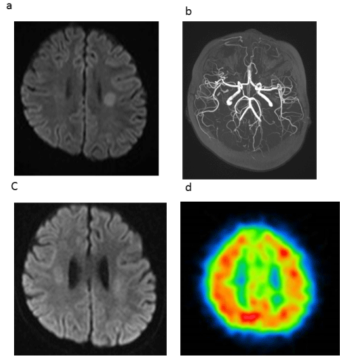
A 10-year-old female child was admitted to our hospital with pre-B-cell ALL, which was positive forCD10, CD19, CD22, CD20, CD24, and HLA-DR. The leukemic blasts had not infiltrated the central nervous system. There was not neurological symptom. Induction therapy was administered for 5 weeks according to the Japan Pediatric Leukemia Study Group (JPLSG) ALL B-12 protocol, with two intrathecal injections of three drugs: 12 mg of MTX, 30 mg of cytarabine, and 10 mg of prednisolone. Complete remission was achieved with this induction therapy. Consolidation therapy, which consisted of cytarabine, cyclophosphamide, mercaptopurine, l-asparaginase, and two intrathecal injections of MTX, cytarabine, and prednisolone, was commenced on day 36 after the induction therapy was completed. The fourth intrathecal injection of MTX, cytarabine, and prednisolone was given on day 45. Paralysis of the right side developed on day 57, and DWI revealed a high-signal lesion in the left centrum semiovale (Figure 1A). Magnetic resonance angiography (MRA) revealed no obstructions or malformations of the cerebral arteries (Figure 1B). The patient’s platelet count was 1.5×10¹⁰/L. The prothrombin time international normalization ratio was 1.00. The activated partial thromboplastin time was 31.3s. Fibrinogen was 448 mg/dL. No bacteria or viruses were cultured from the cerebrospinal fluid (CSF). No myelin basic protein or oligoclonal band was detected in the CSF. Anticoagulant and antiplatelet therapies were performed. The physical symptoms resolved by day 58, but paralysis of the left side and dysarthria developed suddenly on day 59. The signal of the left-sided cerebral lesion was decreased, and a new lesion was detected on the right side (Figure 1C). No defect was detected with singlephoton emission computed tomography (SPECT) (Figure1D). All the phenomena had disappeared by day 60. There were not abnormalities of laboratory analysis during paralysis developing. Remaining JPLSG ALL-12 protocol which included that nine time of MTX intrathecal injections and four rounds of high-dose MTX therapy were done for our patient, but paralysis was never developed again.

    Figure 1:  Analysis of the brain with MRI and SPECT.
a. Diffusion-weighted image (DWI) showed restricted diffusion in the left
centrum semiovale during the period of right-sided paralysis.
b. Axial MR angiography (MRA) revealed no occlusion or malformation of the
brain vessels.
c. DWI obtained 2 days after the first episode. At this time, the right-sided
paralysis had resolved, but left-sided paralysis and dysarthria had developed.
DWI showed subtle decreased intensity in the left centrum semiovale, and
demonstrated a new lesion in the right centrum semiovale.
d. SPECT showed no defect area.
Discussion
There are previous reports of transient encephalopathy induced by MTX that have shown high-signal brain lesions on DWI [1,3- 6,8,10,13,14]. MTX-induced encephalopathy is categorized as acute, subacute, or chronic [1,3-6,8,10, 13, 14]. However, it is unclear how this encephalopathy is induced by MTX.
In our patient, paralysis of the right side occurred 12 days after the intrathecal administration of MTX (Figure 1A). Therefore, subacute MTX-induced encephalopathy is consistent with the presentation of our patient [1], and was detected as a high-signal lesion on DWI, as in previous reports [3-5,8,10,11,13]. Second episodes of subacute MTX-induced encephalopathy after subsequent rounds of intrathecal MTX have also been reported previously [3-5,8,10, 11,13]. However, our patient’s right-sided paralysis recovered 14 days after intrathecal injection, but left-sided paralysis and dysarthria developed almost immediately. The high-signal lesion on the left side decreased and a new area of high signal appeared on the right (Figure 1A and C). Thus, our patient’s case was characterized by chronological and spatial changes in the lesions detected on DWI, and the synchronous development of central nervous system disorders after MTX intrathecal injection. These phenomena are consistent with the pathogenesis of multiple sclerosis (MS).
MS has a high component of neuronal, axonal, and myelin loss [15].These changes develop in three steps: the immune dysregulation of myelin; the infiltration of the blood-brain barrier by dendritic cells, macrophages, and Th1 and Th17 lymphocytes; and the activation of proinflammatory cytokines and the production of oxygen and nitric oxide by the infiltrating cells [15]. This progressive pathogenesis produces myelin basic protein and an oligoclonal band in the CSF of MS patients [15]. We consider that in our patient, MTX induced the dysregulation of the immunological mechanism, as occurs in the development of MS, but without the induction of myelin basic protein or the oligoclonal band. Therefore, the symptoms of our patient are interesting in terms of the development of subacute MTX-induced encephalopathy.
Transient ischemic attack (TIA) also produces reversible neurological disorders. However, SPECT images obtained in our patient showed no defect induced by vascular insufficiency (Figure 1D). This result indicates that there was no vascular insufficiency at the time of the patient’s neurological dysfunction. Her blood coagulation capacity was normal and there was no suggestion of hypercoagulation. These results confirmed that a TIA did not occur in our patient.
References
- Vezmar S, Becker A, Bode U, Jaehde U. Biochemical and clinical aspects of methotrexate neurotoxicity. Chemotherapy. 2003; 49: 92-104.
- Allen JC and Rosen G. Transient cerebral dysfunction following chemotherapy for osteogenic sarcoma. Ann Neurol. 1978; 3: 441-444.
- Aradillas E, Arora R, Gasperino J. Methotrexate-induced posterior reversible encephalopathy syndrome. J Clin Pharm Ther. 2011; 36: 529-536.
- Dicuonzo F, Salvati A, Palma M, Lefons V, Lasalandra G, De Leonardis F, et al. Posterior reversible encephalopathy syndrome associated with methotrexate neurotoxicity: conventional magnetic resonance and diffusion-weighted imaging findings. J Child Neurol. 2009; 24:1013-1018.
- Haykin M, Gorman M, Van Hoff J, Fulbright R, Baehring J. Diffusion-weighted MRI correlates of subacute methotrexate-related neurotoxicity. J Neurooncol. 2006; 76: 153-157.
- Iliskili IM and Ensefalopati PR. Intrathecal Methotrexate-Induced Posterior Reversible Encephalopathy Syndrome (PRES). Turk J Hematol. 2014; 31:109-110.
- Inaba H, Khan RB, Laningham FH, Crews KR, Pui CH, Daw NC. Clinical and radiological characteristics of methotrexate-induced acute encephalopathy in pediatric patients with cancer. Ann Oncol. 2008; 19:178-184.
- Küker W, Bader P, Herrlinger U, Heckl S, Nägele T. Transient encephalopathy after intrathekal methotrexate chemotherapy: diffusion-weighted MRI. J Neurooncol. 2005; 73: 47-49.
- Rollins N, Winick N, Bash R, Booth T. Acute methotrexate neurotoxicity: findings on diffusion-weighted imaging and correlation with clinical outcome. Am J Neuroradiol. 2004; 25:1688-1695.
- Vagace JM, Caceres-Marzal C, Jimenez M, Casado MS, de Murillo SG, Gervasini G. Methotrexate-induced subacute neurotoxicity in a child with acute lymphoblastic leukemia carrying genetic polymorphisms related to folate homeostasis. Am J Hematol. 2011; 86: 98-101.
- Kawakita A, Yazawa A, Kumano A, Ohta G, Kometani H, Suzuki K, et al. Recurrent Methotrexate-Induce Transient Subacute Encephalopathy in a patient with Malignant Lymphoma. (Japanese)Japanese J Pediatric Hematology Oncology. 2014; 51:18-23.
- Quinn CT, Bottiglieri T, Griener JC, Farrow A, Hum M, Kamen BA. Elevation of Homocysteine and Excitatory Amino Acid Neurotransmitters in the Cerebrospinal Fluid of Children Receiving Methotrexate for Leukemia 671. Pediatr Res. 1997; 41: 114-114.
- Bond J, Hough R, Moppett J, Vora A, Mitchell C, Goulden N. ‘Stroke-like syndrome’caused by intrathecal methotrexate in patients treated during the UKALL 2003 trial. Leukemia. 2013; 27: 954-956.
- Salkade PR, Lim TA. Methotrexate-induced acute toxic leukoencephalopathy. Journal of cancer research and therapeutics. 2012; 8: 292.
- Grigoriadis N, Pesch V. A basic overview of multiple sclerosis immunopathology. European Journal of Neurology. 2015; 22: 3-13.
Citation: Ando Y, Sato Y, Kuwashima S, Fukushima K, Okuya M, Imataka G, et al. Subacute Encephalopathy with Transhemispheric Transition of a Focal Lesion Following Intrathecal Methotrexate Injection in Pre-B-Cell Acute Lymphoblastic Leukemia: A Case Report. Ann Hematol Oncol. 2016; 3(9): 1112. ISSN : 2375-7965