Clinical Image
Ann Hematol Oncol. 2017; 4(7): 1158.
Aspergillus Invading Metastatic Cavity!
Biswas B*, Dabkara D and Mukhopadhyay S
Department of Medical Oncology, Tata Medical Center, India
*Corresponding author: Bivas Biswas, Department of Medical Oncology, Consultant Medical Oncologist, Tata Medical Center, 14 MAR (EW), New town, Rajarhat, Kolkata, India
Received: May 29, 2017; Accepted: June 12, 2017; Published: June 26, 2017
Clinical Image
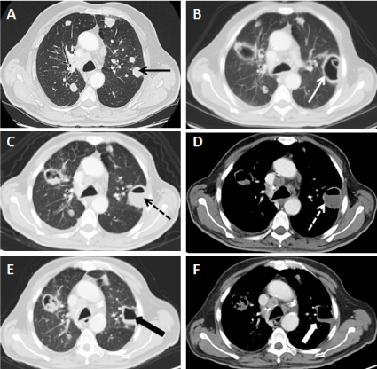
A 61-year, male with clear cell carcinoma of right kidney and multiple, bilateral solid metastatic lung nodules (Figure A) was started on pazopanib 800 mg/day in May’2016. CT chest after 3 months showed decrease in number and size of lung metastases with few showing cavitations (Figure B). His symptom reappeared with cough and hemoptysis since Jan’2017. CT chest showed cavitary lesions in both lungs with increase in wall thickness and some lesion shows new air-fluid levels (Figure C and D). Histopathology of lung biopsy revealed lung parenchyma infiltrated by colonies of fungal hyphae showing acute angle branching and septae resembling those of aspergillus species. His symptoms improved after starting on oral voriconazole. CT chest in May’2017 (Figure E and F) showed cavitary lesions in both lungs with reduction in size of cavitary lesions with reduction in wall thickness and air-fluid level. He is doing well on pazopanib with stable disease.
