Case Report
Ann Hematol Oncol. 2018; 5(1): 1187.
Chronic Myeloid Leukemia Presenting with both Hemophagocytic Syndrome and Pure Red Cell Aplasia: A Unique Case
Horny HP¹*, Alpay N², Spiekermann K² and Müller S¹
¹Institute of Pathology, University Hospital Munich, Germany
²University of Munich, Germany
*Corresponding author: Horny HP, Institute of Pathology, University Hospital Munich, Munich, Germany
Received: November 15, 2017; Accepted: January 23, 2018; Published: February 05, 2018
Abstract
The association of chronic phase chronic myeloid leukemia (CML-CP) and hemophagocytic syndrome with aplastic anemia (pure red cell aplasia or chronic erythroblastophthisis, respectively) is extremely uncommon and has not been reported as a presenting feature at initial diagnosis.
Keywords: Chronic myeloid leukemia; Aplastic anemia; Hyperleukocytosis
Case Presentation
A 27 year-old chinese woman presented with hyperleukocytosis of > 400 G/L while the platelet count was only slightly elevated with 420 G/L. A marked normochromic-normocytic anemia, (hemoglobin 8 g/dL) associated with a significantly elevated reticulocyte count (36 promille of RPI) were noted. Regarding a pronounced splenomegaly, CML was suspected and accordingly molecular studies revealed the BCR-ABL1 fusion gene. Bone marrow cytological findings revealed a virtual absence of erythroblasts but other findings confirmed the diagnosis of CML. Therapy with hydroxycarbamid was started and after significant drop of the leukocyte count, the tyrosine kinase inhibitor nilotinib was administered.
Bone marrow histology
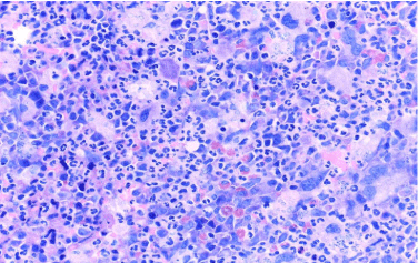
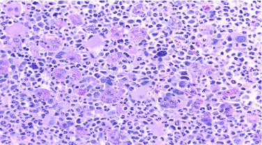
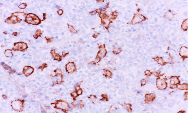
Morphological evaluation of a routinely processed core biopsy of the iliac crest (after mild decalcification in edetic acid for 6 hours and subsequent embedding in paraffin wax) revealed an extremely hypercellular bone marrow with subtotal depletion of fat cells (100% cellularity). The picture was dominated by a markedly increased and fully maturing granulocytopoiesis (Figure 1). The number of loosely scattered pleomorphic megakaryocytes was slightly increased. Typical micromegakaryocytes were seen while megakaryocytic clusters were missing. Erythroid nests were not detected. A great number of large activated histiocytes/macrophages was detected, all showing signs of pronounced hemophagocytosis (Figure 2). A granulomatous reaction, however, was absent. Immunohistochemical investigation confirmed the complete aplasia of erythrocytopoiesis by using antibodies against three erythroid antigens, namely glykophorin C, E-cadherin and CD71 (transferrin receptor). Antibodies against stem-cell related antigens like CD34, CD117 and TdT showed no increase of blast cells. As expected, E-cadherin was strongly expressed by all activated histiocytes that coexpressed other macrophage-related antigens like CD68 and CD163 (Figure 3). Numbers of monocytes expressing CD14 and mast cells expressing tryptase both were not increased.

    Figure 1:  Extreme hypercellularity of the bone marrow (100%) with markedly
increased maturing granulocytopoiesis and subtotal depletion of fat cells is
shown. Note the loosely scattered megakaryocytes including small immature
forms. At first glance this is a typical case of CML-CP. Giemsa.

    Figure 2:  Higher magnification reveals abundant large macrophages with
signs of markedly increased hemopgagocytosis, mainly erythrophagocytosis.
This is never seen in typical CML-CP. PAS.

    Figure 3:  Immunohistochemical staining with an antibody against E-cadherin
shows both aplasia of erythroblasts and specific anular staining of the
macrophages which do not express this antigen in normal states.
ABC method; anti-E-cadherin.
Diagnosis
BCR-ABL1+ CML-CP with pure red cell aplasia and signs of a hemophagocytic syndrome.
Discussion
The association of CML with pure red cell aplasia (PRCA) is highly uncommon. PRCA is usually observed during the course of the disease and after cytoreductive therapy [1-4] or the application of tyrosine kinase inhibitors [5]. Even a case of PRCA preceding overt CML has been reported [6]. PRCA in patients with CML is regarded as an adverse prognostic factor associated with incipient blast crisis [7]. The presented case is unique regarding both the presentation of PRCA even at diagnosis and its association with a hemophagocytic syndrome. Almost always, hemophagocytic syndromes are associated with signs of a hemolytic anemia and subsequent massive erythroid hyperplasia. Altogether, the histomorphological presentation of this case seems to be unique but the pathophysiological interrelationship of the complex morphological findings cannot be elucidated.
References
- Tukic L, Malesevic M, Krstic C, Lakic-Trajkovic Z, Cik Z. [Erythrocyte cell aplasia in chronic myeloid leukemia--coincidence or pathogenic link].[Article in Croatian]. Med Pregl. 1990; 43: 32-36.
- Lion T, Gaiger A, Henn T, Geissler K, Lechner K, Haas OA. Pure red cell aplasia in a case of Ph negative BCR/ABL rearranged CML with t(12;14) (q23;p11). Cancer Genet Cytogenet. 1991; 15; 56: 189-195.
- Karti S, Yilmaz M, Sonmez M, Akdogan R, Ersoz S, Ucar F, et al. Philadelphia negative, Bcr-Abl positive chronic myeloid leukemia associated with pure red cell aplasia. J Exp Clin Cancer Res. 2003; 22: 341-342.
- Yasuyama M, Kawauchi K, Takei K, Ogasawara T, Ohkawa S. [Pure red cell aplasia occurring during the course of chronic myelogenous leukemia]. [Article in Japanese]. Rinsho Ketsueki. 2004; 45: 66-71.
- Poudyal BS, Tuladhar S, Gyawali B. TKI-induced pure red cell aplasia: first case report of pure red cell aplasia with both imatinib and nilotinib. ESMO Open. 2016; 24; 1: e000058.
- Haas OA, Hinterberger W, Mörz R. Pure red cell aplasia as possible early manifestation of chronic myeloid leukemia. Am J Hematol. 1988; 27: 20-25.
- Mijovic A, Rolovic Z, Novak A, Biljanovic-Paunovic L, Tomin D. Chronic myeloid leukemia associated with pure red cell aplasia and terminating in promyelocytic transformation. Am J Hematol. 1989; 31: 128-130.