Clinical Image
Ann Hematol Oncol.2019; 6(3): 1237.
Langerhans Cell Histiocytosis in a Five-Month-Old Infant
Chi MH1¹, Cheng CD¹ and Yeh TC²*
¹Department of Pediatrics, Hsinchu Cathay General Hospital, Taiwan
²Division of Hematology & Oncology, Mackay Children’s Hospital, Taiwan
*Corresponding author: Yeh TC, Division of Hematology & Oncology, Mackay Children’s Hospital, 92 Sec. 2, Chung-San North Road, Taipei, Taiwan
Received: January 04, 2019; Accepted: February 11, 2019;Published: February 18, 2019
Keywords
Langerhans cell histiocytosis; Enlarged phalangeal bone; Granulation tissue
Clinical Image
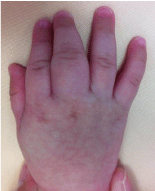
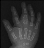
A well-nourished 5-month-old boy presented with a 4-week history of swelling in his right middle finger. Physical examination did not find hepatosplenomegaly or lymphadenopathy, but an enlarged phalangeal bone (Figure 1). Laboratory examinations were within normal limits. X-ray examination revealed an isolated expansile lytic bone lesion without cortical destruction or periosteal reaction from metaphysis or diaphysis in the proximal phalanx of the right middle finger (Figure 2). Fine needle aspiration cytology was performed suggestive of an inflamed granulation tissue with atypical cells displaying irregular nuclei and ample cytoplasm. Immunohistochemical study of the atypical cells showed positive staining of CD1a, CD5, CD68 (KP1), Langerin, S-100 protein, and negative staining of CD20. The findings confirmed the diagnosis of Langerhans cell histiocytosis. The patient did not receive any treatment, but routine follow-up. One year later, the patient was asymptomatic and in good health.

Figure 1: Langerhans cell histiocytosis.

    Figure 2:  X-ray imaging of the right middle finger showing an isolated
expansile lytic bone lesion in the proximal phalanx of the right middle finger.
- Siegel RL, Miller KD, Jemal A. Cancer Statistics, 2017. CA: a cancer journal for clinicians. 2017; 67: 7-30.
- The BH, Yong SL, Sim WW, Lau KB, Suharjono HN. Evaluation in the predictive value of serum Human Epididymal Protein4 (HE4), Cancer Antigen125 (CA125) and a combination of both in detecting ovarian malignancy. Hormone Molecular Biology and Clinical Investigation. 2018; 35: 1868-1891.
- Hailstorm I, Reycraft J, Hayden-Ledbetter M, Ledbetter JA, Schumer M, McIntosh M, et al. The HE4 (WFDC2) protein is a biomarker for ovarian cancer Carcinoma. Cancer Res. 2003; 63: 3695-3700.
- Montag nana M, Lippi G, Ruzzenente O, Bresciani V, Danese E, Scevarolli S, et al. The utility of serum HE4 in patients with a pelvic mass. Journal of Clinical Laboratory Analysis. 2009; 23: 331-335.
- Park Y, Lee JH, Hong DJ, Lee EY, Kim HS. Diagnostic performances of HE4 and CA125 for the detection of ovarian cancer from patients with various gynecologic and non-gynecologic diseases. Clin Biochem. 2011; 44: 884- 888.
- Granato T, Porpora MG, Longo F, Angeloni A, Mangarano L, Anastasi A. HE4 in the differential diagnosis of ovarian masses. Clin Chim Acta. 2014; 446: 147-155.
- Jacob F, Meier M, Caduff R, Goldstein D, Pochechueva T, Hacker N, et al. No benefit from combining HE4 and CA125 as ovarian tumor markers in a clinical setting. Gynecologic Oncology. 2011; 121: 487-491.