Clinical Image
Ann Hematol Oncol. 2019; 6(4): 1243.
A Case of Pure Erythroid Leukemia with a Rare Complex Karyotype
Yufei Mao, Qian Jiang* and Chaoming Mao*
1Department of Nuclear and Laboratory Medicine, The Affiliated Hospital of Jiangsu University, China
*Corresponding author: Chaoming Mao or Qian Jiang, Department of Nuclear and Laboratory Medicine, The Affiliated Hospital of Jiangsu University, 438 Jiefang Road, Zhenjiang 212001, China
Received: March 05, 2019; Accepted: March 06, 2019;Published: March 13, 2019
Clinical Image
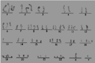
An 86-year-old man presented with an over-1-month history of dizziness and a 1-week history of weakness, poor appetite, nausea, with no lymphadenopathy, hepatomegaly, and splenomegaly. Blood tests revealed a hemoglobin concentration of 50 g/L, a platelet count of 12×109/L. White blood cell count was 3.1×10⁹/L. Bone marrow smears showed erythroblastic proliferation with 95.0% erythroid, of which 40.5% were pronormoblasts. The pronormoblast had a large body, tumorous protuberances, oil-painted blue cytoplasm with different sizes of vacuoles. Binucleated and multinucleated pronormoblasts were easily found. Some immature erythrocytes have giant juvenile changes, with fine and loose chromatin and one or more nucleoli (Figure 1A). Primitive cells of 4% and immature granulocytes were presented in the peripheral blood smears. The count of 100 white blood cells showed one early normoblast, one polychromatic normoblast and one orthochromatic normoblast (Figure 1B). Cellular histochemical stain revealed that Peroxidase (POX) (Figure 1C) and Specific Esterase (SE) (Figure 1D) stains were negative. The positive rate of Periodic Acid-Schiff (PAS) was 21% with a score of 39 points in precursor cells. Positive granules, light red and deep red positive clumps presented in pronormoblasts (Figure 1E). Flow cytometric analyses revealed that CD71, CD235a, CD13 and CD117 were expressed on the surfaces of these pronormoblasts, but not HLA-DR, CD34, CD33, CD14, CD64, CD5, CD7, CD10, CD19, CD20 and CD22. MPO, CD79a and CD3 were also negative in the cytoplasm of these cells. Cytogenetic tests presented with complex karyotypes: analysis of 20 metaphase cells showed 58-68 chromosomes (+1, +2, +8, +10, +15, +16, +18, +19, +20, +21, +22, +6q-, 11q+) (Figure 2).

Figure 1:

Figure 2:
A diagnosis of Pure Erythroblastic Leukemia (PEL) for the patient was made. Ten days after admission, the patient died. PEL is a kind of acute leukemia that is specific to erythroid immature cells. It can occur at any age and is an extremely rare and aggressive form of acute leukemia whose biology remains poorly characterized. PEL becomes the only type of acute erythroid leukemia in the recently published 2016 revision to the World Health Organization (WHO) classification of myeloid neoplasms.