Clinical Image
Ann Hematol Oncol. 2019; 6(7): 1259.
Giant Platelets
Van Der Hagen VAE, Van Gammeren A and Ermens AAM*
Department of Clinical Chemistry, Amphia Hospital, Netherlands
*Corresponding author: Ermens AAM, Department of Clinical Chemistry, Amphia Hospital, Netherlands
Received: April 12, 2019; Accepted: July 04, 2019;Published: July 11, 2019
Clinical Image
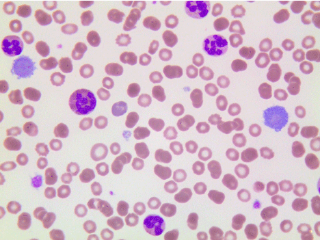
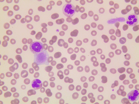
A 71-year-old male diagnosed with MDS/MPD NOS three months before, was hospitalized with increasing leukocyte count (90x109/L) and low platelets (67x109/L). The blood smear showed extraordinary large and giant platelets (Figures 1 and 2). Bone marrow aspirate indicated no increased blast count. A few days later the patient suffered a subdural hematoma and died several weeks later.
Under normal conditions, giant platelets (8-20 μM in diameter) are only seen in blood smears of (premature) neonates.
They can also be present in several rare inherited diseases with thrombocytopenia such as Bernard-Soulier syndrome and May- Hegglins anomaly which usually demonstrate a uniformly increased platelet volume.
More frequently, large platelets are found in enhanced sequestration (e.g. ITP, TTP) and in myeloproliferative and myelodysplastic disorders.
The elevated volume of these giant platelets may cause spurious counts of leukocytes (falsely increased) and platelets (falsely decreased). Especially the latter can be of clinical importance in case of severe thrombocytopenia.

Figure 1: Giant Platelets 1.
