Case Presentation
Ann Hematol Oncol. 2019; 6(9): 1268.
Electronic Medical Records “Cut and Paste”: A Cautionary Tale
Adam T and Moylan E*
Department of Medical Oncology, Liverpool Hospital, Australia
*Corresponding author: Moylan E, Department of Medical Oncology, Liverpool Hospital, Elizabeth St, Liverpool, Sydney, NSW 2170, Australia
Received: October 15, 2019; Accepted: November 01, 2019; Published: November 08, 2019
Abstract
A case is presented of a 66 year old man following a first seizure, on a background of Chronic Lymphocytic Leukaemia (CLL) and hypogammaglobulinaemia treated with ibrutnib and Intravenous Gammaglobulin (IVIg). An excised melanoma in-situ was incorrectly documented in the patient medical records as “melanoma”. This incorrect diagnosis was propagated throughout the patient’s electronic medical record and was included in the referral for diagnostic imaging. Computed Tomography (CT) scan of the brain was performed and was reported to show multiple cerebral subcortical enhancing lesions with associated vasogenic oedema suggestive of metastases. The patient subsequently underwent neurosurgical intervention resulting in the diagnosis of invasive cerebral aspergillosis. This case highlights how incorrectly propagated information can seriously compromise optimal patient management.
Keywords: Invasive cerebral aspergillosis; Chronic lymphocytic leukaemia; CLL; Ibrutinib; Melanoma
Case Presentation
A 66 year old man presented with a first episode of seizure and mild expressive dysphasia. His medical background included a 6-year history of Chronic Lymphocytic Leukaemia (CLL) currently on Ibrutinib and hypogammaglobulinaemia on (IVIg), peripheral vascular disease, hypertension and asthma. He also had an abnormal skin lesion excised from the upper back 18 months earlier. Histopathology confirmed melanoma in-situ with clear surgical margins. Due to the incorrect propagation of information including multiple episodes of “cutting and pasting” of the medical history, this was documented on many subsequent occasions in the patient’s clinical records as being “melanoma”.
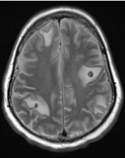
Upon presentation to hospital a Computed Tomography (CT) scan of the brain was performed; this showed multiple cerebral subcortical enhancing lesions with associated vasogenic oedema suggestive of metastases; the left frontal and parietal lesions were hyperdense in precontrast CT, raising the possibility of haemorrhagic metastases. The patient proceeded to have complete staging CT scans of the chest, abdomen and pelvis; these showed widespread lymphadenopathy and splenomegaly consistent with a known diagnosis of CLL, an incidental finding of an enlarged thyroid gland, degenerative changes at the C5-6 vertebrae with associated moderate canal and neural exit foramen narrowing, right upper lobe of lung subsegmental collapse/consolidation, a 5mm pulmonary nodule and subtle bony lytic lesions in both iliac wings and the L4 vertebral body. He went on to have a Magnetic Resonance Imaging (MRI) scan of the brain (Figure 1) which echoed the cerebral CT findings. As the history of melanoma was incorrectly provided, metastatic melanoma was thought to be the most likely aetiology of many of the reported scan abnormalities.

    Figure 1: T2 MRI sequence showing multiple subcortical rim enhancing
lesions with surrounding vasogenic oedema.
Due to the uncertain diagnosis and ultimate correction of the incorrectly propagated history of invasive melanoma, he underwent neurosurgical intervention (temporo-parietal craniotomy). An abnormal area of tissue was identified, described as “capsule-like” with egress of purulent material under high pressure upon manipulation. Based on the appearance of the abnormality, the differential diagnosis of an abscess versus necrotic tumour was considered and frozen samples were taken and sent to the laboratory for microscopy and histology.
Microscopy showed fungal elements and aspergillus fumigatus complex was later isolated from the tissue samples. The infectious diseases team were consulted and the patient was commenced on the appropriate anti-fungal treatment (Voriconazole).
Discussion
One of the issues that this case highlights is how inappropriate propagation of information through “cutting and pasting” patient information can markedly alter patient management.
In this particular case, the patient had a melanoma in-situ excision and re-excision with clear surgical margins had been performed, in keeping with recommended guidelines. The prognosis of melanoma in-situ is excellent with a very low risk of loco-regional or metastatic disease recurrence [1,2]. However, at some point in time it was incorrectly documented in the patient’s electronic medical records that he had a diagnosis of melanoma. This incorrect diagnosis continued to be copied and pasted by different medical practitioners over several months and thus became part of the patient’s “past medical history”. The natural history of melanoma and the risk of brain metastases from melanoma is related to disease stage as well as well as other prognostic factors [3].
When a patient aged in their 60s presents with a new episode of seizure the differential diagnosis is quite broad and can include; space occupying lesion (benign, malignant), acute ischaemic or haemorrhagic stroke, subdural haematoma, brain abscess, concurrent medical illness, metabolic disturbance, medication-related, substance abuse or as a first presentation of epilepsy. The initial work-up of blood investigations and a CT scan of the brain are routine and can narrow down the differential diagnosis. The radiologist interpreting the scans relies on the clinical history provided as well as the radiological appearance and characteristics of the abnormality to guide them in making their assessment. In this case, an incorrectly documented history of melanoma would have influenced the radiologist into thinking that metastatic melanoma was a likely diagnosis. The neurosurgeon that was initially consulted made a decision not to proceed with neurosurgical intervention based on the incorrect history of invasive melanoma. Only a thorough review of the previous histological reports enabled a broader differential diagnosis to be entertained and a diagnostic neurosurgical procedure was performed.
In this case, the focus on metastatic melanoma clouded the actual diagnosis and delayed neurosurgical intervention. Given the long-term immunosuppression related to chronic lymphocytic leukemia and Ibrutinib therapy, the risk of invasive fungal infections such as Aspergillosis should have been a diagnosis of exclusion [4]. Clearly, omitting this from the differential diagnosis on the basis of misinformation could have had catastrophic implications for the patient given the significant morbidity and mortality associated with this disease [5], especially if the appropriate anti-fungal treatment had not been promptly commenced.
References
- Bichakjian C, Halpern A, Johnson T. Guidelines of care for the management of primary cutaneous melanoma. J Am Acad Dermatol. 2011; 65: 1032.
- Balch C, Buzaid A, Soong S. Final version of the American Joint Committee on Cancer staging system for cutaneous melanoma. J Clin Oncol. 2001; 19: 3635-3648.
- Balch C, Gershenwald J, Soong S. Final version of 2009 AJCC melanoma staging and classification. J Clin Oncol. 2009; 27: 6199.
- Arthurs B, Wunderle K, Hsu M. Invasive aspergillosis related to ibrutinib therapy for chronic lymphocytic leukaemia. Respir Med Case Rep. 2017; 21: 27-29.
- Marzolf G, Sabou M, Lannes B. Magnetic Resonance Imaging of Cerebral Aspergillosis: Imaging and Pathological Correlations. PLoS One. 2016; 11: e0152475.