Clinical Image
Ann Hematol Oncol. 2020; 7(6): 1305.
Histoplasma Capsulatum in Peripheral Blood Smear
Priti Chatterjee*, Piali Mandal, Sunita Sharma and Jagdish Chandra
Department of Pathology, Lady Hardinge Medical College, Delhi University, India
*Corresponding author: Priti Chatterjee, Department of Pathology, Lady Hardinge Medical College, Delhi University, India
Received: May 21, 2020; Accepted: June 30, 2020; Published: July 07, 2020
Clinical Image
A 7 year old female patient, known case of B cell ALL on maintenance phase therapy presented with chief complaints of fever and bleeding per rectum for one week.
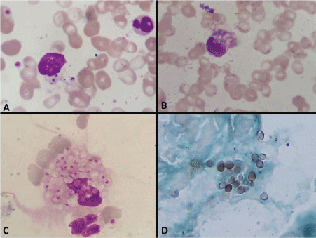
On clinical examination the patient was febrile and had pallor. There was bilateral cervical lymphadenopathy, hepatomegaly and petechiae. Laboratory investigations showed haemoglobin of 9.3 gm/dl, total count of 3500 cells/mm3 and platelet count of 38,000/ uL. The peripheral blood smear showed thrombocytopenia and normocytic normochromic anemia. Few monocytes showed presence of intracellular yeast like organisms measuring 2-4u which were surrounded by a pseuducapsule. Multiple phagocytic vacuoles were also present in the monocytes. Occasional extracellular organism was also seen. These features were morphologically compatible with histoplasma capsulatum. Bone marrow aspirate also showed presence of similar organisms which were positive for PAS and Silver stains. Hence a diagnosis of disseminated histoplasmosis was made. It is extremely rare to see histoplasma in peripheral blood smears (Figure 1).

    Figure 1: A: Monocyte with yeast form of histoplasma in the cytoplasm
in peripheral smear. Wright-Giemsa 1000x. B: Histoplasma along with
phagocytic vacuoles in a monocyte. Wright-Giemsa 1000x. C: Bone marrow
aspirate with intracellular yeast forms of Histoplasma. Wright-Giemsa 1000x.
D: Bone marrow aspirate with histoplasma. GMS stain 1000x