Case Presentation
Austin J HIV/AIDS Res. 2016; 3(2): 1024.
Classic Kaposi’s Sarcoma in a 75-Year-Old Woman
Puente Martín M De la1, Guarino MF2, Garde JB2 and Gómez-Pavón J1*
1Geriatrics Department, Central Hospital of the Red Cross, Madrid, Spain
2Dermatology Unit, Central Hospital of the Red Cross, Madrid, Spain
*Corresponding author: Javier Gómez Pavón, Geriatrics Department, Central Hospital of the Red Cross, Madrid, Spain
Received: March 28, 2016; Accepted: April 18, 2016; Published: April 19, 2016
Case Presentation
Kaposi’s Sarcoma (KS) is a systemic disease of predominantly cutaneous involvement, due to the proliferation of endothelial cells stimulated by various cytokines, which occurs as a result of infection by human herpes virus 8 (HHV8) together with other immune and environmental factors. First described in 1872, the reported cases are scarce until the emergence of the HIV [1] pandemic, when its incidence increased exponentially. There are currently four clinical forms of KS recognized: Classical KS (typical appearance over 60 years of age), endemic KS, KS and KS iatrogenic associated with HIV.
We present the case of a 75-year-old woman who was admitted to the acute geriatric unit proceeding from the nursing home where she had lived for the last three years for a biopsy of skin lesions of indeterminate evolution in time and respiratory infection associated with bronchospasm. Personal history consists of hypertension, dyslipidaemia, morbid obesity, and ischemic heart disease of AMI type nine years ago, chronic lung disease, long-standing depressive disorder, degenerative Alzheimer’s dementia in a stage (Global Deterioration Scale 5) and stenosis of the lumbar canal. The baseline situation is severe dependence of ABVDs (Barthel Index 10/100) and mild cognitive impairment (GDS 5). She follows usual treatment with digoxin, omeprazole, dicoumarin, furosemide, acetaminophen, trazodone, bupropion, and inhaled bronchodilators.
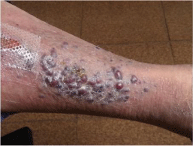
At admittance, some signs stand out at physical examination: prolonged expiration with tachypnea and wheezing in both lung fields. Right lower limb presents lilaceouspapular lesions (Figure 1a). Both analytical, with blood count, basic biochemistry with ions, liver enzymes, vitamin B12, TSH and C-reactive protein, such as chest X-ray showed no significant alterations and HIV serology was negative.

    Figure 1a:  Macules and papules isolated and grouped purplish red hue in
the right leg.
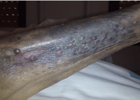
During admission, systemic antibiotic therapy and bronchodilator therapy with intravenous methyl prednisolone was prescribed, in descending pattern, with resolution of respiratory symptoms. The biopsy of skin lesions showed proliferation of spindle cells arranged in intersecting beams and forming vascular slits, occupied by red blood cells; Immunohistochemistry was positive for CD31, CD34, factor VIII, D2-40 and HHV-8. At discharge she returned to her habitual nursing home. After discussion of the case with the oncology department, expectant treatment was initially decided given comorbidity and functional status of the patient. Reevaluated one month later, in which no topical or systemic treatment was administered, a greater involvement was found in the right lower limb with progression to the contra lateral lower limb, without other symptoms or injuries or notable impact on overall health. (Figure 1b).

Figure 1b: Progression of injuries of the previous image with the presence of hematic extravasation and involvement of both lower limbs.
In the elderly population the classic KS is the one which typically appears; more frequently in men and Mediterranean population of Eastern Europe or their descendants [2]. In Sardinia and Sicily it is found the highest incidence rate, while in Spain the rate is intermediate compared to other countries, with an estimate of four cases per million inhabitants [3]. These geographical differences correspond to differences in the prevalence of HHV-8 infection [4].
KS etiology is complex; being the main causes HHV8 infection (necessary but not sufficient) and some degree of immunosuppression in the patient [5]. In a study in Italy, it was found a strong and independent association with diabetes and the use of oral corticosteroids [6]. Tobacco has also been pointed as a protective factor against disease [7].
Clinically, classic KS is characterized by the appearance of macules, papules, nodules and purple plaques, red-blueish, typically in the lower limbs, of varying size and number and that can ulcerate and bleed. Visceral involvement in classic KS is estimated in less than 10%, below that found in KS associated with HIV, although some series indicates, however, that the involvement occurs more frequently, although it has important clinical and prognostic impact [8-9].
The course of classic KS is usually benign, being rarely a cause of death. There is no consensus on what the optimal treatment is; there are local therapies (mainly radiotherapy, with good results, and alternatives such as cryotherapy and surgery) and systemic (chemotherapy along with experimental therapies) with mixed results, and whose indication has been extrapolated from KS treatment of HIV.
Therefore, given the characteristics of the elderly, the fact that treatments are not without side effects (eg, alopecia, gastrointestinal symptoms and myelosuppression, in the case of some chemotherapeutic agents) and usually indolent course of the disease, it is frequent to consider the possibility of the monitoring of therapeutic abstention injuries [10], such as this patient, who remains clinically and functionally stable, with progression of skin lesions but no evidence of involvement at other levels. In any case the therapeutic decision will be individualized and will be determined by comorbidity and functional status.
References
- Wabinga HR, Parkin DM, Wabwire-Mangen F, Mugerwa JW. Cancer in Kampala, Uganda, in 1989-91: changes in incidence in the era of AIDS. Int J Cancer. 1993; 54: 26-36.
- Kaldor JM, Coates M, Vettom L, Taylor R. Epidemiological characteristics of Kaposi's sarcoma prior to the AIDS epidemic. Br J Cancer. 1994; 70: 674-676.
- Parkin DM, Whelan SL, Ferlay J, Raymond L, Young J. Cancer incidence in five continents. IARC Scientific Pub. No. 143. Lyon: International Agency for research on Cancer. 1997.
- Angeloni A, Heston L, Uccini S, Sirianni MC, Cottoni F, Masala MV, et al. Age-specific seroprevalence of Human Herpesvirus 8 in relatives of patients with classic Kaposi’s sarcoma from Sardinia. J Infect Dis. 1998; 177: 1715-1718.
- Iscovich J, Boffetta P, Franceschi S, Azizi E, Sarid R. Classic kaposi sarcoma: epidemiology and risk factors. Cancer. 2000; 88: 500-517.
- Anderson LA, Lauria C, Romano N, Brown EE, Whitby D, Graubard BI, Li Y. Risk factors for classical Kaposi sarcoma in a population-based case-control study in Sicily. Cancer Epidemiol Biomarkers Prev. 2008; 17: 3435-3443.
- Goedert JJ, Vitale F, Lauria C, Serraino D, Tamburini M, Montella M, et al. Risk factors for classical Kaposi's sarcoma. J Natl Cancer Inst. 2002; 94: 1712-1718.
- Cottoni F, Masala MV, Piras P, Montesu MA, Cerimele D. Mucosal involvement in classic Kaposi's sarcoma. Br J Dermatol. 2003; 148: 1273-1274.
- Kolios G, Kaloterakis A, Filiotou A, Nakos A, Hadziyannis S. Gastroscopic findings in Mediterranean Kaposi's sarcoma (non-AIDS). Gastrointest Endosc. 1995; 42: 336-339.
- Di Lorenzo G. Update on classic Kaposi sarcoma therapy: new look at an old disease. Crit Rev Oncol Hematol. 2008; 68: 242-249.