
Case Report
Austin J Infect Dis. 2022; 9(2): 1067.
Presentation of Clinical Cases of Rabbit Viral Hemorrhagic Disease Type 2 in Rabbit Units in Havana
Reyes MP*
Department of Epidemiology, National Center for Animal and Plant Health, Cuba
*Corresponding author: M Peláez Reyes, Department of Epidemiology, National Center for Animal and Plant Health, Cuba
Received: July 02, 2022; Accepted: August 01, 2022; Published: August 08, 2022
Abstract
This work shows the study of clinical cases of Rabbit Viral Hemorrhagic Disease type 2, which showed symptoms and lesions never present before in Cuba.
Keywords: Rabbit viral hemorrhagic disease; Mortality; Lethality; Septicemia
Case Presentation
Rabbit Viral Hemorrhagic Disease (RHCV), also called viral septicemia or Chinese plague, is a highly contagious and in most cases fatal disease; it is an internationally notifiable disease, as established by the World Organization for Animal Health (OIE) in the Terrestrial Animal Health Code [2].
What could be considered the great crisis for the rabbit was the appearance of the Viral Hemorrhagic Disease. This disease was first described in China in 1984. The disease soon spread throughout Europe (France, Spain, Portugal, Italy, Austria and the British Isles, among others), Asia (China, Korea), Africa and the Middle East (Tunisia, Saudi Arabia, and Israel) and America (United States and Mexico). In 1993, it affects Cuba. On the other hand, in 2010 an antigenic variant of the virus was described, which has been called RHDV type b, and which shows significant differences with the previously described groups, also known as type 2 [3].
This is an extremely contagious and often fatal disease for lagomorphs caused by the RHDV virus. It is caused by a member of the Lagovirus genus and the Caliciviridae family. Serotype 2 or RHDV2 (identified in France in 2010) has almost 75-80% lethality in a period of two to three days in animals from 10-15 days of age. Death is the result of generalized circulatory dysfunction associated with disseminated intravascular coagulation and necrotizing hepatitis lesions, the liver being considered the main site of virus replication. Signs of the disease are often not seen until sudden death and a bloodstained nose occur, caused by internal bleeding associated with liver necrosis. Infected animals may also develop a fever, lose their appetite, be nervous, or have rapid breathing, resulting in congested and swollen lungs [4].
According to Cima, G. [1], research indicates the increasing mortality rates in Europe and the results of a recent study suggest that RHDV-2 has become more lethal. In one study, experimental infection of New Zealand white rabbits showed similar pathogenicity between RHDV-2 and RHDV strains, killing 70% to 90% of susceptible adult rabbits. In addition, he reports that the disease, whether due to classic strains of RHDV or RHDV-2, often kills rabbits with no.
Outward signs of the disease, and often the only signs of the disease are sudden death and blood-stained noses caused by internal bleeding,” states the APHIS information. “Infected rabbits may also develop a fever, be reluctant to eat, or show respiratory or nervous signs.”
Case History
In the month of June of the year 2021, Cuba reports the involvement of the Rabbit Viral Hemorrhagic Disease Type 2 for the first time, since in previous years it was affected by Type 1. Nine provinces from all regions of the country were affected. The main effects were caused by the mortality that ranged from 20 to 80 percent and the stamping-out carried out to stop the progression of the disease.
From the clinical point of view, a strong terminal shriek was observed in general, quickly followed by collapse and death (Figure 1); a lot of bleeding in the cavities of the chest and abdomen (Figure 2); The necropsy showed hepatic involvement (Figure 3), as well as intense hemorrhages in various internal organs (Figure 4 and 5). Unlike the clinical cases observed for type 1, in type 2 no bleeding from the nose was observed. It should be noted that the disease was confirmed by the Real Time Polymerase Chain Reaction (RT-PCR) technique, at the National Center for Agricultural Health (CENSA) and its sequencing.
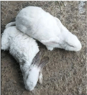
Figure 1:

Figure 2:

Figure 3:
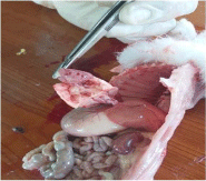
Figure 4:
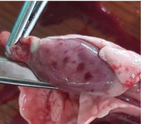
Figure 5:
References
- Cima G. Rabbit hemorrhagic disease virus serotype 2 spreading among wild rabbits, hares. JAVMA News. 2020.
- OIE. CAPÍTULO 13.2. ENFERMEDAD HEMORRÁGICA DEL CONEJO. Código Sanitario para los Animales Terrestres. 2019.
- Sonsoles Pacho, Elías Dahdouh, Mónica Suárez. SANIDAD Y BIOSEGURIDAD, Enfermedad hemorrágica Viral del conejo. 2018.
- THERYA. La enfermedad hemorrágica viral del conejo impacta a México y amenaza al resto de Latinoamérica. 2020: 11.