Research Article
Austin J Med Oncol. 2014;1(2): 4.
Applications of a Capacitor-Based Respiratory Position Sensing Device: Implications for Radiation Therapy
Weng Y1, Westover MB2, Speier C3, Sharp G3, Bianchi MT2 and Westover KD4*
1UT Southwestern Medical School, Dallas, TX, USA
2Department of Neurology, Massachusetts General Hospital, Boston, MA, USA
3Division of Radiation Physics, Department of Radiation Oncology, Massachustets General Hospital, Boston, MA, USA
4Department of Radiation Oncology, UT Southwestern Medical School, Dallas, TX, USA
*Corresponding author: Westover, KD, UT Southwestern Medical Center, 5323 Harry Hines Blvd, L4.268, Dallas, TX, USA, Tel: 75390-9038
Received: August 26, 2014; Accepted: October 22, 2014; Published: October 24, 2014
Abstract
Respiratory motion may significantly affect the outcome in a number of medical imaging techniques and some radiation therapy applications. 4-dimensional computed tomography (4DCT) and respiratory gating technology, which account for the dynamics of respiration, are expensive and often unavailable in smaller radiation treatment centers. Here we evaluate the ability of an inexpensive, technology comprised of two capacitors placed next to the skin to provide real-time respiratory phase information. Three subjects were simultaneously monitored by the new capacitor-based device (CBD) and a commercially available Real time Position Management (RPM) system by Varian. All respiratory phases detected by the RPM system were also detected by the CBD. Automatically detected peaks were not significantly different in timing when comparing RPM and CBD-derived respiratory amplitudes. The anatomic locations of the CBD were varied to evaluate the change in signal quality across the abdomen and thorax. CBD signals were reliable on the abdomen and lower thorax but degraded when recorded from the upper thorax. We also used computed tomography (CT) to assess the imaging characteristics of CBD and found that there were minimal artifacts. We therefore conclude that CBD respiratory amplitude measurements may be useful for tracking respiratory movements as part of a number of advanced radiation therapy technologies including 4DCT image resorting, adaptive radiation therapy and gated radiation therapy.
Keywords: Respiratory gating; 4DCT; RPM
Introduction
Measuring respiratory phase is important in several types of medical imaging, especially in circumstances where respiratory motion can degrade image quality, or when treatment optimization requires real-time knowledge of respiratory phase. Examples include 4-dimensional computed tomography (4DCT), gated positron emission tomography (Gated PET) and several MRI-based methods [1–4]. Respiratory phase information is also commonly used when delivering gated therapeutic radiation to minimize radiation treatment volumes and enable dose escalation [5,6].
Several systems have been developed to acquire respiratory information including the real time position management (RPM) system and Vision RT system [7,8]. The widely used RPM system utilizes an infrared marker affixed to the supine patient’s abdomen which rises and falls with respiration. The marker is detected by a camera mounted near the patient’s feet and respiratory amplitudes are extracted in real time by automated image processing algorithms. The Vision RT system consists of a camera mounted above the patient couch and relies on extraction of visual cues from rapid sequence imaging of the patient’s thorax surface to construct respiratory amplitudes. In both of these systems, tumor motion observed in rapid sequence imaging has been shown to correlate with respiratory output from these systems [7,9]. Thus, radiation oncologists routinely use real-time respiratory effort information when designing radiation plans that are expected to be impacted by respiration.
There is also now interest in using multiple respiratory signal inputs to improve tumor motion or deformation models. The RPM system is not designed to provide multiple inputs. The Vision RT system may be used to provide multiple respiratory signals but is complex, relying on real time image processing. Capacitor-based technology has long been used to sense changes in position and to obtain motion information because physical stresses on a capacitor alter its capacitance due to geometric changes. In this study, we employed a capacitor-based medical device (CBD) to monitor the patient’s chest wall position during respiration. Two versions of the CBD were used: 1) a lycra shirt with two small, adjacent capacitors supported by a printed vinyl layer (Figure 1a); 2) the capacitors were adapted to fit on an adhesive bandage allowing for placement of the sensor on multiple locations of the body (Figure 1b). The change of capacitance over time has been shown to correlate with the respiratory cycle. The advantage that CBD has over the two existing methods discussed previously is that it is inexpensive and utilizes an electronic readout, which is computationally inexpensive to analyze. This enables multiple respiratory signals monitoring via the strategic and simultaneous placing of CBD sensors in different anatomic locations.
Capacitor-based respiratory sensors.
(A) Two co-planar capacitors embedded in vinyl are printed onto a lycra shirt. The change in capacitance can be detected using electronics (not shown) attached to leads that end at the bottom of the shirt.
(B) The same general approach as in panel A was adapted to an adhesive bandage platform.
Figure 1:Capacitor-based respiratory sensors.
(A) Two co-planar capacitors embedded in vinyl are printed onto a lycra shirt. The change in capacitance can be detected using electronics (not shown) attached to leads that end at the bottom of the shirt.
(B) The same general approach as in panel A was adapted to an adhesive bandage platform.
Here we present an analysis of respiratory signals obtained from the CBD and directly compare them to respiratory signals simultaneously obtained using the Varian RPM system. The appropriateness of the CBD signal for use in 4DCT image resorting is also assessed.
Material and Methods
Capacitor-based medical device (CBD)
We employed two versions of the CBD developed by Nyx Devices in Boston, MA. One utilized a lycra shirt as the support matrix while the other used an adhesive bandage. For both, the capacitors consisted of a layer of silver amalgam deposited onto the fabric surface and this was then covered by a layer of vinyl. Leads were connected to custom circuitry which detected the capacitance at a rate of 5 Hz.
Respiratory measurements
Three healthy volunteers were tested in the current study. All were male in their 20’s or 30’s with BMI < 25. Subjects did not receive training or instruction prior to respiratory signal measurements. Subjects were placed on a CT simulator equipped with a Varian RPM system. Subjects were simultaneously outfitted with a CBD imprinted shirt from Nyx Devices (capacitors located over the right costal margin) and an RPM infrared reflector box on the upper abdomen. Respiratory information from both devices was simultaneously recorded for approximately 5 minutes at a rate of 30 Hz for the RPM trace and 5 Hz for the CBD. Subjects were instructed to breathe normally except at an arbitrary time point during each recording where they were asked to take a series of short rapid breaths, hold their breath, and then take several more short rapid breaths before resuming normal breathing. For one subject respiratory data was also measured with a small adhesive CBD placed sequentially at each of 3 locations: the mid abdomen at the level of the navel, costal margin and mid thorax adjacent to the right nipple.
Imaging
The shirt version and the adhesive bandage version of the CBD were imaged with a General Electric Light Speed RT 16-Slice CT simulator at a 2.5 mm slice thickness. The CBD shirt was placed on a foam phantom and the adhesive bandage version was placed in a small water bath at the time of imaging.
Peak detection algorithm
- All signal processing was performed in Matlab (MathWorks, Natick, MA). To enable direct comparison with the RPM data, we linearly interpolated the CBD data so that the time interval between data points would be the same for both the RPM and the CBD datasets while preserving the integrity of the latter. The datasets were baseline corrected and then aligned point-by-point based on optimization of the root mean squared deviation (RMSD). Peaks were automatically detected using a simple recursive algorithm, which was designed to process data sequentially using a sliding window. A pseudo code and an executable Matlab code is provided in Supplementary Information, but a summary is provided belowData is processed sequentially across time. A sliding window average across 0.25 sec is used to determine if there is an upward or downward trend. Typically, the min value (which keeps track of local minimum values) is updated for a downward trend and the max value (which keeps track of local maximum values) are updated for an upward trend.
- The presence of a trough and a possible peak is assumed if a downward-to-upward change is detected. The max value is updated until the apex of the peak is reached (or upward-to-downward change is detected).
- The peak is recorded if the difference between the apex and trough exceeds an empirically determined threshold.
- Thereafter, the min value is updated until a trough is reached (or downward-to-upward change is detected).
- This process is repeated until the entire data is analyzed.
Although our data processing has been done on Matlab, this algorithm could be easily implemented on a data acquisition platform such as NI Labview for simultaneous data acquisition and processing in real-time.
Results
Variability of respiratory signals between subjects
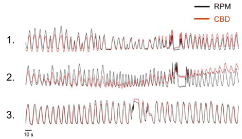
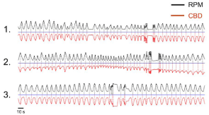
Figure 2 shows aligned respiratory traces from the RPM and CBD shirt systems for the three subjects. The RPM and CBD signals for subjects 1 and 3 showed exceptional qualitative agreement on visual inspection. The CBD signal for Subject 2 exhibited more noise and baseline drift than subjects 1 and 2, but inspiration and expiration were readily discernable and similar to the RPM signal. To better visualize the data in figure 2 for peak time locations, we have plotted the RPM and an inverted CBD trace in figure 3. An automated peak detection algorithm allowed for assignment of peak times. Calculation of the differences between CBD and RPM peaks revealed a per-cycle average difference in peak times of 22, 79 and 67 milliseconds for the three subjects. This corresponded to a 0.3%, 1.6% and 1.0% difference in timing between the CBD and RPM measurements within the respiratory cycle (see Table 1).
Comparison of respiratory traces from CBD shirt.
Superimposed respiratory traces from the CBD (red) and RPM systems (black) . RMS aligned respiratory traces from approximately 5 minutes of breathing activity are displayed from the three volunteers. Qualitative agreement between the locations of respiratory peaks (point of maximum amplitude) is apparent.Figure 2:Comparison of respiratory traces from CBD shirt.
Superimposed respiratory traces from the CBD (red) and RPM systems (black) . RMS aligned respiratory traces from approximately 5 minutes of breathing activity are displayed from the three volunteers. Qualitative agreement between the locations of respiratory peaks (point of maximum amplitude) is apparent.
Comparison of automatically detected peaks.
Baseline corrected, aligned and normalized traces from the RPM (black) and CBD shirt (red) are shown for each of the three subjects. The CBD trace has been inverted to better visualize comparisons of peak locations between methods.Figure 3:Comparison of automatically detected peaks.
Baseline corrected, aligned and normalized traces from the RPM (black) and CBD shirt (red) are shown for each of the three subjects. The CBD trace has been inverted to better visualize comparisons of peak locations between methods.
    Subject
    No. CBD Peaks
    No. RPM Peaks
    Average difference in peak times (ms)
    Mean IPI (s)
    Percent difference RPM vs. CBD


    1
    49
    49
    21.8
    6.47
    0.34%


    2
    56
    56
    78.6
    4.76
    1.65%


    3
    39
    41
    -67.5
    6.47
    1.04%
Table 1: Variability of respiratory signals between subjects.
Variability of respiratory signal based on anatomical location of CBD
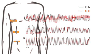
Simultaneous RPM and CBD respiratory traces were recorded for subject 2 using the adhesive bandage version of the CBD and the standard RPM setup. CBD signals were recorded with the CBD device at each of three anatomic locations as shown in Figure 4. The RPM setup was the same as in the between-subjects testing. RPM and CBD traces were aligned and superimposed. The RPM and CBD signal were visually similar when the CBD was on the abdomen and lower thorax but the signal degraded when the device was placed on the upper thorax.
CBD respiratory signal by anatomic location.
Superimposed traces from RPM (black) and CBD (red) systems where the location of the CBD was varied as shown (mid abdomen, costal margin, mid thorax). Signals are qualitatively similar for measurements taken at the abdomen and costal margin. However, the CBD signal becomes noisier on the thorax.Figure 4:CBD respiratory signal by anatomic location.
Superimposed traces from RPM (black) and CBD (red) systems where the location of the CBD was varied as shown (mid abdomen, costal margin, mid thorax). Signals are qualitatively similar for measurements taken at the abdomen and costal margin. However, the CBD signal becomes noisier on the thorax.
Imaging characteristics of the CBD
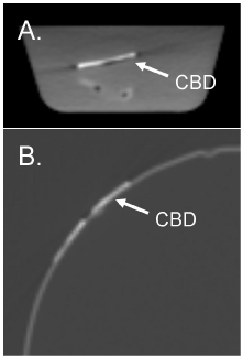
The CBD is comprised of two small capacitors composed of a layer of silver embedded in a vinyl support matrix. This sensor configuration can be mounted on either a shirt or adhesive bandage, as tested here. Both formats were imaged by CT to assess for device-related CT artifacts that might impact on volumetric radiation planning. The adhesive bandage version of the CBD was imaged while submerged in a small water bath so as to approximate a comparison between the CBD and tissue-equivalent material. Using standard body CT windowing (level = 40 HU, window = 400 HU) minimal imaging artifact was observed (Figure 5a). The shirt version of CBD was placed on a low density foam phantom and imaged. Using standard lung CT windowing (level = -600 HU, window = 1600 HU) minimal imaging artifact was observed (Figure 5b).
CT images of the CBD revealed minimal artifact.
(A) Adhesive bandage version of the CBD was immersed in a water bath and imaged by CT. The bandage is supported in the water by a small plastic stand. The CT windowing level approximates a typical setting for viewing abdominal CT images (level = 40 HU, window = 400 HU). (B) The lycra shirt version of the CBD was placed over a foam phantom and imaged by CT. CT windowing approximates typical settings for viewing thoracic CT images (level = -600 HU, window = 1600 HU).Figure 5:CT images of the CBD revealed minimal artifact.
(A) Adhesive bandage version of the CBD was immersed in a water bath and imaged by CT. The bandage is supported in the water by a small plastic stand. The CT windowing level approximates a typical setting for viewing abdominal CT images (level = 40 HU, window = 400 HU). (B) The lycra shirt version of the CBD was placed over a foam phantom and imaged by CT. CT windowing approximates typical settings for viewing thoracic CT images (level = -600 HU, window = 1600 HU).
Conclusions
4DCT and respiratory gating technology are generally not available at smaller free-standing radiation therapy centers, yet approximately 1/3 of patients receiving radiation in the United States are treated in these settings. Respiratory monitoring with gating is routinely used at larger centers for treatment of mobile targets [5] and to reduce dose to nearby normal tissues as in the treatment of left-sided breast cancers [10]. The availability of an inexpensive system, such as the CBD, to gather respiratory phase information may allow more radiation therapy centers to offer these services to patients.
The CBD may also be useful as a research tool for tumor motion management. Accurate modeling of tumor deformation and displacement throughout the respiratory cycle may allow delivery of therapeutic radiation with smaller margins, which will spare normal tissues. While CBD respiratory signals were generally similar when comparing signals from the costal margin and abdomen, they did show differences on the upper chest. We speculate that respiratory signal differences across anatomical locations may be useful for construction of multi-parametric tumor motion models, although proving this is beyond the scope of this study. If feasible, these models would be useful in radiation delivery techniques where radiation treatment tolerances are tight and, therefore, motion management is important.
These data demonstrate that a simple device composed of two small capacitors placed next to the skin at an appropriate anatomic location can produce reliable respiratory effort amplitudes similar in quality to that obtained from the commercially available RPM system. Because the temporal location of respiratory peaks measured by the CBD and RPM systems generally showed an error of ~1%, differences in phase assignment between the two methods are minimal. Thus for the purposes of image resorting in construction of 4DCT data sets and for respiratory gating at the time of therapeutic radiation delivery, signals recorded from CBDs placed on the abdomen or costal margin are interchangeable with the those derived from the RPM system.
CT imaging of the CBD showed few CT artifacts. It is anticipated that a production model of the device will use less high-z material and should therefore produce even less x-ray scatter. Thus, the CBD would likely not interfere with CT from an imaging perspective. However, the reliability of the CBD system during radiation exposure will need to be tested to ensure that the electronic components of the CBD can withstand exposure. Indeed, diagnostic and therapeutic energies will need to be considered separately.
Conflicts of Interest Notification
MTB is listed as an inventor on the provisional patent for the capacitor-based device evaluated in this manuscript. All other authors have no conflicts of interest relating to the work reported in this manuscript.
References
- Bailes DR, Gilderdale DJ, Bydder GM, Collins AG, Firmin DN. Respiratory ordered phase encoding (ROPE): a method for reducing respiratory motion artefacts in MR imaging. J Comput Assist Tomogr 1985; 9: 835-838.
- Nehmeh SA, Erdi YE, Pan T, Pevsner A, Rosenzweig KE, et al. Four-dimensional (4D) PET/CT imaging of the thorax. Med Phys 2004; 31: 3179-3186.
- Nehrke K, Börnert P, Manke D, Böck JC. Free-breathing cardiac MR imaging: study of implications of respiratory motion--initial results. Radiology 2001; 220: 810-815.
- Rietzel E, Chen GTY, Choi NC, Willet CG. Four-dimensional image-based treatment planning: Target volume segmentation and dose calculation in the presence of respiratory motion. Int J Radiat Oncol Biol Phys 2005; 61:1535-1550.
- Keall PJ, Mageras GS, Balter JM, Emery RS, Foster KM, et al. The management of respiratory motion in radiation oncology report of AAPM Task Group 76. Med Phys 2006; 33: 3874-3900.
- Wagman R, Yorke E, Ford E, Giraud P, Mageras G, et al. Respiratory gating for liver tumors: use in dose escalation. Int J Radiat Oncol Biol Phys 2003; 55: 659-668.
- Hughes S, McClelland J, Tarte S, Lawrence D, Ahmad S, et al. Assessment of two novel ventilatory surrogates for use in the delivery of gated/tracked radiotherapy for non-small cell lung cancer. Radother Oncol 2009; 91: 336-341.
- Pan T, Lee TY, Rietzel E, Chen GTY. 4D-CT imaging of a volume influenced by respiratory motion on multi-slice CT. Med Phys 2004; 31: 333-340.
- Ford EC, Mageras GS, Yorke E, Rosenzweig KE, Wagman R, et al. Evaluation of respiratory movement during gated radiotherapy using film and electronic portal imaging. Int J Radiat Oncol Biol Phys 2002; 52: 522-531.
- Pedersen AN, Korreman S, Nyström H, Specht L. Breathing adapted radiotherapy of breast cancer: reduction of cardiac and pulmonary doses using voluntary inspiration breath-hold. Radother Oncol 2004; 72: 53-60.


