Mini Review
Austin Med Sci. 2016; 1(3): 1012.
Cardiac Function and Hemodynamics Assessed by Speckle Tracking Echocardiography: Evolution from Structure to Function
Ryuhei Tanaka*
Department of Cardiology, Murakami Memorial Hospital, Japan
*Corresponding author: Ryuhei Tanaka, Department of Cardiology, Murakami Memorial Hospital, 3-23 Hashimoto Gifu 500-8523, Gifu Prefectural General Medical Center and Gifu Heart Center, Japan
Received: August 29, 2016; Accepted: September 20, 2016; Published: September 21, 2016
Abstract
Echocardiography provides information regarding cardiac structure and function and is the useful cardiovascular examination. Evolution of echocardiography has been performed including that of M-mode, twodimensional, Doppler, stress, transesophageal, contrast, three-dimensional, intracardiac and Speckle Tracking Echocardiography (STE).
Using STE, time-Left Atrial (LA) volume curve is easily obtained and LA phasic volume and function can be measured. It is reported that a novel index named KT index obtained from the time-LA volume curve using STE accurately predicts pulmonary capillary wedge pressure. This novel index may have incremental utility and discriminative value in routine clinical practice.
The complex mechanics of the heart must be best represented by realtime one-beat 3- Dimensional STE (3D-STE) with high volume rates in the examination of LV performance including LV layer function because of cyclic changes of LV in multiple dimensions. LV systolic function including strain and Strain Rate (SR) and diastolic function including relaxation assessed by tau and stiffness may be noninvasively evaluated using 3D-STE.
The alterations of LV structure function and deformation including torsion associated with HTN, especially alterations of torsion in three myocardial layers, may be examined by 3D-STE. Therefore, cardiac function including torsion, tau and stiffness assessed by echocardiography, especially by STE, will be described in this short commentary.
Keywords: Speckle tracking echocardiography; Pulmonary capillary wedge pressure; Cardiac function
Abbreviations
AF: Atrial Fibrillation; EF: Ejection Fraction; E/e’: Ratio of transmittal inflow velocity to mitral annular tissue Doppler velocity; HF: Heart Failure; HHF: Hypertensive Heart Failure; IVRT: Isovolumic Relaxation Time; PCWP: Pulmonary Capillary Wedge Pressure; STE: Speckle Tracking Echocardiography; LA: Left Atrium; LAEF: Left Atrial Emptying Function; SR: Strain Rate; LV: Left Ventricle; MR: Mitral Regurgitation; 3D: Three-Dimension
Introduction
Echocardiography noninvasively provides information regarding cardiac morphology, function and hemodynamics and is the most frequently performed cardiovascular examination after electrocardiography and chest X-ray [1]. In 1953, Hellmuth Hertz started his practical work with medical ultrasound at university of Lund. The first clinical applications of M-mode echocardiography were concerned with the assessment of the mitral valve from the shapes of the corresponding waveforms [2]. A technique for estimating the size of the Left Atrium (LA), right and Left Ventricle (LV) was developed using pulsed reflected ultrasound [3,4]. Evolution of echocardiography has been performed in half a century including that of M-mode, two-dimensional, Doppler, stress, transesophageal, contrast, three-dimensional, intracardiac and Speckle Tracking Echocardiography (STE).
As the progression of echocardiography, not only cardiac structure but also cardiac function and hemodynamics have been examined [5-14]. Using STE, emerging attention has been brought to LA that is very complex due to close coupling with LV and affected by LV properties. LA volume and function are thought to reflect LV diastolic function and may serve as strong and useful predictors for cardiovascular outcomes. LV Ejection Fraction (EF) was usually used as an index of LV shortening parameter and systolic function, which is load dependent. The ratio of transmitral inflow velocity to mitral annular tissue Doppler velocity (E/e’) is usually used as an index of LV filling pressure and diastolic function, which has several limitations [15,16]. Therefore, echo parameters of LV systolic function that is less load dependent and diastolic function that has less limitation are needed. It is of importance to accurately evaluate the cardiac function and hemodynamics as well as structure in the treatment of cardiac diseases. In this short commentary, cardiac function assessed by echocardiography, especially by STE, is described.
LA Function Assessed by STE
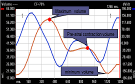
Using two- or Three-Dimensional STE (3D-STE), time-LA volume curve is easily and accurately obtained and LA phasic volume and function can be measured (Figure 1) [5-8, 10-14].

    Figure 1:  Representative image of the time-left atrial volume curve. Red lines
are the time-left atrial volume curves, blue lines are the dV/dt curves.
Hirose et al. demonstrated that reduced active LA Emptying Function (LAEF) (booster pump function) assessed by STE independently predicts the risk of new-onset Atrial Fibrillation (AF), suggesting a stronger association between LA functional remodeling and AF than between LA size and AF [5].
Kawasaki et al. showed that a novel index (KT index: log10 active LAEF / minimum LA volume index) obtained from the time-LA volume curve using STE predicts Pulmonary Capillary Wedge Pressure (PCWP) and that KT index is a more accurate and useful predictor of PCWP than E/e’, LA volume, function or other combinations of LA function and volume [10]. In their paper PCWP was estimated at 10.8 – 12.4xKT index. This novel index may have incremental utility and discriminative value in routine clinical practice [11].
Kawase et al. demonstrated that PCWP estimated by KT index was the useful and reliable echocardiographic parameter to predict PCWP in moderate to severe Mitral Regurgitation (MR) patients with sinus rhythm including both primary and secondary MR and might be also useful in MR patients with AF [12]. Thus, PCWP by KT index may have an incremental value in clinical setting to decide therapeutic strategy in MR because Heart Failure (HF) with MR increases.
Kawasaki et al. demonstrated that the elevated PCWP before AF ablation but not the enlarged LA size assessed by STE was the best predictor of AF recurrence after ablation, suggesting a strong relation between LV filling pressure and the progression of LA remodeling responsible for AF [13]. The echocardiographic parameter of PCWP assessed by KT index could be of interest and useful to improve candidate selection for successful outcomes after AF ablation.
Stratification of the risk of HF in patients as well as healthy subjects using PCWP is important for understanding when and why HF develops. Kawasaki et al. evaluated the impact of gender and healthy aging without hypertension on PCWP by KT index [11]. They reported that as age increases, E/A and E/e’ (markers of LV diastolic dysfunction) deteriorated to the same extent in males and females. PCWP was maintained due to compensation by an increase in active LAEF in both males and females. In contrast, the compensation for LV diastolic dysfunction by an increase in active LAEF in the females tended to be more gradual than males. The parameters that indicated LV diastolic dysfunction deteriorated with advancing age. The compensation in female septuagenarians and octogenarians were weaker than in male septuagenarians and octogenarians. This may explain why HF with preserved LVEF occurs more frequently in females than in males.
LV Systolic and Diastolic Function Assessed by STE
The heart is a complex mechanical organ that undergoes cyclic changes in multiple dimensions [17]. Therefore, the complex mechanics of the heart seem to be best represented by real-time 3D-STE with high volume rates in the examination of LV performance including LV layer function.
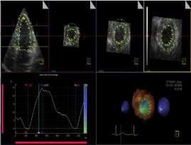
Saeki et al. reported that LV Strain and phasicstrain Rate (SR) at 3 myocardial layers could be measured using automated one-beat realtime 3D-STE with high volume rates [9] (Figures 2-4). They measured LV strain, SR during systole as an index of systolic function that was reported to be closely related to LV contractility, SR during Isovolumic Relaxation (IVR) as an index closely related to relaxation and E/e’ as an index of LV filling pressure in patients with Hypertension (HTN) and found that endocardial SR during systole in HTN with concentric and eccentric hypertrophy decreased compared with that in controls despite no reduction in both LVEF and epicardial SR. LV endocardial SR during IVR decreased even in normal geometry, and it was further reduced in concentric remodeling and hypertrophy despite no reduction in epicardial SR. LV phasic SR assessed by 3D-STE may be a useful index to detect early decreases in systolic function and to predict subclinical layer dysfunction in HTN.

    Figure 2:  Full volume acquisition and automated measurement of left ventricle
by 3D-STE with high volume rates. The upper panel shows the automatic
tracing of the endocardial and epicardial borders of a four slice display for fullvolume
acquisition: one apical four-chamber view and three short axis views.
The left lower panel shows the time-left ventricular strain curve.

    Figure 3:  Left ventricular phasic strain rate by 3D-STE. Left panel shows
strain rate at endocardium and right panel shows strain rate at epicardium.

    Figure 4:  The time-left ventricular torsion curve. Upper line shows timetorsion
curve at endocardium, mid line shows time-torsion curve at mid-wall
and lower line shows time-torsion curve at epicardium.
It is important to estimate PCWP and LV relaxation represented by time constant of LV pressure decline (Tau) in patients with heart disease. Tau measured by left heart catheterization is reported to be deteriorated even in the early stage of HTN. However, echocardiographic parameters to predict Tau are not yet elucidated. Kawasaki et al. reported that PCWP is accurately estimated by the novel KT index [10]. IVR Time (IVRT) was reported to be measured as the time between the end of LV outflow wave and the beginning of inflow wave by Doppler echo [18]. Thus, Tau by echo can be obtained using the formula: Tau = IVRT / (ln 0.9 x systolic blood pressure – ln PCWP) [19]. Kawamura et al. demonstrated that Tau estimated by the noninvasive method using STE has an excellent correlation with Tau obtained by cardiac catheterization (r=0.76, p<0.001) [20]. Bland-Altman analysis revealed a good agreement between Tau by echo and Tau by catheterization.LV relaxation represented by Tau obtained by invasive method may be noninvasively and accurately estimated by echocardiography in various cardiac diseases.
Furthermore, LV myocardial stiffness could be estimated as diastolic stress / strain [21] using 3D-STE [22], where diastolic stress is calculated as 0.334 x PCWP x LV end-diastolic dimension / {end-diastolic thickness (1 + end-diastolic thickness / end-diastolic dimension)} [23]. Ono et al. Demonstrated that LA strain and phasic function were decreased and LA phasic volume was increased in Hypertensive HF (HHF) associated with increased PCWP, LA and LV stiffness and LV diastolic stress and that the relation between LA properties and LV properties in HTN and HHF could be noninvasively assessed by STE [22].
LV systolic function including strain and SR and LV diastolic function including relaxation assessed by Tau and stiffness could be noninvasively evaluated using 3D-STE.
Lv Torsion as a Novel Systolic Function Assessed by 3d-Ste
LV deformation is characterized by torsion as well as longitudinal, radial and circumferential motion and LV torsional deformation was reported to be a sensitive index for LV performance, and thus provides a useful index of myocardial mechanics but difficult to measure [24]. LV is composed of three myocardial layers and the orientation of myofibers changes across LV wall from a right-handed helix in the subendcardium, circumferential orientation in the midwall to a left-handed helix in the subepicardium [25,26].
LV torsion was defined as a difference between apical and basal rotation divided by long axis length for every instant in time. LV torsion caused by inner and outer oblique muscle plays an important role in squeezing blood out of heart and may contribute a part of LVEF. LV outer oblique muscle that produces counterclockwise rotation plays a predominant role in torsion than the inner muscle that produces clockwise rotation [25]. Reduced subendocardial function could result in less endocardial opposition to the dominant epicardium and thus enhances torsion [25,27]. Therefore, LV remodeling such as hypertrophy, fibrosis and dilation caused by HTN may result in significant alterations of torsion. However, assessment of LV torsion by echocardiography has been methodologically challenging. Only tagged cardiac magnetic resonance provides accurate information on the slice rotation and the slice distance at the same time in clinical setting [25].
The prevalence of HF increases with age, and HTN that is one of the most important risk factors for HF also increases with age [27-29]. LV wall thickness is increased in response to elevated blood pressure as a compensatory mechanism to reduce LV wall stress in HTN [23,29]. However, alterations of LV structure, function and deformation including torsion associated with HTN, especially alterations of torsion in three myocardial layers, were not examined by 3D-STE [9,25]. Yoshizane and Minatoguchi et al. examined LV torsion in HTN with 3D-STE and found that LVEF in HHF decreased associated with LV dilation and reduced LV strain and SR at subepicardium and that LVEF in HTN with LVH was preserved accompanied with increased torsion despite reduced LV strain and SR at subendocardium [30]. LV torsion may therefore represent a compensatory mechanism to maintain adequate stroke volume despite reduced systolic function at subendocardium, suggesting that deterioration of torsion caused by the insult of outer oblique muscle and LV dilation may lead to HHF. Furthermore, LV rotational renewal like restore of twist or torsion has been recently demonstrated in a surgical series of patients affected by ischemic cardiomyopathy. These could be very important information of the usefulness of LV torsion in ventricles with impaired mechanics and give a new field of research and clinical application.
Conclusion
LA function, LV systolic function such as strain and SR that are closely related to contractility, diastolic function such as tau and stiffness and hemodynamics are able to be noninvasively estimated by novel 3D-STE. Evolution of echocardiography enables us to comprehensively and noninvasively evaluate not only cardiac structure but also cardiac function and hemodynamics. The use of the recently developed high frequency and volume rates 3D-STE systems may represent a new platform for ultrasound-based assessment in cardiac function with high temporal and spatial resolutions.
References
- Gowda RM, Khan IA, Vasavada BC, Sacchi TJ, Patel R. History of the evolution of echocardiography. Intern J Cardiol. 2004; 97: 1-6.
- Edler I, Rindstrom K. The history of echocardiography. Ultrasound Med Biol. 2004. 30. 1565-1644.
- Popp RL, Wolfe SB, Hirata T, Feigenbaum H. Estimation of right and left ventricular size by ultrasound: A study of the echoes from the interventricular septum. Am J Cardiol. 1969; 24: 523-530.
- Hirata T, Wolfe SB, Popp RL, Helmen CH, Feigenbaum H. Estimation of left atrial size using ultrasound. Am Heart J. 1969; 78: 43-52.
- Hirose T, Kawasaki M, Tanaka R. Left atrial function assessed by speckle tracking echocardiography as a predictor of new-onset non-valvular atrial fibrillation: results from a prospective study in 580 adults. Eur Heart J Cardiovasc Imaging. 2012; 13: 243-250.
- Kojima T, Kawasaki M, Tanak R. Left atrial global and regional function in patients with paroxysmal atrial fibrillation has already been impaired before enlargement of left atrium: velocity vector imaging echocardiography study. Eur Heart J Cardiovasc Imaging. 2012; 13: 227-234.
- Warita S, Kawasaki M, Tanaka R, Ono K, Kojima T, Hirose T, et al. Effects of pitavastatin on cardiac structure and function and on prevention of atrial fibrillation in elderly hypertensive patients. Circulation Journal. 2012; 76: 2755-2762.
- Onishi N, Kawasaki M, Tanaka R, Sato H, Saeki M, Nagaya M, et al. Comparison between left atrial features in well-controlled hypertensive patients and normal subjects assessed by three-dimensional speckle tracking echocardiography. Journal of Cardiology. 2014; 63: 291-295.
- Saeki M, Sato N, Kawasaki M, Tanaka R, Nagaya M, Watanabe T, et al. Left ventricular layer function in hypertension assessed by myocardial strain rate using novel one-beat real-time three-dimensional speckle tracking echocardiography with high volume rates. Hypertension Research. 2015; 38: 551-559.
- Kawasaki M, Tanaka R, Ono K, Minatoguchi S, Watanabe T, Iwama M, et al. A novel ultrasound predictor of pulmonary capillary wedge pressure assessed by the combination of left atrial volume and function: a speckle tracking echocardiography study. Journal of Cardiology. 2015; 66: 253-262.
- Kawasaki M, Tanaka R, Ono, Minatoguchi S, Watanabe T, Arai M, et al. Impacts of healthy aging and gender on pulmonary capillary wedge pressure estimated by the Kinetics-tracking index using two-dimensional speckle tracking echocardiography. Hypertension Research. 2016; 39: 327-333.
- Kawase Y, Kawasaki M, Tanaka R, Nomura N, Fujii Y, Ogawa K, et al. Noninvasive estimation of pulmonary capillary wedge pressure in patients with mitral regurgitation: a speckle tacking echocardiography study. Journal of Cardiology. 2015; 67: 192-198.
- Kawasaki M, Tanaka R, Miyake T, Matsuoka R, Kaneda M, Minatoguchi S, et al. Estimated pulmonary capillary wedge pressure assessed by speckle tracking echocardiography predicts successful ablation in paroxysmal atrial fibrillation. Cardiovascular Ultrasound. 2016; 14: 6.
- Nagaya M, Kawasaki M, Tanaka R, Onishi N, Sato N, Ono K, et al. Quantative validation of left atrial structure and function by two-dimensional and threedimensional speckle tracking echocardiography: a comparative study with three-dimensional computed tomography. Journal of Cardiology. 2013; 62: 188-194.
- Mullens W, Borowski AG, Curtin RJ, Thomas JD, Tang WH. Tissue Doppler imaging in the estimation of intracardiac filling pressure in decompensated patients with advanced systolic heart failure. Circulation. 2009; 119: 62-70.
- Firstenberg MS, Levine BD, Garcia MJ, Greenberg NL, Cardon L, Morehead AJ, et al. Relationship of echocardiographic indices to pulmonary capillary wedge pressures in healthy volunteers. J Am Coll Cardiol. 2000; 36: 1664- 1669.
- Abraham TP, Dimaano VL, Liang HY. Role of tissue Doppler and strain echocardiography in current clinical practice. Circulation. 2007; 116: 2597- 2609.
- Myreng Y, Smiseth OA. Assessment of left ventricular relaxation by Doppler echocardiography. Comparison of isovolumic relaxation time and transmitral flow velocities with time constant of isovolumic relaxtion. Circulation. 1990; 81: 260-266.
- Scalia GM, Greenberg NL, McCarthy PM, Thomas JD, Pieter M, Vandervoort, et al, Circulation. 1997; 95: 151-155.
- Kawamura I, Kawasaki M, Tanaka R. Non invasive estimation of time constant of left ventricular pressure decline as an index of relaxation by speckle tracking echocardiography: validation study by cardiac catheterization. 2016; 27-31.
- Mirsky I. Assessment of diastolic function: suggested methods and future considerations. Circulation. 1984; 69: 836-841.
- Ono K, Kawasaki M, Tanaka R. Relation between left atrial properties and left ventricular properties in hypertension: two- and three-dimensional speckle tracking echocardiography study. ESC. 2016; 27-31.
- Grossman W, Jones D, McLaurin LP. Wall stress and patterns of hypertrophy in the human left ventricle. J Clin Invest. 1975; 56: 56-64.
- Notomi Y, Setser RM, Shiota T, Miklovic MGM, Weaver JA, Popovic ZB, et al. Assessment of ventricular torsional deformation by Doppler tissue imaging. Circulation. 2005; 111: 1141-1147.
- Yoneyama K, Gjesdal O, Choi EU, Wu CO, Hundley WG, Gomes AS, et al. Age, gender and hypertension-related remodeling influences left ventricular torsion assessed by tagged cardiac magnetic resonance in asymptomatic individuals: The multi-ethnic study of atherosclerosis. Circulation. 2012; 126: 2481-2490.
- Streeter DD, Spotnitz HM, Patel DP, Ross J, Sonnenblick EH. Fiber orientation in the canine left ventricle during diastole and systole. Circ Res. 1969; 24: 339-347.
- Takeuchi M, Nakai M, Kokumai M, Nishikage T, Otani S, Lang RM. et al. Agerelated changes in left ventricular twist assessed by two-dimensional speckletracking imaging. J Am Soc Echo cardiogr. 2006; 19: 1077-1084.
- Ceia F, Fonseca C, Mota T, Morais H, Matias F, Sousa AD, et al. Prevalence of chronic heart failure in southwestern Europe: The epica study. Eur J Heart Fail. 2002; 4: 531-539.
- Drazner MH, The progression of hypertensive heart disease. Circulation. 2011; 123: 327-334.
- Yoshizane T, Kawasaki M, Tanaka R. Assessment of left ventricular systolic function and torsion at 3 myocardial layers in hypertension by real-time threedimensional speckle tracking echocardiography with high volume rates. ESC. 2016; 27-31.