Clinical Image
Austin Med Sci. 2018; 3(3): 1029.
A Rare Case of Hepatic Sub Capsular Hematoma
Alawad AAM*
1Department of Hepatobiliary Surgery and Organ Transplantation, King Abdulaziz Medical City, Saudi Arabia
*Corresponding author: Awad Ali M. Alawad, Department of Hepatobiliary Surgery and Organ Transplantation, King Abdulaziz Medical City, Riyadh, Saudi Arabia
Received: August 27, 2018; Accepted: September 04, 2018; Published: September 11, 2018
Clinical Image
A 39-year-old male, presented with sudden onset of right upper quadrant. His past medical history is unremarkable. On physical examination, the patient looked ill and was not pale or jaundiced. He was hemodynamically stable and he had no signs suggestive of peritonitis. He had a normal renal, liver and coagulation screen.
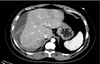
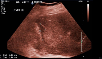
An abdominal ultrasound revealed fluid collection around the right and left liver lobes suggestive of a hematoma. The CT scan of the abdomen showed 16 x 10 x 3 cm large hepatic sub capsular hematoma and no focal lesion (Figure 1). A diagnosis of hepatic sub capsular hematoma was made. The patient’s symptoms resolved with conservative management. A follow up ultrasonography was done after 6 months and showed near complete resolution of the hematoma (Figure 2).

    Figure 1: Abdominal computed tomography revealed 16 x 10 x 3 cm large
hepatic sub capsular hematoma and no focal lesion.
