
Research Article
Austin Med Sci. 2021; 6(1): 1047.
Advantages of Application of Whole-Body Low-Dose Computed Tomography in Multiple Myeloma
Chen J*, Li M, Gao Z, Liu S, Wang J and Ye Z*
Department of Radiology, Tianjin Medical University Cancer Institute and Hospital, China
*Corresponding author: Jian Chen and Zhaoxiang Ye, Department of Radiology, Tianjin Medical University Cancer Institute and Hospital, National Clinical Research Center for Cancer, Key Laboratory of Cancer Prevention and Therapy, Tianjin Clinical Research Center for Cancer, China
Received: May 17, 2021; Accepted: June 05, 2021; Published: June 12, 2021
Abstract
Aim: This study was designed to investigate the application of whole-body low-dose computed tomography in the examination of multiple myeloma.
Method: 40 patients with multiple myeloma admitted to our hospital were prospectively selected as the study subjects. All patients were pathologically confirmed and/or clinically diagnosed with multiple myeloma. Patients were randomly divided into two groups: Group A (n=20) received whole-body lowdose CT scan with SAFIR iterative reconstruction algorithm; Group B (n=20) underwent whole body conventional dose CT scan combined with conventional reconstruction algorithm. The image quality was scored subjectively, and the objective evaluation indexes (including CT value and noise of neck, chest, abdomen, pelvic cavity and lower extremities, signal-to-noise ratio and image quality index) were measured and recorded, and the radiation dose was recorded. Mann-Whitney U test (to evaluate the subjective score) and t test (to evaluate the objective evaluation index and radiation dose) were used to compare the differences of the above indexes between group A and group B.
Result: All the images met the diagnostic requirements. There was no statistical significance in the scores between group A and group B (P>0.05). Significant differences in CT value, noise and SNR of neck, chest, abdomen, pelvis and lower extremities between group A and group B (P<0.05) were identified. For the image quality index (figure of merit, FOM), the FOM of chest, abdomen and pelvis was not statistically significantly changed (P<0.05). The radiation dose of group A decreased by 56.77% (3.06/5.39) compared to group B with a statistically significant difference (P<0.05). The Kappa values of subjective scores of the two groups showed no statistically significant difference (respectively, 0.68 and o.69, P>0.05).
Conclusion: Compared to conventional CT examination, whole-body lowdose CT scan combined with SAFIR iterative reconstruction algorithm can effectively reduce noise, reduce X-ray radiation dose, and obtain ideal image quality in multiple myeloma examination, which has a certain application value.
Keywords: Whole-body low-dose CT scan; Whole-body conventional dose CT; Radiation dose; Multiple myeloma; Reconstruction algorithm
Introduction
Multiple myeloma is a malignant proliferative disease of plasma cells, which is the most common primary tumor with bone involved [1]. Mahnken was the first to use multi-slice spiral CT scan (MDCT) to examine patients with multiple myeloma [2]. Compared to radiography, CT showed higher sensitivity in the presence of fractures and in assessing the risk of vertebral collapse. However, the drawback of Mahnken’s study was the high dose of radiation (23mSv-36mSv) that must be used in order to obtain good images. The radiation hazards brought by conventional CT to multiple myeloma patients have attracted more and more attention. It is the research direction of many scholars to effectively reduce the radiation dose without affecting the diagnostic efficacy and evaluation of multiple myeloma patients. Horger has introduced whole-body, low- dose, multi-detector Computed Tomography (WBLD-MDCT) technique in clinical practice [3]. Compared to conventional CT, the radiation dose (7.5mSv-4.1mSv) of patients with multiple myeloma was significantly reduced by 16-slice scanner with a tube voltage of 120 kVp and four different energy parameters (40, 50, 60, 70 mAs). This study shows that WBLD-MDCT is suitable for the diagnosis of osteolytic changes and the assessment of fracture risk in multiple myeloma patients. The scan length of all patients in the study was 1530.6mm, which could only extend from the top of the skull down to the knee and could not fully cover the entire body of the patients. With the update of equipment, the scanning scheme was improved in this study. The scanning length of all patients reached 1970mm, which could be scanned from the top of the skull to the tip of the toe at one time, that is, the whole body of the patient, and there would be no omission for all the lesions of the patient. The purpose of this study was to investigate the value of Whole-Body Low-Dose CT (WBLDCT) in the detection of multiple myeloma.
Materials and Methods
Research objects
A total of 40 patients who planned to undergo CT scan to evaluate multiple myeloma multiple bone destruction in our hospital from January 2016 to June 2018 were collected. Inclusion criteria: a, confirmed multiple myeloma patients; b, The Body mass index (BMI) of the patients was between 20 and 25; c, Height of the patient ≤1970mm; d, Informed consent of patients. Exclusion criteria: a, prior treatment; b, unable to maintain supine position; c, Pregnant women; d, patients with poor compliance; patients who could not tolerate the examination. Patients were randomly divided into group A and group B, group A (n=20) was low-dose CT imaging group, and group B (n=20) was routine dose scanning group.
Inspection methods
All subjects underwent whole body CT scan using Somatom Definition AS (Siemens Germany, Forchheim, Germany) 64-slice 128-slice CT. Scanning position: patient supine with hands close to the sides of the body. Scanning range: top of head to sole; Into the bed: foot first; Scanning direction: head to foot. In group A, a Sinogram Affermed Iterative Reconstruction (SAFIR) technique with whole-body low-dose CT scanning was used and the SAFIRE index is 3. The scanning conditions were as following: voltage was 100 kVp, Quality reference mAs was 70 mAs, and automatic tube current modulation technology (CARE DOSE4D) is adopted. In group B, whole-body conventional dose CT scanning was performed with conventional reconstruction algorithm. The scanning conditions were as following: voltage was 120 kVp, Quality reference mAs was 70 mAs, and automatic tube current modulation technology was used. Other scanning parameters of A and B were the same: scanning layer thickness was 1.5mm, reconstruction layer thickness was 5mm, screw pitch was 0.6, collimator was at 128*0.6 and FOV (Field of View) was 650mm.
Objective evaluation
Objective evaluation and data measurement: CT values and Standard Deviation (SD) of the neck (the 6th cervical vertebra level sternocleidomastoid muscle), chest (the pulmonary trunk level transverse process spine muscle), abdomen (the portal vein level transverse process spine muscle), pelvic (gluteus maximus muscle), and lower extremities (the femoral medial vastus muscle) were measured respectively. SD was the Objective Image Noise (OIN). The Signal-to-Noise Ratio (SNR) and the Figure of Merit (FOM) of neck, chest, abdomen, pelvic cavity and lower extremities were calculated. SNRn=HUn/SDn, FOM = (SNR2/ED) [4]. n stands for muscle, and ED is the Effective Dose.
Subjective evaluation
The subjective image quality evaluation was carried out by “blind method”. The scores were determined by two radiologists with more than 10 years of working experience, according to Horger [3] scoring criteria. The evaluation included: fracture risk, lesion boundary, sharpness of small lesion (<5mm) contour, cavernous bone trabeculae, etc. The image quality was rated on a 4-point scale. Image quality is very good for a score of 1: all osteolytic lesions and spongy bone trabeculae had clear boundaries and no edge artifacts. The image quality was good, which made a score of 2: all lesions and cavernous bone trabecular structure boundaries were clear, and there were mild artifacts, small lesions (<5mm) blurred outline. General image quality gave a score of 3: the contours of the small lesions were blurred. Especially, the circumference of the anatomical structure was increased, and the absorption of X-ray was increased, such as the shoulder and pelvic cavity, with significant artifacts. Poor image quality made a score of 4: the boundary between the lesion and the spongy bone trabecula was not clear, and there were serious artifacts, which was making it difficult to determine the boundary. A score at ≤3 is considered to meet the diagnostic requirements.
Radiation dose
The dose parameters of the subject, including CT Dose Index- Volume (CTDIvol), Dose Length Product (DLP), and the effective dose were automatically calculated and generated by CT machine [5].
Statistical analysis
SPSS24.0 statistical software was used for statistical analysis. Kolmogorov-Simov was used to test whether measurement data conform to normal distribution. The measurement data conforming to the normal distribution is expressed by x±s, while the enumeration data is expressed by frequency. Kappa test was used to evaluate the consistency of image quality scored by 2 physicians. The consistency was evaluated by Kappa: Kappa ≥0.75 meant very good consistency, Kappa ≥0.4 and <0.75 meant good consistency, and Kappa<0.4 meant poor consistency. T test was used to compare the difference of objective evaluation indexes and radiation dose between the two groups of image quality. Mann-Whitney U test was used to compare the differences in subjective scores. P<0.05 was considered statistically significant.
Results
Image quality of whole-body low-dose CT met diagnostic requirements
Subjective scoring: The two physicians had good consistency in scoring the images of group A and group B. The consistency of Kappa values of subjective scores in the two groups was 0.68 and 0.69, respectively, and the difference was not statistically significant (P>0.05). All the images met the diagnostic requirements, as shown in Table 1. Mann-Whitney U test was used to compare the average image scores between group A and group B, and the difference was not statistically significant (Mann-Whitney U was 1699.500, P = 0.425>0.05). To better visualize and compare the quality of CT imaged obtained from the two methods, representative CT images from two multiple myeloma patients with similar BMI (Body mass index) receiving whole-body low-dose CT and conventional CT, respectively, were illustrated. Although the conventional CT needed much higher effective radiation dose than whole-body low-dose CT (EDwhole-body low-dose:EDconventional = 4.62 msv: 9.27 msv), both methods gave clear and high quality images in skull, neck, abdomen, thoracic and pelvic CT scanning (Figure 1A and B). Subjective evaluation scores of the image quality from both patients were 1, which suggested very good image qualities of the CT images by both methods. MPR sagittal and coronal reconstruction of patients scanned by both whole-body low-dose CT and conventional CT also gave CT images at high qualities from skull to lower extremities (Figure 2A and 2B).
    Group
    Case number
    Score by    physician A
    Score by    physician B
    Kappa


    1
    2
    3
    4
    x ± s
    1
    2
    3
    4
    x ± s


    A
    20
    17
    2
    1
    0
    1.23±0.57
    16
    2
    2
    0
    1.33±0.66
    0.68


    B
    20
    17
    2
    1
    0
    1.23±0.57
    16
    3
    1
    0
    1.33±0.65
    0.69
Table 1: Subjective evaluation of the CT images.
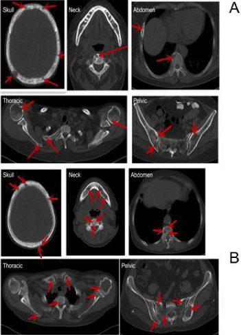
    Figure 1: CT images of two patients with multiple myeloma from whole-body
low-dose CT (A) and conventional CT (B). (A) The patient was a 66-year-old
woman with multiple myeloma. BMI=23.24, effective radiation dose of wholebody
low-dose CT scan was 4.62 mSv, subjective evaluation score of the
image quality was 1 (very good). (B) The patient was a 61-year-old woman
with multiple myeloma. BMI=24.58, effective radiation dose of conventional
CT scan was 9.27 mSv, subjective evaluation score of the image quality was
1 (very good). The red arrows indicated destructed bone areas.
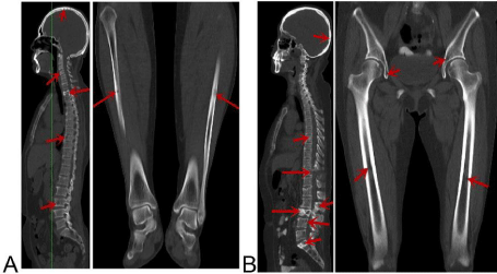
    Figure 2: MPR sagittal and coronal reconstruction images of patients scanned
by whole-body low-dose CT (A) and conventional CT (B). (A) MPR sagittal
and coronal reconstruction images clearly showed bone destruction in the
skull, cervical spine, thoracic spine, lumbar spine, and fibula by whole-body
low-dose CT. (B) MPR sagittal and coronal reconstruction images clearly
showed bone destruction in the skull, thoracic spine, lumbar spine, sacrum,
ilium and femur by conventional CT. The red arrows indicated destructed
bone areas.
Objective evaluation: FOM (figure of merit) was used to quantify the quality of the CT images. Among the FOM indexes, only the FOM of neck and lower extremity had slight statistical differences (P<0.05), while the FOM of other parts was not significantly changed (P>0.05) (Figure 3 and Table 3). These data suggested that both whole-body low-dose CT and conventional CT could generate diagnosis required CT images.
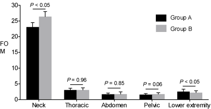
    Figure 3: Comparison of CT image quality from patients scanned by wholebody
low-dose CT (Group A) and conventional CT (Group B). Note that
only the FOM of neck and lower extremity CT had slight significance and all
others showed no significance, suggesting comparable CT image quality of
wholebody low-dose CT (Group A) and conventional CT (Group B).
Objective evaluation of the whole-body low-dose CT
To objectively evaluate the whole-body low-dose CT in multiple myeloma, CT values of the muscles from different body parts were quantified (Table 2). Compared to conventional CT (Group B), whole-body low- dose CT showed lower CT values in all the muscles we evaluated with the exception of vastus medialis (Figure 4A). In addition, CT noise of the major body parts, including neck, thoracic, abdomen, pelvic lower extremity CT, by whole-body low-dose CT (Group A) was higher (Figure 4B) and SNR of these body parts was significantly lower (Figure 4C).
Group
Case number
Value of thoracic spinous
Value of thoracic value of abdominal transverse spinal transverse spinal muscle
Value of pelvic gluteus maximus
Value of vastus
medialis CTmuscle CT (HU)
muscle CT (HU) CT (HU)
CT (HU)
(HU)
A
20
50.78±2.32
40.72±2.24
44.51±2.33
38.15±1.72
43.60±2.63
B
20
55.42±2.81
47.97±2.01
48.42±2.81
42.95±1.62
45.04±2.33
t
4.76
9.45
3.81
11.19
1.73
P
<0.05
<0.05
<0.05
<0.05
0.1
Group
Case
Noise of neck CT
Noise of thoracic
Noise of abdomen
Noise of pelvic
Noise of lower
number
(HU)
CT (HU)
CT (HU)
CT (HU)
extremity CT (HU)
A
20
4.58±0.23
10.15±0.52
14.85±0.67
13.49±1.04
12.13±0.97
B
20
3.76±0.27
9.60±0.50
13.45±1.83
11.43±0.99
10.66±0.77
t
10.34
3.02
3.14
7.19
5.17
P
<0.05
<0.05
<0.05
<0.05
<0.05
Group
Case number
SNR of neck
SNR of thoracic
SNR of abdomen
SNR of pelvic
SNR of lower extremity
A
20
11.10±0.79
4.02±0.28
3.00±0.15
2.84±0.24
3.61±0.34
B
20
14.77±1.03
5.01±0.30
3.67±0.54
3.78±0.33
4.25±0.39
t
13.38
8.63
5.75
10.65
4.94
P
<0.05
<0.05
<0.05
<0.05
<0.05
Table 2: Objective evaluation indexes of the CT.
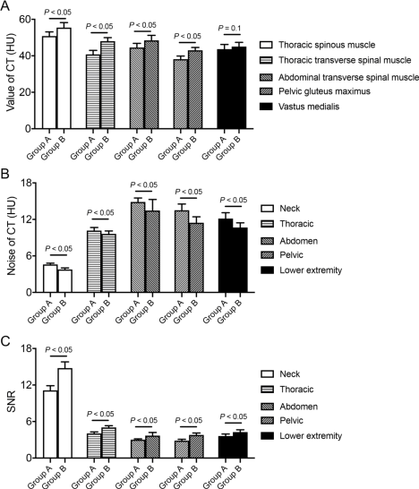
    Figure 4: Objective evaluation indexes of the two groups by different CT
methods. (A) Bar plots quantification and comparison of CT values of different
muscles. Whole-body low-dose CT group showed significantly reduced CT
values in the muscles compared to conventional CT. Visualization of CT
noise (B) and SNR (C) in different body parts in whole-body low-dose CT
and conventional CT.
Reduced radiation dose to patients by whole-body lowdose CT
As radiation worsens the outcome of multiple myeloma, radiation produced by CT is another parameter that the physician in this field should consider. The radiation produced by whole-body low-dose CT (group A) was much lower than that of conventional CT (group B) (Figure 5). Specifically, the CT dose index-volume (CTDIvol) in group A and group B was 2.11±0.48 mGy and 3.24±0.54 mGy, respectively, and the difference had a statistical significance (P<0.05) (Figure 5A and Table 3). The Dose Length Product (DLP) of group A and group B was 359.98±73.48 mGy·cm and 563.72±101.72 mGy·cm, respectively, with a statistical significance (P<0.05) (Figure 5B and Table 3). The effective radiation (ED) doses of group A and group B were 5.39±1.10 msv and 8.45±1.53 msv, respectively. Compared to group B, the radiation dose of group A was decreased by 56.77% (3.06/5.39) (Figure 5C And Table 3). These data confirmed that whole-body low- dose CT produced much less radiation compared to conventional CT.
    CTDIvol
    DLP
    ED
    FOM
    FOM
    FOM
    FOM
    FOM    (lower


    Group
    (mGy)
    (mGy·cm)
    (mSv)
    (neck)
    (thoracic)
    (abdomen)
    (pelvic)
    extremity)


    A
    2.11±0.48
    359.98±73.48
    5.39±1.10
    23.09±1.39
    3.06±0.56
    1.73±0.36
    1.59±0.34
    2.58±0.72


    B
    3.24 ±0.54
    563.72±107.72
    8.45±1.53
    26.40±1.60
    3.07±0.65
    1.76±0.72
    1.77±0.43
    2.23±0.59


    t
    12.23
    21.64
    21.59
    10.52
    0.05
    0.2
    2.03
    2.18


    P
    <0.05
    <0.05
    <0.05
    <0.05
    0.96
    0.85
    0.06
    <0.05


    Note: CTDIvol: CT dose index-volume; DLP: Dose Length Product;    ED: Effective Dose; FOM: Figure of Merit.
Table 3: Comparison of CT image quality from patients scanned by wholebody
low-dose CT (Group A) and conventional CT (Group B). Note that
only the FOM of neck and lower extremity CT had slight significance and all
others showed no significance, suggesting comparable CT image quality of
wholebody low-dose CT (Group A) and conventional CT (Group B).
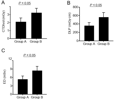
    Figure 5: Comparison of radiation dose between two CT methods.
Quantification and comparison of radiation dose of whole-body low-dose CT
and conventional CT represented by CTDIvol (A), DLP (B) and ED (C).
Discussion
The main clinical manifestations of multiple myeloma are “CRAB” symptoms, namely hypercalcemia, renal damage, anemia and bone destruction, and osteolytic lesions are the main feature. 90% of patients have osteolytic lesions along with the disease development, and 70~80 % of multiple myeloma patients have osteolytic lesions at the time of diagnosis [6]. Therefore, it is necessary to evaluate the degree of bone lesions correctly [1]. In addition to CT, X-ray, ECT, PET/CT and MR are the common examination methods for MM patients, but all these methods have certain limitations. X-ray imaging is a two-dimensional imaging, limited in the diagnosis of early and small lesions, low sensitivity to osteolytic lesions, high rate of missed diagnosis, has not been used as a recommended examination method for multiple myeloma bone lesions [7]. ECT examination requires injection of radioactive isotopes, which remain in the patient’s body for a period of time and have a certain potential damage to themselves and others, and the preparation and examination time is long. PET/ CT examination is long, expensive, and has low spatial resolution, resulting in false negative results. MRI is time-consuming, expensive, and has many limitations in its application. multiple myeloma is usually multiple and may affect every bone component of the body, so a proper assessment of the progression of the disease in multiple myeloma patients requires full-body imaging [8]. CT has high sensitivity and resolution for cortical and trabecular bone to assess fracture risk and stability, and to understand overall expansion and potential complications in patients with multiple myeloma [9]. However, the main disadvantage of conventional CT is that the radiation dose is too high. Therefore, here we propose whole-body low-dose computed tomography (WBLDCT) and our study is the first trial in China. WBLDCT greatly reduces the amount of radiation (similar to X-ray) while retaining high sensitivity and resolution. WBLDCT can show a variety of pathological changes of multiple myeloma at the same time: bone marrow involvement (especially bones of the four limbs), medullary lesions, soft tissue involvement degree and the dissolved osseous changes (Figure 1). Other lesions of multiple myeloma outside the bones such as pulmonary infection could also be diagnosed. So WBLDCT is significantly necessary to help the comprehensive treatment of multiple myeloma patients and is critical for guiding the determination of disease stage, chemotherapy, surgery and pathology. Horger believed that WBLDCT can replace X-ray as the imaging examination method at first diagnosis [9], and San Gerardo Hospital in Monza even chose WBLDCT as the preferred examination method at baseline for multiple myeloma patients [10]. CT (including WBLDCT) or PET/CT indicated one or more osteolytic lesions (=5 mm in diameter), which was defined as the diagnostic criteria for multiple myeloma bone disease at the 2015 IMWG meeting [11, 12]. In this study, images obtained from both WBLDCT scan and conventional CT scan met the diagnostic requirements, and the subjective scores of the two groups were consistent with each other. The difference in image scores was not statistically significant, suggesting that WBLDCT can provide lowdose and reliable whole-body CT scan with image quality for multiple myeloma patients and meet the needs of clinical treatment.
The rapid development of modern CT instruments and the application of advanced reconstruction algorithms make it possible to obtain better image quality with lower radiation dose. Radiation hazards have attracted more and more attention and reducing the radiation dose received by patients has always been the goal pursued by the majority of scholars. In CT examination, the most effective way to reduce the radiation dose is to reduce the voltage, so this study adopted a low voltage of 100 kVp. WBLDCT needs only plain CT, and does not need injection of contrast medium, so there would be no contrast medium related side effects. On the basis of the lower tube voltage 100 kVp combined with SAFIRE iterative reconstruction algorithm, WBLDCT not only effectively reduces the patient radiation dose and damage caused by multiple examinations and radiation doses, but also provides high quality images diagnosis needs, so that lesions in patients with multiple myeloma and bone destruction can be detected and accurate treatment judgment can be made [8]. WBLDCT provides a fast, low-dose radiation, high image quality, whole-body CT scan for patients with multiple myeloma, and is a reliable method for the evaluation of multiple myeloma bone disease [13]. All the images in this study met the diagnostic requirements. Compared with conventional CT, the radiation dose of WBLDCT was reduced by 56.77%, and the length of one scan reached as long as 1970mm, and it only took 20 seconds to complete the examination for patients. Due to the low dose scan used in group A, the ideal image quality can be obtained with reduced effective radiation dose of patients during CT examination. In conclusion, WBLDCT scan has important application value in the examination of multiple myeloma.
There are still some limitations in this study: 1. the two groups of cases are not the same individual, which may cause some individual differences; 2. Patients with BMI>25 were not included in the study; 3. Further study needs to be performed to determine whether 80 kVp can be used for patients with BMI<20.
Conclusion
To summarize, compared to conventional CT, whole-body low-dose CT scan combined with SAFIR iterative reconstruction algorithm in multiple myeloma examination can effectively reduce noise, reduce X-ray radiation dose and obtain ideal image quality, which has certain clinical application values.
Acknowledgment
This research was supported by the National Natural Science Foundation of China (no. 81601492), Tianjin Science and Technology Major Project (no. 12ZCDZSY15500), Public Science and Technology Research Funds Projects of NHFPC of the P.R. China (no. 201402013), National Key R&D Program of China (2016YFE0103000).
Data Availability
The datasets in the present study are available from the corresponding author on reasonable request.
Ethical Approval
The studies involving human subjects were reviewed and approved by the Medical Ethics Review Committee of Tianjin Medical University Cancer Institute and Hospital. All patients participating in our observational cohort study provided written informed consent before entering the study.
Authors’ Contributions
Jian Chen, and Zhaoxiang Ye designed the study. Jian Chen carried out the analysis and wrote the manuscript. Meng Li, Zhipeng Gao, and Shichang Liu participated in the coordination of the study and interpretation of results. Shichang Liu, and Jialin Wang participated in manuscript writing. Jian Chen, and Meng Li collected data. Jian Chen and Zhipeng Gao participated in figure typesetting. All authors read and approved the final manuscript.
References
- Gulla A, Anderson KC. Multiple myeloma: the (r)evolution of current therapy and a glance into future. Haematologica. 2020;105: 2358-2367.
- Mahnken AH, Wildberger JE, Gehbauer G, Schmitz-Rode T, Blaum M, Fabry U, et al. Multidetector CT of the spine in multiple myeloma: comparison with MR imaging and radiography. AJR Am J Roentgenol. 2002; 178: 1429-1436.
- Horger M, Claussen CD, Bross-Bach U, Vonthein R, Trabold T, Heuschmid M, et al. Whole-body low- dose multidetector row-CT in the diagnosis of multiple myeloma: an alternative to conventional radiography. Eur J Radiol. 2005; 54: 289-297.
- Schindera ST, Nelson RC, Yoshizumi T, Toncheva G, Nguyen G, DeLong DM, et al. Effect of automatic tube current modulation on radiation dose and image quality for low tube voltage multidetector row CT angiography: phantom study. Acad Radiol. 2009; 16: 997-1002.
- Tsalafoutas IA, Metallidis SI. A method for calculating the dose length product from CT DICOM images. Br J Radiol. 2011; 84: 236-243.
- Palma BD, Guasco D, Pedrazzoni M, Bolzoni M, Accardi F, Costa F et al. Osteolytic lesions, cytogenetic features and bone marrow levels of cytokines and chemokines in multiple myeloma patients: Role of chemokine (C-C motif) ligand 20. Leukemia. 2016; 30: 409-416.
- Kropil P, Fenk R, Fritz LB, Blondin D, Kobbe G, Modder U, et al. Comparison of whole-body 64-slice multidetector computed tomography and conventional radiography in staging of multiple myeloma. Eur Radiol. 2008; 18: 51-58.
- Ippolito D, Besostri V, Bonaffini PA, Rossini F, Di Lelio A, Sironi S. Diagnostic value of Whole-Body Lowdose Computed Tomography (WBLDCT) in bone lesions detection in patients with Multiple Myeloma (MM). Eur J Radiol. 2013; 82: 2322-2327.
- Horger M, Kanz L, Denecke B, Vonthein R, Pereira P, Claussen CD, et al. The benefit of using whole- body, low-dose, nonenhanced, multidetector computed tomography for follow-up and therapy response monitoring in patients with multiple myeloma. Cancer. 2007; 109: 1617-1626.
- Zacchino M, Bonaffini PA, Corso A, Minetti V, Nasatti A, Tinelli C, et al. Interobserver agreement for the evaluation of bone involvement on Whole Body Low Dose Computed Tomography (WBLDCT) in Multiple Myeloma (MM). Eur Radiol. 2015; 25: 3382-3389.
- Tabit CE, Chung WB, Hamburg NM, Vita JA. Endothelial dysfunction in diabetes mellitus: molecular mechanisms and clinical implications. Rev Endocr Metab Disord. 2010; 11: 61-74.
- International Myeloma Working G. Criteria for the classification of monoclonal gammopathies, multiple myeloma and related disorders: a report of the International Myeloma Working Group. Br J Haematol. 2003; 121: 749-757.
- Cretti F, Perugini G. Patient dose evaluation for the Whole-Body Low-Dose Multidetector CT (WBLDMDCT) skeleton study in Multiple Myeloma (MM). Radiol Med. 2016; 121: 93-105.