Research Article
Austin J Microbiol. 2019; 5(1): 1027.
Extracelluler Enzymes, Pathogenicity and Biofilm Forming in Staphylococci
Sahin R*
Microbiology Laboratory, Mersin City Hospital, Mersin, Turkey
*Corresponding author: Sahin R, Microbiology Laboratory, Mersin City Hospital, Mersin, Turkey
Received: August 23, 2019; Accepted: September 27, 2019; Published: October 04, 2019
Abstract
The pathogenicity of S.aureus strains are related with features like its adherence, various toxins, enzymes, structural and extracellular factors. In our study, the relationship between biofilm formation and lipase, protease, urease activity were investigated in S. aureus strains isolated from various clinical specimens sent to our microbiology laboratory. Congo red agar was used to detect biofilm production. The lipolytic activity of all strains was evaluated on Tween 20 agar. The proteolytic activity of the strains was evaluated by Skim Milk Agar. Christensen Urea agar was used to determine the urease activities of all strains. Slime factor and biofilm formation are pathogenity factors as well. 101 (57.7%) of 175 clinical isolates were negative for biofilm formation while 74 (42.3%) samples were positive according to phenotypic assessment of colony morphology on CRA. The relationship between biofilm formation and lipase, protease and urease activity of all the isolates are researched by using Spearman’s correlation coefficient. There was an evident relation between biofilm formation with lipase activity (r=0.195, p=0.10) while protease (r=0.001, p=0.99) and urease (r=0.06, p=0.4) activity were not found related.
Keywords: S. aureus; Biofilm; Lipase; Protease; Ürease; Pathogenicity
Introduction
What is biofilm?
A biofilm is a compacted assemblage of microorganisms enclosed in a matrix primarily composed of polysaccharide, and attached on a surface. Biofilms have been found on a variety of surfaces such as indwelling medical devices, industrial water system pipes or aquatic systems in the natural environment. The microbial organisms growing in a biofilm are physiologically distinct from their planktonic counterparts [1,2]. Biofilm formation has been recognized as a protective mode of cell growth which allows for survival in hostile environments, and also under certain circumstances, such as nutrient deprivation. Biofilm dispersal in the form of clumps plays an important role in helping the cells to colonize new niches [3]. At present, the general resistance of biofilms has been explained by several possible mechanisms [4,5]. First, the biofilm matrix might react with superoxides, neutralized charged metals or dilute antimicrobial agents to generate sub-lethal concentrations. Moreover, resistant phenotypes referred to as “persisters”, which have been found in a biofilm, contribute to the resistance. Whether these are indeed a Stoodley unique resistant phenotype or are simply the most resistant cells remains unclear [5,6].
Biofilm development
The process of biofilm formation Recent advances have been made to show that biofilm development experiences a multiple-stage and differentiated process rather than a simple, uniform step. Five sequential regulated stages have been proposed for biofilm formation [6,7]. During the first two stages, the cells are loosely adhered to surfaces. Further, the attached cells aggregate together and form micro-colonies; subsequently mature biofilm develops on surfaces in stages three and four [7,8]. Then, under certain circumstances, the biofilm cells are shed off, return to the mobile mode characterized in stage five [8]. The cells eventually attach to a surface when conditions are appropriate, start a new cycle of biofilm formation [1,9-12].
Biofilm formation in Staphylococcus aureus
Biofilm formation in S. aureus experiences a similar process to that of S. epidermidis; it begins with the initial reversible bacterial adherence to a surface by some non-specific adhesion, followed by an irreversible bacterial specific attachment mediated mainly by an array of MSCRAMMS [13,14]. Then a mature biofilm is developed characterized by multilayered bacterial cells stuck together and producing Extracellular Polymeric Substances (EPS)
[15]. In circumstances such as nutrient deprivation, or under heavy shear forces, detachment of clumps of the biofilm bacteria occurs [3,7]. The released bacterial clumps start to attach to new niches, and initiate a new cycle of biofilm formation [3]. Polysaccharide Intercellular Adhesin (PIA) mediated biofilm formation in S. aureus. Polysaccharide intercellular adhesin was initially purified from S. epidermidis. It was identified in S. aureus later and shown to have a similar function. Since the structure of the N-acetylglucosamine residues in S. aureus is shown totally succinylated, it was designated as Poly-N-Succinyl β-1, 6-Glucosamine (PNSG) [16]. Polysaccharide intercellular adhesin has been defined as an important virulence factor for S. epidermidis pathogenicity in various foreign-body animal infection models [17]. Biofilm production has been shown to play a major role in the pathogenesis of infection caused by Staphylococcus aureus [17,18]. The biofilm formation is the leading cause of the pathogenesis of S. aureus associated with biomaterial infections [17,18]. In S. aureus Polysaccharide Intercellular Adhesin (PIA) was encoded by icaA and icaD genes [16,18,19]. Production of PIA Biofilms are communities of microorganisms that are attached to each other and/or a biotic or abiotic surface, are embedded in a self-produced extracellular matrix, and show markedly reduced susceptibility to antimicrobial agents [7]. It is estimated that the majority of chronic infections and most device-related infections are biofilm-associated [2,3,17,18]. However, biofilm infections are difficult to diagnose and extremely difficult to treat [19].
S.aureus pathogenicity
Staphylococcus aureus is a virulent pathogen that is currently the most common cause of infections in hospitalized patients [9]. The success of S. aureus as a pathogen and its ability to cause such a wide range of infections are the result of its extensive virulence factors [9]. The structural characteristic of biofilms that has the greatest impact on the outcome of chronic bacterial infections, such as native valve endocarditis, is the tendency of individual microcolonies to break off and/or detach when their tensile strength is exceeded [8]. Urease is needed in the urea cycle and in the metabolism of amino acids to degrade urea to form CO2 and NH3 [17]. The resulting ammonium and/or ammonia (depending of the pH of the cells) is toxic for the host cells and might accumulate in and outside the bacterial cells [14,15]. Bacterial proteases secreted into an infected host may exhibit a wide range of pathogenic potentials. Staphylococci, in particular Staphylococcus aureus are known to produce several extracellular proteases, including serine-, cysteine- and metalloenzymes [18,19]. In our study, the presence of lipase, protease and urease enzymes in S. aureus strains isolated from various clinical specimens sent to our microbiology laboratory were investigated. The properties of adherence depend on properties such as various toxins, enzymes, structural and extracellular factors. Slime factor production and biofilm formation are also pathogenicity factors. Staphylococci have been shown to be able to adhere to medical devices. S.aureus and S.epidermidis are the most frequently isolated agents associated with medical device-related infections [15-17]. Staphylococcus aureus is a virulent pathogen that is currently the most common cause of infections in hospitalized patients [9,12].
Extracelullar enzymes of Staphylococcus aureus
Staphylococcus species secrets many extracellular active substances, such as coagulase, hemolysin, nuclease, phosphatase, lipase, proteases, fibrinolysin, enterotoxins and toxin shock syndrome toxin [20,21]. These proteins are known as virulence factors that cause disease in animal and animals [21]. The report, it was verified for the production of Lipase from among the 25 isolates, 15(60%) of isolates produce the Lipase production [22]. Most of the known Staphylococcal lipases are produced by pathogenic members of the genus, i.e. Staphylococcus aureus and S.epidermidis. Lipase interferes with the phagocytosis of the infectious lipase- producing S. aureus cells by host granulocytes, thus indicating a direct involvement of lipase in pathogenesis [20,21]. The success of S. aureus as a pathogen and its ability to cause such a wide range of infections are the result of its extensive virulence factors [2]. The structural characteristic of biofilms that has the greatest impact on the outcome of chronic bacterial infections, such as native valve endocarditis, is the tendency of individual microcolonies to break off and/or detach when their tensile strength is exceeded [3]. Lipolytic activity was determined by using the method [9]. Urease is needed in the urea cycle and in the metabolism of amino acids to degrade urea to form CO2 and NH3 [4]. The resulting ammonium and/or ammonia (depending of the pH of the cells) is toxic for the host cells and might accumulate in and outside the bacterial cells [4,5]. Proteolytic activity was assayed [10]. The overnight broth culture was spoted into 1% skim milk agar and incubated at 37°C for overnight. After incubation period, the clear zone of hydrolysis was observed. The presence of a transparent zone around the colonies indicated protease activity Bacterial proteases secreted into an infected host may exhibit a wide range of pathogenic potentials. Staphylococci, in particular Staphylococcus aureus, are known to produce several extracellular proteases, including serine-, cysteine- and metalloenzymes [6,7]. Their insensitivity to most human plasma protease inhibitors and, even more, the ability to inactivate some of these make the proteases potentially harmful [6,8]. In our study, the presence of lipase, protease and urease enzymes in S. aureusstrains isolated from various clinical specimens sent to our microbiology laboratory were investigated.
Material and Methods
Biofilm formation and lipase, protease, urease activity were investigated in S. aureus strains isolated from various clinical specimens sent to our microbiology laboratory, Denizli ,Turkey.
Investigation of biofilm formation
Qualitative detection of biofilm formation of these isolates was performed using Congo Red Agar (CRA), according to (Freeman et al., 1989) Isolates were streaked onto the agar to obtain single colonies and incubated overnight at 37°C aerobically and a further 24 hours at room temperature. The interpretation of results followed (Freeman et al., 1989) and (Arciola et al., 2002) [23,24].
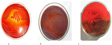
Congo red agar was used to detect biofilm production [23]. Congo reddish agar medium was prepared to contain 10g of agar, 50g of sucrose, 37g of brain-heart infusion vial and 0.8g of Congo red. Cultures made in such a way that a single colony fell on these mediums were incubated overnight at 37°C, followed by incubation of the cultures for 48 hours at room temperature. S. epidermidis ATCC 12228, which does not produce biofilms, and S. epidermidis ATCC 35984, which produces strong biofilms, were used as controls. Cultures cultured in Congo red agar medium at 37°C overnight and after 48 hours incubation at room temperature after 48 hours of reddish-black, rough, dry, transparent colony forming biofilm positive, pinkish-red, flat and central dark (ox-eye view) colony biofilm was considered negative (Figure 1).

    Figure 1: Evaluation of biofilm formation by Congo red agar method [a-biofilm
negative, b-biofilm positive, c-biofilm negative (upper) and biofilm positive
(bottom)].
Screening for extracellular enzymes
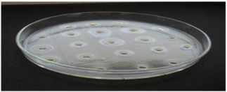
Investigation of lipolitic activities: The lipolytic activity of the isolates was determined onto Tween 20 agar [18,25]. The experiments were carried out in The lipolytic activity of all strains was evaluated on Tween 20 agar (containing 10g peptone, 5g NaCl, 0.1g CaCl2 , 20g agar and 1ml Tween 20) per liter. Produced by incubation at 37°C overnight in Brain heart medium (pH 7.5) containing 10g of peptone, 5g of yeast extract, 5g of NaCl, 1g of K2 HPO4.3H2O. Subsequently, strains diluted 1: 100 in Brain hart medium were inoculated into 20μl of the wells opened with sterile glass pipette onto Tween 20 agar. The plates were evaluated after 72 hours incubation at 37°C. The presence of lipolytic activity was detected around the inoculation by the appearance of halo formation, depending on whether the tween was a line-shaped precipitate (Figure 2) .

Figure 2: Lipolytic activity of S. aureus strains on Tween 20 agar.
Investigation of proteolytic activities: The proteolytic activity of the strains was evaluated by the agar plate method. For this, Skim Milk Agar (SMA) containing 1% skim milk, 1% tryptone, 0.5% yeast extract, 0.5% NaCl and 1.5% agar was used. 20mu.l of supernatant from 1:100 diluted samples were added to sterile diluted wells in SMA. Plates were assessed after being incubated for 48 hours at 37°C. The presence of proteolytic activity was determined by the occurrence of opaque zones (halo formation) around the wells due to casein hydrolysis [26-30].
Urease activity investigation: Christensen Urea agar was used to determine the urease activities of all strains [31]. The 20μl supernatant of the 1:100 diluted samples were taken grown on urea agar after being produced on Brain heart medium. The tubes were evaluated after 24 h incubation at 37°C. Urea hydrolysis in this medium was shown by a change in color from the pale yellow of the fresh medium to an intense red-violet color.
Conclusion
SPSS Ver 10.0 was used for statistical analysis. Spearman’s correlation coefficient was used to evaluate the relationship between biofilm production and enzyme activities of the working clinical isolates. The statistical error margin was accepted as 5%.
S. aureus strains were isolated from various clinical specimens (eye, urine, nasal swab, etc.). Numbers and percentages of biofilm positive and negative specimens were shown on Congo red agar medium (Table 1).
Biofilm
For example, where it
is isolated nNegative
N (%)Positive
N (%)Wound 87
34 (39.1)
53 (60.9)
Blood 25
17 (68.0)
8 (32.0)
Tracheal aspirate 35
31 (88.6)
4 (11.4)
Sputum 10
7 (70.0)
3 (30.0)
Catheter 8
7 (87.5)
1 (12.5)
Other 10
5 (50.0)
5 (50.0)
Total 175
101 (57.7)
74 (42.3)
Table 1: Of the 175 S. aureus strains used in the study, 87 wound (49.7%), 25
blood culture (14.3%), 35 tracheal aspirate (20.0%), 10 sputum (5.7%), 8 catheter
4.6%) and 10 (5.7%) were isolated from various clinical specimens (eye, urine,
nasal swab, etc.). Numbers and percentages of biofilm positive and negative
specimens were shown on Congo red agar medium.
The extracellular enzyme production among the isolates
The relationship between biofilm production and lipase, protease and urease activities of all isolates was investigated [32].
Lipolytic activity of S. aureus strains was showed on Tween 20 agar (Figure 2). Of the clinical isolates, 146 (83.4%) showed lipase activity whereas 29 (16.6%) did not have lipase activity. Lipase (+) was detected in 68 (91.9%) and lipase (-) was detected in 6 (8.1%) in 74 samples of biofilm positive in Congo red agar medium. In biofilm negative 101 samples, 78 (77.2%) were lipase (+) while 23 (22.8%) were lipase (-) (Table 2).
    Biofilm


    Lipase
    Positive
      N (%)
    Negative
      N (%)
    Total
      N (%)


    Positive
    68 (91.9)
    78 (77.    2)
    104 (59.4)


    Negative
    6 (8.1)
    23 (22.    8)
    71 (40.6)


    Total
    74
    101
    175


    There was a good correlation between lipase    activity and biofilm production of the isolates (r=0.195, p=0.10).
Table 2: Lipolytic activity of S. aureus strains on Tween 20 agar.
Of the 175 clinical isolates, 104 (59.4%) showed proteolytic activity whereas 71 (40.6%) showed no proteolytic activity. Protease (+) was detected in 44 (59.5%) and protease (-) was detected in 30 (40.5%) of the 74 samples in which biofilm was positive in Congo red agar medium. Protein (+) was found in 60 (59.4%) and protease (-) was detected in 41 (40.6%) of the biofilm negative 101 samples (Table 3).
    Biofilm


    Protease
    Positive
      N (%)
    Negative
      N (%)
    Total
      N (%)


    Positive
    44 (59.5)
    60 (59.4)
    104 (59.4)


    Negative
    30 (40.5)
    41 (40.6)
    71 (40.6)


    Total
    74
    101
    175


    There was    no correlation between biofilm production and proteolytic activity of the    isolates (r=0.001, p=0.99).
Table 3: Relationship between biofilm formation and protease activity.
144 (82.3%) of the clinical isolates showed urease activity, while 31 (17.7%) had no urease activity. Urease (+) was found in 63 (85.1%) and urease (-) was detected in 11 (14.9%) in 74 samples of biofilm positive in Congo red agar medium. Urease (+) was found in 81 (80.2%) and urease (-) was detected in 20 (19.8%) of the biofilm negative 101 samples (Table 4).
    Biofilm


    Urease
    Positive
      N (%)
    Negative
      N (%)
    Total
      N (%)


    Positive
    63 (85.1)
    81 (80.2)
    144 (82.3)


    Negative
    11 (14.9)
    20 (19.8)
    31 (17.7)


    Total
    74
    101
    175


    There was    no relationship between biofilm production and urease activity of the    isolates (r=0.06, p=0.4).
Table 4: Relationship between biofilm formation and urease activity.
Discussion
Clinical impact of bacterial biofilm. The study of bacteria residing in biofilms as an interactive community has recently gained a great deal of interest, in part, because a number of human diseases are involved in biofilms. Several mechanisms have been proposed to explain why pathogens in biofilms are more virulent than their planktonic counterparts [8]. First, pathogens in biofilms can initiate an infection through the seeding or dispersal of biofilm clumps which contain large numbers of cells. Second, among the phenotypically heterogeneous pathogens within a biofilm, certain virulent phenotypes might survive and spread within biofilm matrix. Finally, the closely related cells within biofilms might initiate quorum sensing networks which regulate virulence gene expressions [9]. Taken together, the dense aggregated virulent organisms within the context of biofilms might be a major contributor to the pathogenesis of bacterial biofilm related infections [10,11].
The success of S. aureus as a pathogen and its ability to cause such a wide range of infections are the result of its extensive virulence factors. Biofilm formation occurs through a series of steps which begins with initial attachment of planktonic bacteria to a solid surface that is present at the air-water/liquid interface. This step is followed by subsequent proliferation and accumulation of the cells in small multilayer cell clusters known as microcolonies. The microcolonies then further proliferate to form giant assemblages of cells enmeshed in an extracellular matrix, which covers entire surfaces, and protects its inhabitants from detrimental effects of all sorts [4-6]. A mature well established biofilm is not a static structure, rather it is highly dynamic in nature, where old cells are constantly being dispersed and new members being recruited for this surfaceassociated community to expand, at all times. The composition of the extracellular matrix is very difficult to ascertain and variable among different bacterial species and even within the same species under different environmental conditions [3]. Despite this fact exopolysaccharides are an essential component of virtually all biofilm structures, providing the necessary matrix in which the bacterial cells are initially embedded [7]. Bacterial cells have to protect themselves from a pH that is too low [13]. Bacterial cells have assumed that the urease activity determined contributes to the persistence of the bacterial cells in the biofilm by counteracting the low pH values caused by the production of lactic acid, acetic acid, and formic acid. Beenken et al. have also reported up-regulation of the urease operon in 7-day-old biofilms [5]. According to Resch et al. urease activity might be an important factor for keeping the biofilm alive [15]. Since excess ammonia would be toxic for the bacterial cells, they should have some mechanism of resistance against this chemical and should also have enzymes or other mechanisms to detoxify this compound. Bacterial proteases secreted into an infected host may exhibit a wide range of pathogenic potentials [15]. Bacterial protease is reported as a pathogenic factor [22]. S. aureus has two enzymes, metallo-protease and serine protease [26-30]. The distribution of both enzymes varies greatly between strains. Researchers report that the protease activity is a pathogenic factor [26,28]. Urease activity is said to be important in the maintenance of biofilm formation. The bacterial cells in the biofilm protect themselves by up-regulation of the urease genes from the pH decreasing by the production of lactic acid, formic acid, which is the result of metabolism [15,16,19]. Coagulase-negative staphylococci were demonstrated to present lipase and protease activities more often than coagulase-positive staphylococci. Staphylococcus aureus produce some of the industrially important extracellular enzymes. Lipase is the most abundant enzyme produced by this bacteria. The lypotic activity of staphylococci was originally observed [10]. The lypotic activity is now known to be the result of the enzymes lipase and esterase which act against water-soluble, water-insoluble glycerol esters and on water-soluble Tween polyoxyethlene esters. So staphylococci split variety of lipid substrates with lipase acting on fat-soluble glycerides and with esterase acting on water-soluble esters [9]. It is known that staphylococci tend to remain in lipoid secretions in the cutaneous habitat of the host organism [10]. Lipids are found ubiquitously on the surface of human skin, and are largely composed of sebum-derived triacylglycerides [11]. When the natural host defence is weakened the opportunistic pathogens invade the host. One of these microorganisms is Staphylococcus epidermidis known as the human cutaneous commensal that lives on the skin of its host which is also able to become an opportunistic pathogen [21,33,34]. It is thought that during the infection process, two secreted lipases support the colonisation and growth of the bacteria by the cleavage of the triacylglycerols derived from the sebum of the skin [21,35,36]. The clinical studies have proven that Staphylococcus aureus that those isolated from the more superficial ones suggesting that lipase activity might be important for nutrition or dissemination of the bacteria [36,37]. The strongest hint that was ever found out about the correlation between lipase activity and pathogenity of staphylococci is the detection of anti-lipase IgG antibodies in patients with the Staphylococcus aureus infections which showed the pathogenetic potential of the extracellular lipase [38,39]. Staphylococcal lipase is encoded by geh which stands for glycerol eser hydrolyse that was identified from Staphylococcus epidermidis strain in the studies aiming to identify extracellular colonization factors important for the persistence of cutaneous bacteria on skin [15,35,37]. The lipase activity was significantly higher in strains isolated from deep or subcutaneous infections, i.e., septicemia, pyomyositis, osteomyelitis, aerobic and anaerobic furunculosis, than in strains from superficial infections, i.e. impetigo, or from nasal mucosa [40]. According to our studies; Biofilm is a pathogenicity factor in Staphylococcus spp. Biofilm production in Staphylococcus spp can be detect with Congo red agar method. No correlation between biofilm formation and urease and protease activities, but there is a correlation between lipase and biofilm production. The previous and our studies in this area, suggest that the formation of lipase and biofilm, which play a role in settlement, may function together in pathogenic strains.
References
- Rodney M Donlan. Biofilms: Microbial Life on Surfaces. Emerg Infect Dis. 2002; 8: 881–890.
- Hall-Stoodley L, Costerton JW, Stoodley P. Bacterial biofilms: from the natural environment to infectious diseases. Nat Rev Microbiol. 2004; 2: 95-108.
- Kaplan JB. Biofilm Dispersal Mechanisms, Clinical Implications, and Potential Therapeutic Uses. J Dent Res. 2010; 89: 205–218.
- Hall-Stoodley L, Stoodley P. Developmental regulation of microbial biofilms. Curr Opin Biotechnol. 2002; 13: 228-233.
- Michael Brandwein, Doron Steinberg, Shiri Meshner. Microbial biofilms and the human skin microbiome. NPJ Biofilms Microbiomes. 2016; 2: 3.
- Juan E González, Neela D Keshavan. Messing with Bacterial Quorum Sensing. Microbiol Mol Biol Rev. 2006; 859–875.
- Bogino PC, Oliva Mde L, Sorroche FG, Giordano W. The Role of Bacterial Biofilms and Surface Components in Plant-Bacterial Associations. Int J Mol Sci. 2013; 14: 15838–15859.
- Donlan RM, Costerton JW. Biofilms: survival mechanisms of clinically relevant microorganisms. Clin Microbiol Rev. 2002; 15: 167-193.
- Archer GL. Staphylococcus aureus: a well-armed pathogen. Clin Infect Dis. 1998; 26: 1179-1181.
- Foster TJ, Höök M. Surface protein adhesins of Staphylococcus aureus. Trends Microbiol. 1998; 6: 484-488.
- Hans-Curt Flemming, Thomas R Neu, Daniel J Wozniak. The EPS Matrix: The House of Biofilm Cells. J Bacteriol. 2007; 189: 7945–7947.
- Arciola CR, Stefano Ravaioli, Lucio Montanaro. Polysaccharide intercellular adhesin in biofilm: structural and regulatory aspects. Front Cell Infect Microbiol. 2015; 5: 7.
- Rupp ME, Ulphani JS, Fey PD, Bartscht K, Mack D. Characterization of the importance of polysaccharide intercellular adhesin/hemagglutinin of Staphylococcus epidermidis in the pathogenesis of biomaterial-based infection in a mouse foreign body infection model. Infect Immun. 1999; 67: 2627-2632.
- Lowy FD. Staphylococcus aureus infections. N Engl J Med. 1998; 339: 520- 532.
- Resch A, Rosenstein R, Nerz C, Götz F. Differential gene expression profiling of Staphylococcus aureus cultivated under biofilm and planktonic conditions. Appl Environ Microbiol. 2005; 71: 2663-2676.
- Beenken KE, Dunman PM, McAleese F, Macapagal D, Murphy E, Projan SJ, et al. Global gene expression in Staphylococcus aureus biofilms. J Bacteriol. 2004; 186: 4665-4684.
- Kloos WE, Bannerman TL. Staphylococcus and Micrococcus. In: Manual of Clinical Microbiology (7th Edition.), ASM Press, Washington DC, USA. 1999.
- Wróblewska J, Sekowska A, Zalas-Wiecek P, Gospodarek E. The evaluation of lipolytic activity of strains of Staphylococcus epidermidis and Staphylococcus haemolyticus. Med Dosw Mikrobiol. 2011; 63: 99-103.
- Paul DC, Hill C. Surviving the Acid Test: Responses of Gram-Positive Bacteria to Low pH. Microbiol Mol Biol Rev. 2003; 67: 429-453.
- Ryding U, Renneberg J, Rollof J, Christensson B. “Antibody response to Staphylococcus aureus whole cell, lipase and staphylolysin in patients with S. aureus infections”. FEMS Microbiol. Immunol. 1992; 4: 105-110.
- Farrell AM, Foster TJ, Holland KT. “Molecular analysis and expression of the lipase of Staphylococcus epidermidis”. J Gen Microbiol. 1993; 139: 267-277.
- Maeda H, Yamamoto T. “Pathogenic mechanisms induced by microbial proteases in microbial infections”. Bio Chem. 1996; 377: 217-226.
- Freeman DJ, Falkiner FR, Keane CT. New method for detecting slime production by coagulase negative staphylococci. J Clin Pathol. 1989; 42: 872-874.
- Arciola CR, Campoccia D, Gamberini S, Cervellati M, Donati E, Montanaro L. Detection of slime production by means of an optimised Congo red agar plate test based on a colourimetric scale in Staphylococcus epidermidis clinical isolates genotyped for ica locus. Biomaterials. 2002; 23: 4233-4239.
- Rollof J, Hedström SA, Nilsson-Ehle P. “Lipolytic activity of Staphylococcus aureus strains from disseminated and localized infections”. Acta Pathol Microbiol Immunol Scand [B]. 1987; 95: 109–113.
- Dubin G. Extracellular proteases of Staphylococcus spp. Biol Chem. 2002; 383: 1075-1086.
- Lasa I. Towards the identification of the common features of bacterial biofilm development. Int Microbiol. 2006; 9: 21-28.
- Karlsoon A, Arvidon S. Variatien in Extracellular protease production among clinical isolates of Staphylococcus aureus Due to different levels of Expression of the protease Repressor Sar A. Infection and immunity. 2004; 4239-4246.
- Drapeau GR, Boily Y, Houmard J. “Purification and properties of an extracellular protease of Staphylococcus aureus”. J Biol Chem. 1972; 247: 6720-6726.
- Rice K, Peralta R, Bast D, Azavedo J, McGavin MJ. Description of Staphylococcus serine protease (ssp) operon in Staphylococcus aureus and nonpolar inactivation of sspA-encoded serine protease. Infect Immun. 2001; 69 :159-169.
- Christensen WB. Urea decomposition as a means of differentiating Proteus and paracolon cultures from each other and from Salmonella and Shigella. J Bacteriol. 1946; 52: 461-466.
- Sahin R, Kaleli I. Protease, Lipase, Ürease Activity in Biofilm Forming Strains of S. aureus. J Microbiol Modern Tech. 2018; 3: 203.
- Arvidson SO. “Extracellular enzymes from Staphylococcus aureus. In: Staphhyloccocci and Staphyloccoccal Infections”. 1983; 2: 745-808.
- Arvidson S. “Extracellular enzymes. In: Gram-Positive Pathogens”. Fischetti VA, Novick RP, Ferretti JJ, Portnoy DA, and Rood J. American Society for Microbiology. 2000; 8: 379-385.
- Saising J, Singdam S, Ongsakul M, Voravuthikunchai SP. Lipase, protease, and biofilm as the major virulence factors in staphylococci isolated from acne lesions. BioScience Trends. 2012; 6: 160-164.
- Justna Bien, Olga Sokolova, Przemyslaw Bozko. Characterization of Virulence factors of Staphylococcus aureus: Noval function of known virulence factors that are implicated in activation of Airway Epithelial Proinflammatory Response. Journal of Pathogens. 2011; 1-3.
- Otto M. Staphylococcal Biofilms. Curr Top Microbiol Immunol. 2008; 322: 207–228.
- Neslihan Gundogan, Asi Decren. Protease and lipase activity of Staphyloccus aureus obtained from meat, chicken and meatball samples. Gaziuniversity. journal of secience. 2010; 23: 381-384.
- Hu C, Xiong N, Zhang Y, Rayner S, Chen S. Functional characterization of lipase in the pathogenesis of S. aureus. Biochem Biophys Res Commun. 2012; 419: 617-620.
- Rollof J, Hedström SA, Nilsson-Ehle P. Lipolytic activity of S. Aureus strains from disseminated and localized infections. Acta Pathol Microbiol Immunol Scand B. 1987; 95:109-113.