
Case Report
J Mol Biol & Mol Imaging. 2016; 3(1): 1024.
Incidental Finding of a Hyperfunctioning Parathyroid Gland- Importance of Routine Viewing of the Raw Data in Myocardial Perfusion Imaging
Evbuomwan O*, Matentji P and Vangu MDTH
Department of Nuclear Medicine, Charlotte Maxeke Johannesburg Academic Hospital, University of Witwatersrand, Wits Medical School, South Africa
*Corresponding author: Evbuomwan O, Department of Nuclear Medicine, University of Witwatersrand, Wits Medical School, Area 559, Blue block, Parktown, Postal Code 2193, Johannesburg, South Africa
Received: March 22, 2016; Accepted: April 22, 2016; Published: April 25, 2016
Abstract
The raw projection image of a single-photon emission computed tomography (SPECT) myocardial perfusion study (MPS) identifies quality control problems like motion and a host of other artifacts. Significant and non-significant incidental findings can also be visualized on these raw projection images. Therefore systematic and routine review of these images is required in clinical practice. We present a 68 year old female patient referred to our facility for a myocardial perfusion scan as part of her surgical work up for a right hip replacement. The raw projection images of her scan showed abnormal uptake of tracer in her neck region.
Keywords: Hyperfunctioning parathyroid gland; Incidental finding; Myocardial perfusion study
Case Presentation
A 68 year old female patient was referred to our facility for a myocardial perfusion scan as part of her surgical work up for a right hip replacement. She had a stress and rest MPS done using the radio pharmaceutical Technetium 99m Sestamibi (Tc-99m MIBI). The raw projection images of her scan showed abnormal uptake of tracer in her neck region (Figure 1). A hyperfunctioning parathyroid gland (HPG) was suspected and further investigation was advised. She then had a dual phase parathyroid scintigraphy with Tc-99m MIBI done a week later, which showed focal increased uptake of tracer in the mid portion of the right lobe of the thyroid gland, with retention of this uptake after the thyroid gland had washed out most of the tracer on the delayed image (Figure 2). SPECT and SPECT\CT images localized the abnormal uptake of tracer to the mid portion of the right thyroid lobe with posterior extension (Figure 3). A Tc-99m pertechnetate thyroid scan was subsequently done and showed a normal thyroid gland with no areas of prominent ‘hot’ or ‘cold’ nodules, confirming that the finding on the parathyroid scan was most likely due to a HPG. Serum calcium and parathyroid hormone levels done on the same day of parathyroid imaging were 3.07mmol/L (2.20-2.55mmol/L) and 13.6pmol/L (1.5-7.6pmol/L) respectively. A diagnosis of primary hyperparathyroidism was made and patient was referred to the head and neck surgeons for further management. Excision biopsy of a right superior parathyroid gland was subsequently done. The histology showed a gland weighing 800mg and measuring 20x12x6mm with features suggestive of a hyperplastic parathyroid gland and concluded that a parathyroid adenoma cannot be excluded.
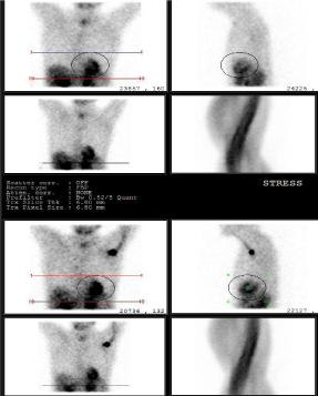
    Figure 1:  Raw projection images of a stress (top) and rest (bottom) SPECT
MPS study using Tc-99m MIBI showing an abnormal focal uptake of tracer
in the neck region.
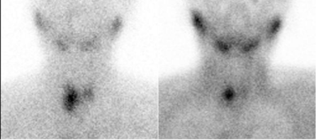
    Figure 2:  Dual phase parathyroid scintigraphy with Tc-99m MIBI showing
focal increased uptake of tracer in the mid portion of the right lobe of the
thyroid gland (left image). The delayed image on the right shows focal
retention in that same location after wash out of tracer in the rest of the gland.
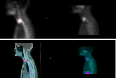
    Figure 3:  SPECT and SPECT\CT images (top and bottom respectively) show
localization of the abnormal uptake of tracer in the neck to the mid portion of
the right thyroid lobe with posterior extension.
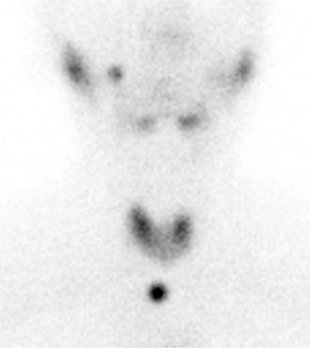
    Figure 4:  Tc-99m pertechnetate thyroid scan showing a normal thyroid gland
with no areas of prominent ‘hot’ or ‘cold’ nodules.
Discussion
In evaluating myocardial perfusion scintigraphy studies, review of the raw projection data is an essential part of the interpretation process [1]. The raw rotating images are used to detect the presence of attenuation and motion artifacts, but a careful examination of these images can provide important information about the presence of significant non-cardiac abnormalities [2]. Display of these images also allows visualization of regions close to the heart in which clinically important incidental findings; like non cardiac pathologies may be found [3]. These non-cardiac findings are usually rare. For instance, Gedik et al. [4] reported 1.2% of non-cardiac incidental findings in their study involving 582 patients. Tc-99m MIBI planar and SPECT scintigraphy are useful in the diagnosis of parathyroid gland disease [5]. Tc-99m MIBI is used routinely, either alone during a dual phase study, or in conjunction with Tc-99m pertechnetate during a dual isotope study for parathyroid scintigraphy pre operatively, like in the case of our patient [6]. Abnormal parathyroid findings have previously been reported on cardiac perfusion studies [1,7]. It is well known that most patients with primary hyperparathyroidism will require surgery [8]. In this case, the incidental finding on the raw data of the MPS resulted in prompt diagnosis and surgical excision of a HPG.
Conclusion
The raw projection image of a SPECT MPS can identify significant non cardiac incidental findings, as this was the case in this patient. We therefore recommend thorough evaluation of the raw data of a SPECT MPS.
References
- Iagaru A, Hachamovitch R, Colletti PM, Wassef H. Demonstration of an ectopic mediastinal parathyroid adenoma on Tc-99m Sestamibi myocardial perfusion scintigraphy. J Nucl Cardiol. 2006; 13: 719-721.
- Raza M, Panjrath GS, Haider A, Jain D. Extracardiac abnormalities on myocardial perfusion imaging. J Nucl Cardiol. 2005; 12: S36.
- Chamarthy M, Travin MI. Altered biodistribution and incidental findings on myocardial perfusion imaging. Semin Nucl Med. 2010; 40: 257-270.
- Gedik GK, Ergun EL, Aslan M, Caner B. Unusual extra cardiac findings detected on myocardial perfusion single photon emission computed tomography studies with Tc-99m sestamibi. Clin Nucl Med. 2007; 32: 920- 926.
- Nguyen BD. Parathyroid imaging with Tc-99m sestamibi planar and SPECT scintigraphy. Radiographics. 1999; 19: 601-614.
- Hindié E, Mellière D, Perlemuter L, Jeanguillaume C, Galle P. Primary hyperparathyroidism: higher success rate of first surgery after preoperative Tc-99m sestamibi-I-123 subtraction scanning. Radiology. 1997; 204: 221- 228.
- Garcia de la Torre N, Wass JA, Turner HE. Parathyroid adenomas and cardiovascular risk. Endocr Relat Cancer. 2003; 10: 309-322.
- Lumachi F, Ermani M, Basso S, Zucchetta P, Borsato N, Favia G. Localization of parathyroid tumours in the minimally invasive era: which technique should be chosen? Population-based analysis of 253 patients undergoing parathyroidectomy and factors affecting parathyroid gland detection. Endocrine-Related Cancer. 2001; 8: 63–69.