Case Report
J Mol Biol & Mol Imaging. 2016; 3(1): 1026.
Ectopic Parathyroid Adenoma Localized by Tc-99m Sestamibi SPECT/CT Localization Prior to Re-operation is Useful
Purbhoo KP¹*, Bombil I² and Mpanya D¹
¹Department of Nuclear Medicine and Molecular Imaging, University of the Witwatersrand, Chris Hani Baragwanath Academic Hospital, Johannesburg, South Africa
²Department of Surgery, University of the Witwatersrand, Chris Hani Baragwanath Academic Hospital, Johannesburg, South Africa
*Corresponding author: Purbhoo KP, Department of Nuclear medicine and molecular imaging, University of the Witwatersrand, P.O Bertsham, Johannesburg 2013, South Africa
Received: August 02, 2016; Accepted: June 29, 2016; Published: August 24, 2016
Abstract
We present a 31-year old male patient with primary hyperparathyroidism (PHPT), complicated by obstructive uropathy due to a staghorn calculus. He had minimally invasive right lower pole parathyroidectomy (MIP), following localization in the right lower pole of the parathyroid gland, on ultrasound. Postsurgical histology confirmed it to be a lymph node. The serum calcium and PTH levels remained elevated post-operative. Tc-99m methoxyisobutyl isonitrile (Sestamibi) planar imaging showed a focus of uptake superior to the upper pole of the right thyroid gland, that was localized lateral to the right true vocal cord on SPECT/CT. Post- operative histology confirmed a parathyroid adenoma.
Keywords: Ectopic parathyroid adenoma; Tc-99m; Sestamibi; Primary hyperparathyroidism; SPECT/CT
Case Presentation
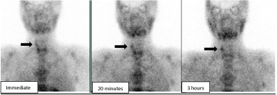
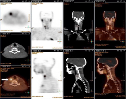
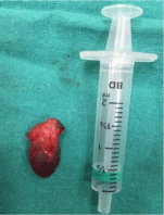
A 31-year old male patient with a diagnosis of PHPT was sent to Nuclear Medicine with persistently raised serum calcium and parathyroid hormone (PTH) levels, following MIP. His hyperparathyroidism was complicated by obstructive uropathy, due to a staghorn calculus, for which he had a double J stent insitu. The ultrasound done prior to surgery showed an enlarged lesion in the right lower pole of the parathyroid gland. Post-surgical histology confirmed it to be a lymph node. No parathyroid gland was demonstrated. The serum calcium and PTH levels remained elevated post-operative. 20 mCi (740MBq) Tc-99m Sestamibi planar imaging (Figure 1) showed a solitary focus of uptake superior to the upper pole of the right thyroid gland, suspicious for a parathyroid lesion. Differential washout of tracer is seen from the immediate to the 3 hour image [black arrows] with a clearer delineation of the lesion on delayed imaging. Single Photon Emission Computed Tomography with CT [SPECT/CT], (Figure 2) demonstrated increased uptake lateral to the right true vocal cord. The patient had removal of the lesion by MIP confirming a parathyroid adenoma (Figure 3). The patients’ serum calcium and PTH levels reverted to the normal range post-surgery.

    Figure 1:  Anterior static Sestamibi images, done immediately, and at 20
minutes and 3 hours show a focal lesion superior and lateral to the right
thyroid lobe.

    Figure 2:  SPECT/CT in tomographic, CT and fused images in the axial, sagittal and coronal plane, showing focal increased uptake lateral to the right hyoid bone
(white arrow).

    Figure 3:  Surgical specimen of the resected ectopic upper pole parathyroid
lesion measuring 15x10x4mm.
Discussion
Ectopic parathyroid adenomas are rare and are reported to account for 5–10% of cases of PHPT [1]. Pre-operative localization of hyperfunctioning parathyroid tissue is an essential component if minimally invasive surgery is scheduled. Parathyroid localization has improved with numerous imaging techniques, including Sestamibi scintigraphy, ultrasonography and four-dimensional CT [2]. Sestamibi scintigraphy, often with complementary ultrasonography, is considered the imaging method of choice for localizing a parathyroid adenoma in the neck [3]. This information facilitates a focused or minimally invasive surgical approach to be used, and therefore may reduce surgical failure rate, complication rates, and operative time [1]. This is even more important in cases of ectopic parathyroid adenomas in which the surgical approach can differ greatly depending on the position of the adenoma [4]. In cases of recurrence of the disease or failed surgery, localization of adenoma by Sestamibi scan is mandatory [5].
SPECT is increasingly used due to the three-dimensional information it provides and the improved sensitivity for the detection and localization of hyperfunctioning parathyroid lesions [2]. SPECT/ CT can further enhance localization by providing better resolution of surrounding structures, and has the added benefit of a more precise localization of ectopic and mediastinal parathyroid lesions [2].
Ultrasound allows anatomical detection of an enlarged parathyroid gland and accurate localization relative to the thyroid gland, although the presence of co-existing nodular thyroid disease reduces the sensitivity and specificity [6]. Deep-seated or ectopic adenomas in the neck are poorly visualized with ultrasound. In a study by Patel et al, ultrasound alone detected 64% of all solitary parathyroid adenomas, versus SPECT/CT (90%) [6]. We routinely do SPECT/CT imaging at 20 minutes post tracer injection for more accurate anatomical localization the lesion. If it is posteriorly located we can confirm a parathyroid lesion, however if it is located anteriorly, it may be due to an intrathyroidal parathyroid lesion or a false positive finding of a thyroid nodule. In this instance Tc-99m Pertechnetate thyroid imaging will be helpful in deciding if a lesion is due to an intrathyroidal lesion versus a thyroid nodule. If the lesion is in an ectopic location, more accurate anatomical localization is obtained. SPECT/CT is more reproducible as compared to ultrasound, which is operator dependent.
The single isotope, dual phase technique is simple and easy to perform [1,7]. It requires a single injection of Sestamibi followed by early and delayed imaging. It takes advantage of the differential washout rates of Sestamibi from the thyroid and the parathyroid glands. On delayed images, parathyroid lesions are easily visualized. This principle is based on the observation that Sestamibi washes out more rapidly from the thyroid than from abnormal parathyroid tissue, where it is retained for a longer period of time. The retention in parathyroid lesions is assumed to be related to the presence of oxyphil cells, which are rich in mitochondria, and are the site of intracellular Sestamibi sequestration [5].
Conclusion
Localization of parathyroid adenomas prior to surgery requires a multimodality approach to aid operative planning. This is particularly important in cases of ectopic parathyroid adenomas, in which the surgical approach has to be modified. The combination of functional imaging using Tc-99m Sestamibi with SPECT or SPECT/CT and anatomical imaging provides accurate surgical localization and therefore greater confidence with surgical planning.
References
- Imene Zerizeret, Arman Parsai, Zarni Win, Adil Al-Nahhasa. Anatomical and functional localization of ectopic parathyroid adenomas: 6-year institutional experience. Nucl Med Commun. 2011; 32: 496-502.
- Michele L. Taubman, Melanie Goldfarb, John I. Lew. Role of SPECT and SPECT/CT in the surgical treatment of primary hyperparathyroidism. International Journal of Molecular Imaging. 2011; 1-7.
- Yodphat Krausz, Lise Bettman, Luda Guralnik, Galina Yosilevsky, Zohar Keidar, Rachel Bar-Shalom, et al. Technetium-99m-MIBI SPECT/CT in Primary Hyperparathyroidism. World J Surg. 2006; 30: 76–83.
- AACE/AAES Task Force on Primary Hyperparathyroidism. The American association of clinical endocrinologists and the American association of endocrine surgeons position statement on the diagnosis and management of primary hyperparathyroidism. Endocr Pract. 2005; 11: 49-54.
- David Taieb, Elif Hindie, Gaia Grassetto, Patrick M. Colletti, Domenico Rubelloet. Parathyroid Scintigraphy. Clin Nucl Med. 2010; 37: 568-574.
- CN Patel, HM Salahudeen, M Lansdown, AF Scarsbrook. Clinical utility of ultrasound and 99mTc sestamibi SPECT/CT for preoperative localization of parathyroid adenoma in patients with primary hyperparathyroidism. Clinical Radiology. 2010; 65: 278–287.
- Vani Vijaykumar, Matthew E Anderson. Detection of ectopic parathyroid adenoma by early Tc-99m sestamibi imaging. Ann Nucl Med. 2005; 19: 157–159.