Research Article
J Mol Biol & Mol Imaging. 2020; 6(1): 1032.
High Resolution Genotyping of Bacillus anthracis Isolated from the Georgia- Azerbaijan Border Territory
Khmaladze E1, Su W2, Sidamonidze K1, Tevdoradze T1, Imnadze P1, Nikolich MP2,3 and Malania L1*
¹National center for Disease Control and Public Health, Georgia
²Walter Reed Army Institute of Research, USA
³US Army Medical Research Unit, Georgia
*Corresponding author: Malania L, National Center for Disease Control and Public Health, Tbilisi, 0152, Georgia
Received: May 18, 2020;; Accepted: October 09, 2020; Published: October 16, 2020
Abstract
B. anthracis is a member of a highly diverse group of endospore-forming bacteria. B. anthracis is considered one of the bacteria with a high degree of genetic homogeneity which makes it difficult to discriminate among the bacterial strains (Rume FI et al.).
Here we present study on genetic characteristics of environmental B. anthracis isolated from the Georgia-Azerbaijan border territory. We examined the genetic diversity of 62 B. anthracis isolates from Georgia-Azerbaijan border by B. anthracis MLVA-25 assay to better understand the dynamic of anthrax in this area. It was found these B. anthracis isolates were conserved. Elucidation of the molecular characteristics and relationships between Georgian and Azerbaijani B. anthracis strain populations will aid in the identification and tracking of strains and their origins.
Introduction
Anthrax is a bacterial zoonosis caused by Bacillus anthracis, a spore-forming, soil-borne bacterium with a remarkable ability to persist in the environment. Found on nearly every continent, the disease is considered a re-emerging zoonotic disease, and despite the development of anthrax vaccines for animals and humans, the disease continues to be endemic in many countries, including Georgia and Azerbaijan.
B. anthracis is a member of a highly diverse group of endosporeforming bacteria. The genus Bacillus contains at least 51 described species and many other species of uncertain taxonomic status. B. anthracis has been classified as a “Category A” organism by the US Centers for Disease Control and Prevention (CDC) and is considered a potential weapon for bioterrorism.
Animal transmission of B. anthracis is classically defined as ingestion of soils, contaminated plants, or contaminated water, as well as mechanical transmission by flies [1-3]. The toxins produced by the vegetative form of the bacterium are associated with virulence and differ from the toxins produced by other Bacillus species. Due to the environmental stability of spores, B. anthracis can remain viable in soil for many years and because its persistence does not necessarily depend on animal reservoirs [4], B. anthracis is extremely difficult to eradicate from endemic areas. Anthrax has recently reemerged as a veterinary and human public health concern in Georgia. In addition to expanding geographically, the number of reported human cases (457) from 2010-2018 has increased nearly three-fold over the previous three years and more than 40-fold since 1985-1987 [5,6]. Moreover, approximately 2,000 B. anthracis foci are registered in Georgia, of which more than 20% are active. Recent research has shown new areas of emergence and clustering around urban centers in Georgia, which contrasts with the normal pattern of high infectivity associated with rural agriculture or remote areas [5,6]. In Azerbaijan, B. anthracis has been responsible for large outbreaks of anthrax in humans and livestock. Although reporting is more infrequent in Azerbaijan than in Georgia, anthrax has persisted for decades across a wide geographic expanse. The disease is considered to be sporadic in Azerbaijan although it is bordered by countries that are hyperendemic [7], and recent evidence has shown that distribution of anthrax in Azerbaijan has undergone changes in its occurrence and spatial distribution [5]. Given the recent re-emergence of the disease in Georgia, there is growing concern over the status of the disease in Azerbaijan. Recent human outbreaks have raised concerns over the re-emergence of the disease, especially given the relatively few reported livestock cases in relation to these human cases. Although the region is experiencing an increased number of anthrax cases, few scientific efforts have been made to link trans-boundary outbreaks of anthrax using molecular characterization and geographic modeling.
In general, the geographic distribution of B. anthracis has been shown to be constrained by a combination of environmental factors such as temperature and precipitation [8,9]. While soil pH of 6.0 or above (alkaline) and factors such as rich organic content and high calcium levels are generally considered to be suitable conditions for the persistence of B. anthracis [4]. Research has also suggested that specific ecological affinities may contribute to the geographic variation in the distribution of genotypes [9].
Elucidation of the molecular characteristics and relationships between Georgian and Azerbaijani B. anthracis strain populations will aid in the identification and tracking of strains and their origins. In addition, genetic data may provide a mechanism for a retrospective epidemiological trace-back and for studying transmission dynamics, both of which will contribute to in-depth knowledge of the distribution and ecology of B. anthracis in Georgia, Azerbaijan, and globally.
Here we present study on genetic characteristics of environmental B. anthracis isolated from the Georgia-Azerbaijan border territory.
Biosafety and biosecurity overview
All of the studies presented in this paper were adhered to the biosafety recommendations contained in the 5th edition of the Biosafety in Microbiological and Biomedical Laboratories (BMBL) Manual published jointly by the CDC and NIH (relevant text from the BMBL Manual is reproduced below for each pathogen).
Safety issues
Safety at sample collection sites was ensured by the use of standard PPE, including: Tyvek coveralls, boots (12 inches high), N95 respirators, safety goggles, disposable hats, and double gloves (inside pair taped to the sleeves of the coverall). Staff at the collection sites have been using this PPE for many years, and are skilled in the safe handling of potentially infectious material.
Materials and Methods
Bacillus anthracis strain culture and inactivation
In this study, we analyzed 62 B. anthracis strains isolated from soil samples collected in the Georgia-Azerbaijan border territory. Bacillus anthracis isolates from pure cultures were grown on 5% Sheep Blood Agar (SBA) plates (Eliava Media Production, Georgia) at 37oC for 24 hours. Several loops of culture were transferred to 1.5mL micro centrifuge tubes and heat-inactivated in an autoclave at 121oC for 20 minutes [13].
DNA isolation and sterility testing
Sterile genomic DNA was extracted by using QIAamp DNA Mini Kits (Qiagen, USA) according to the manufacturer’s instructions.
The sterility of samples was checked by pipetting 5% of the final volume of the DNA and incubating at 37oC in the same growth media used in bacterial culturing. To confirm sterility, at day 3 and day 7, 5μL of isolated DNA was placed on 5% SBA and incubated at 37oC. If no growth was observed after 72 hours at either time point, then the preparation was considered sterile. Primary and secondary containers were decontaminated with 1% sodium hypochlorite for 30 minutes, and stored at -20oC. After surface decontamination, sterile samples could be handled safely under biosafety level 1 or 2 containment.
Before genotyping, DNA quality and concentration of samples was measured using a NanoDrop 2000 instrument (Thermo Fisher Scientific, USA).
Genotyping using Multiple Locus Variable number tandem repeat Analysis
Genetic subtyping of B. anthracis using Multiple Locus Variable number tandem repeat Analysis (MLVA) was performed by a 25-marker MLVA genotyping scheme (MLVA-25) described previously [14] but adapted to a 5-dye Applied Biosystems platform (ABI 3130xl). The 25 PCR primer pairs were divided into four groups: Multiplex-A (eight loci including CG-3, Bams-44, Bams- 3, vrrB-2, Bams-5, Bams-15, Bams-1 and vrrC-1), Multiplex-B (six loci including Bams-13, vrrB-1, Bams-28, vrrC-2, Bams-53 and Bams-31), Multiplex-C (five loci including Bams-25, vrrA, Bams-21, Bams-34 and Bams-24), and Multiplex-D (six loci including Bams- 51, Bams-22, Bams-23 , Bams-30, pXO-1 and pXO-2). Multiplex PCR was performed in a 15-μl reaction volume combining 1×PCR buffer, 0.2mM each of four deoxynucleoside triphosphates, 5mM MgCl2, 1 U of Platinum Taq DNA polymerase with optimized concentrations of pre-mixed primers. The PCR conditions were as follows: initial denaturation at 95oC for 5 min, and then 35 cycles of 95oC for 30 s, 55oC for 30 s, and 72oC for 60 s, with a final extension of 72oC for 5 min and 4oC hold. After amplification, 2μL of each PCR reaction was diluted 100-fold in 198μL of molecular grade water. A denaturation solution/sizing standard solution was prepared from 18.7μL of Hi-Di formamide and 0.3μL of 1200 LIZ size standard; 19μL of the resulting solution and 1μL of the diluted multiplex samples were added to the wells of an ABI platform-compatible plate, e.g., MicroAmp Optical 96-well Reaction Plate (Life Technologies). Samples were denatured in a GeneAmp PCR System 9700 (Applied Biosystems) for five minutes at 95oC and then placed on ice for three to five minutes. PCR products for the 25 loci were resolved by capillary electrophoresis on an ABI Prism 3130xl automated fluorescent capillary DNA sequencer (Applied Biosystems). Amplicons were sized using ROX (carboxy- X-rhodamine)-labeled molecular ladder Liz 1200 (MapMaker 1000; Bioventures Inc., Murfreesboro, TN, USA) and Gene Mapper software version 4.0 (Applied Biosystems).
Data analysis
For each bacterial strain, amplicons from each of the 25 VNTR loci were normalized according to the expected fragment sizes across the diversity of B. anthracis. The higher expected fragment size was used if the difference between the actual fragment size and the expected fragment size was greater than half of the repeat length for that locus; otherwise the next lower expected fragment size was taken if the difference between the actual fragment size and the expected fragment size was less than half a repeat length. The resulting data were analyzed with Bionumerics software package version 7.6 (Applied- Maths, Saint-Martens-Latem, Belgium) as a character dataset. Clustering analysis was done using the categorical coefficient and Unweighted Paired Group Method Arithmetic Average (UPGMA) to generate similarity matrices and dendrograms from this MLVA dataset. Dendrograms are presented with percent similarity.
Calculation of discriminatory power. The discriminatory power of B. anthracis MLVA-25 was presented using the Hunter-Gaston diversity index. Diversity index (Hunter-Gaston) of each locus was calculated using Bionumerics software.
Results
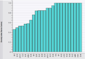
To evaluate the discriminatory power of the selected loci, the Hunter and Gaston discrimination index was calculated for each of the 25 loci used in this study. Among these 62 isolates, the Diversity index value of each locus ranged from 0.46 to 1.00 (Figure 1). Among the 25 loci, there were ten loci for which the allelic diversity index was 1.00, while the allelic diversity index was lowest (0.46) for Bams 3.

    Figure 1: The diversity index value of each locus in 62 isolates from Georgia-
Azerbaijan border.
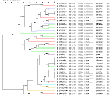
We examined the genetic diversity of 62 B. anthracis isolates from Georgia-Azerbaijan border by B. anthracis MLVA-25 assay to better understand the dynamic of anthrax in this area. It was found these B. anthracis isolates were conserved. Their similarity was 91.6% (Figure 2). When a cutoff value of 93% similarity was applied to define the MLVA cluster, there were two MLVA strain clusters (genotypes) in this collection of 62 isolates. Cluster A is represented by 33 isolates while the cluster B contains 29 isolates. Each cluster has several sub-clusters. Cluster A strains came from three regions (Gardabani, Lagodekhi and Marneuli) while cluster B came from seven regions (Gardabani, Lagodekhi, Rustavi, Sagarejo, Sighnaghi, Axmeta and Dedofliwyaro). Two regions (Gardabani and Lagodekhi) were represented by both cluster A and B. Marneuli only had cluster

Figure 2: Dendrogram based on MLVA-25 of B. anthracis isolates.
Discussion
B. anthracis is considered one of the bacteria with a high degree of genetic homogeneity which makes it difficult to discriminate among the bacterial strains [15]. The high genetic homogeneity is most caused by its spore survival capacity which has allowed B. anthracis to multiply a relatively limited number of times during its evolution. MLVA is a standard assay for bacterial genotyping and has proved to be useful for molecular typing for B. anthracis [16]. There are different MLVA assays used to characterize and differentiate B. anthracis strains on different instrument platforms [17,18,14]. To obtain an increased power of discrimination, a 25-locus MLVA assay was applied to characterize 62 strains of B. anthracis collected in the Georgia-Azerbaijan border region. Of these 25 loci tested, ten loci had a diversity index of 1.00 to provide enough discriminatory power to differentiate B. anthracis isolates to strain level.
According to the results of the MLVA-25 genotyping done in this study, there were two MLVA strain types (genotypes) in this collection of 62 isolates. Looking at geographic distribution, three regions (Gardabani, Lagodekhi and Marneuli) had strains from cluster A while seven regions (Gardabani, Lagodekhi, Rustavi, Sagarejo, Sighnaghi, Axmeta and Dedofliwyaro) had strains from cluster B. Only two regions (Gardabani and Lagodekhi) have both cluster A and B. This distribution suggests that B. anthracis has circulated the most in restricted area. Their rate of spread is relatively slow. It might be due to diversity of environmental conditions and economic activity in these territories [19]. Both Georgia and Azerbaijan have reported sporadic and focal outbreaks of B. anthracis cases in animals and human beings [20-23]. Thus active surveillance is important to prevent and control B. anthracis outbreaks. Based on our knowledge, this is first report of high resolution genetic characterization of B. anthracis circulating in the Georgian-Azerbaijan border region. This provides an initial snapshot of the distribution of B. anthracis genetic subtypes in this area, as well as an important tool for active surveillance to monitor the B. anthracis circulated in the Georgian- Azerbaijan border region in the future [24-29].
Acknowledgment
The work was made possible by support provided by the US Defense Threat Reduction Agency (CBR/GG-27 project) through the Cooperative Biological Engagement Program in Georgia and Azerbaijan. The findings, opinions and views expressed herein belong to the authors and do not reflect an official position of the Department of the Army, Department of Defense of the US Government or any other organization listed.
References
- Blackburn JK, Hadfield TL, Curtis AJ, Hugh-Jones ME. Spatial and Temporal Patterns of Anthrax in White-Tailed Deer, Odocoileus virginianus, and Hematophagous Flies in West Texas during the Summertime Anthrax Risk Period. Annals of the Association of American Geographers. 2014; 104: 939- 958.
- Fasanella A, Adone R, Hugh-Jones M. Classification and Management of Animal Anthrax Outbreaks Based on the Source of Infection. Annali Dell’Istituto Superiore Di Sanita. 2014; 50: 192-195.
- Turner WC, Imologhome P, Havarua Z, Kaaya GP, Mfune JKE, Mpofu IDT, et al. Soil Ingestion, Nutrition and the Seasonality of Anthrax in Herbivores of Etosha National Park. Ecosphere. 2013; 4: 13.
- Hugh-Jones M, Blackburn J. The Ecology of Bacillus anthracis. Molecular Aspects of Medicine. 2009; 30: 356-367.
- Kracalik I, Malania L, Tsertsvadze N, Manvelyan J, Bakanidze L, Imnadz P, et al. Human Cutaneous Anthrax, Georgia 2010-2012. Emerging Infectious Diseases. 2014; 20.
- Kracalik IT, Malania L, Tsertsvadze N, Manvelyan J, Bakanidze L, Imnadze P, et al. Evidence of Local Persistence of Human Anthrax in the Country of Georgia Associated with Environmental and Anthropogenic Factors. PLOS Neglected Tropical Diseases. 2013; 7: e2388.
- Kracalik I, Abdullahyev R, Asadov K, Ismayilov R, Baghirova M, et al. E. Changing Patterns of Human Anthrax in Azerbaijan during the Post-Soviet and Preemptive Livestock Vaccination Eras. PLOS Neglected Tropical Diseases. 2014; 8.
- Blackburn JK, McNyset KM, Curtis A, Hugh-Jones ME. Modeling the Geographic Distribution of Bacillus anthracis, the Causative Agent of Anthrax Disease, for the Contiguous United States using Predictive Ecologic Niche Modeling. The American Journal of Tropical Medicine and Hygiene. 2007; 77: 1103-1110.
- Mullins J, Lukhnova L, Aikimbayev A, Pazilov Y, Van Ert M, Blackburn JK. Ecological Niche Modelling of the Bacillus anthracis A1. a Sub-Lineage in Kazakhstan. BMC Ecology. 2011; 11: 32.
- Kracalik IT, Blackburn JK, Lukhnova L, Pazilov Y, Hugh-Jones ME, Aikimbayev A. Analysing the Spatial Patterns of Livestock Anthrax in Kazakhstan in Relation to Environmental Factors: a Comparison of Local (Gi*) and Morphology Cluster Statistics. Geospatial Health. 2012; 7: 111-126.
- Joyner TA, Lukhnova L, Pazilov Y, Temiralyeva G, Hugh-Jones ME, Aikimbayev A, et al. Modeling the Potential Distribution of Bacillus anthracis under Multiple Climate Change Scenarios for Kazakhstan. PLOS One. 2010; 5: e9596.
- Khmaladze E, Birdsell DN, Naumann AA, Hochhalter CB, Seymour ML, Nottingham R, et al. Phylogeography of Bacillus anthracis in the Country of Georgia shows Evidence of Population Structuring and is Dissimilar to other Regional Genotypes. PLOS One. 2014; 9: e102651.
- Kukhalashvili T. Anthrax Foci in Georgia. Tbilisi, Georgia: National Center for Disease Control and Public Health (NCDC); 2007.
- Lista F, Faggioni G, Valjevac S, Ciammaruconi A. Genotyping of Bacillus anthracis strains based on automated capillary 25-loci multiple locus variable number tandem repeats analysis. BMC Microbiology. 2006; 6: 33.
- Rume FI, Affuso A, Serrecchia L, Rondinone V. Genotype analysis of Bacillus anthracis strains circulating in Bangladesh. PLOS one. 2016; 11: 1-13.
- Belkum AV. Tracing isolates of bacterial species by multilocus variable number of tandem repeat analysis (MLVA). FEMS Immunology and Medical Microbiology. 2007; 49: 22-27.
- Stratilo CW and Bader DE. Genetic diversity among Bacillus anthracis soil isolates at fine geographic scales. Applied and Environmental Microbiology. 2012; 78: 6433-6643.
- Keim P, Kalif A, Schupp J, Hill K, Travis SE, Richmond K, et al. Molecular Evolution and Diversity in Bacillus anthracis as Detected by Amplified Fragment Length Polymorphism Markers. Journal of Bacteriology. 1997; 179: 818-824.
- Smith K, DeVos V, Bryden H, Price L, Hugh-Jones M, Keim P. Bacillus anthracis Diversity in Kruger National Park. Journal of Clinical Microbiology. 2000; 38: 3780-3784.
- Navdarashvili A, Doker TJ, Geleishvili M, Haberling DL. Epidemiology and infection. 2016; 144: 76-87.
- Kracalik I, Malania L, Imnadze P, Blackburn JK. Human anthrax transmission at the Urban-Rural interface, Georgia. American Journal of Tropical Medicine and Hygiene. 2015; 93: 1156-1159.
- Shikhaliyeva S, Ismayilova R. Jahanova J. Isolation of Bacillus anthracis from soil in selected high-risk area of Azerbaijan in 2017. Journal of Prevention and Infection Control. 2018; 4: 48.
- Blackburn JK, Kracalik IT, Fair JM. Applying Science: Opportunities to inform disease management policy with cooperative research within a one health framework. Frontiers in public health. 2016; 3: 276.
- Williams RR WH, Stroup JR, Park M, Poindexter BE. Effects of Freezing and Laboratory Procedures on the Recovery of Bacteria from Ground Beef. J Food Sci. 1980; 45: 757-759.
- Christensen DR, Hartman LJ, Loveless BM, Frye MS, Shipley MA, Bridge DL, et al. Detection of Biological Threat Agents by Real-Time PCR: Comparison of Assay Performance on the R.A.P.I.D., the LightCycler, and the Smart Cycler Platforms. Clinical Chemistry. 2006; 52: 141-145.
- Hoffmaster AR, Fitzgerald CC, Ribot E, Mayer LW, Popovic T. Molecular Subtyping of Bacillus anthracis and the 2001 Bioterrorism-Associated Anthrax Outbreak, United States. Emerging Infectious Diseases. 2002; 8: 1111-1116.
- Stockwell D. The GARP Modelling System: Problems and Solutions to Automated Spatial Prediction. International Journal of Geographical Information Science. 1999; 13: 143-158.
- Klee SR, Ozel M, Appel B, Boesch C, Ellerbrok H, Jacob D, et al. Characterization of Bacillus anthracis-like bacteria isolated from wild great apes from Cote d’Ivoire and Cameroon. Journal of bacteriology. 2006; 188: 5333-5344.
- Keim P, Price LB, Klevytska AM, Smith KL, Schupp JM, et al. Multiple-locus variable number tandem repeat analysis reveals genetic relationships within Bacillus anthracis. Journal of Bacteriology. 2000; 182: 2928-2936.