
Research Article
Austin J Musculoskelet Disord. 2024; 11(1): 1065.
Ice Cream Cone Pelvic Reconstruction after Peri-Acetabular Tumour Resection - Institutional Experience and Description of Implant Fatigue Failure
Ermias Hailemeskel; Viswanath Jayasankar; Ewen Fraser; Ashish Gulia; Ashish Mahendra; Sanjay Gupta*
Department of Trauma & Orthopaedic, Glasgow Royal Infirmary, Glasgow, Scotland
*Corresponding author: Sanjay Gupta Department Trauma & Orthopaedic, Glasgow Royal Infirmary, 84 Castle Street, Glasgow, G4 0SF, Scotland. Email: sanjaygupta@doctors.org.uk
Received: November 27, 2023 Accepted: January 05, 2024 Published: January 12, 2024
Abstract
Aims: Reconstruction following resection of tumours involving the peri-acetabular area is challenging. Achieving acceptable long-term functional status remains difficult and complications are common. The aim of this study was to describe our experience of using the Stanmore ice-cream cone implant after major pelvic tumour resection and present 2 unique cases of implant fatigue failure.
Methods: Ten patients with primary pelvic tumours involving the peri-acetabular region were treated with resection (type 2/2+3) and reconstruction using the Stanmore ice-cream coned pelvic implant between 2010 and 2019 at our centre. Retrospective analysis of our database and patient records was carried out to identify outcomes and complications.
Results: Seven male and three female patients with a mean age of 46 years (range- 17-74 years) were treated with reconstruction using the Stanmore ice-cream cone implant. Chondrosarcoma (n=7) was the most common diagnosis. At a mean follow up of 67 months, 7 patients were alive without disease, 1 patient developed metastatic disease and 2 patients died of recurrent/metastatic disease. Dislocation occurred in 2 patients and 1 patient developed deep infection requiring re-operation for local control. Two patients developed fatigue failure of the implant, which has not been described in the literature. No revision was performed at the time of study.
Conclusion: Reconstruction after peri-acetabular tumour resection remains challenging with high rates of complications. Surgical techniques and implant options are evolving but the ideal implant is yet to be established. Patients should be advised about potential complications including the need for further treatment related to implant failure.
Keywords: Pelvis tumour resection; Reconstruction; Stanmore coned pelvis; Implant fatigue failure
Introduction
Approximately 10% of bone sarcomas occur in the pelvis. The complex 3D anatomy of the pelvis and proximity of tumours to major vessels and nerves make surgical management particularly challenging. Extensive surgery is required to achieve an acceptable oncological margin and postoperative complications are frequent [1-3].
Surgical management of pelvic tumours has undergone a paradigm shift/evolved over recent decades. Hind-quarter amputation, which used to be the mainstay of management haslargely been abandoned and is currently reserved for select indications [2-4]. Limb preserving internal hemipelvectomies are the standard of treatment and the classification system by Enneking and Dunham serves as guide for extent of resection based on the anatomic segment(s) of the pelvis involved by the tumour (resections type I-IV) [1].
A range of reconstructive options have been described following internal hemipelvectomies [2]. While Biological reconstruction /no reconstruction has proved to be a viable option following some type of resections (type I, type III, type I+IV) [5], resections involving the periacetabular region (type II) benefit from reconstruction to restore force transmission and weight bearing along anatomic axes [3,6,7].
Design of endoprosthesis used for peri-acetabular resection have also evolved over the past few decades [3,6]. The Stanmore hemi pelvic stemmed coned implant (ice cream cone implant) is one of the models that was first introduced in 2003. Inspired by the Mckee Farrar stemmed acetabular prosthesis design, the ice cream coned implant was marketed as an off the shelf option for reconstruction of peri acetabular defects following tumour resection. The conical stem design is partly coated with hydroxyapatite and has cutting flutes which provide axial and rotational stability. Additional stability is gained by putting antibiotic-containing cement around the implant and inserting screws/pins in different directions into the remaining Ilium. It also has the advantage of an offset stem which allows for better loading and positioning in the iliolumbar bar and the reaming for the stem is in line with that of the implant thus providing a more secure fit of the stem in the remaining ilium [8,10].
Initial description of short-medium term results of 27 patients by Fisher et al., 2019 [9], has shown an overall complication rate of 37% (infection-11.1%, dislocation-14.8%, loosening-4.4%) and a reoperation rate of 18.5%. Refinements in surgical techniques, namely computer-aided navigation, use of large femoral heads and hypotensive epidural anaesthesia) have resulted in improved complication rates like dislocation and infection [10-12].
The Stanmore ice cream coned pelvic implant has also been utilised in revision arthroplasty with major acetabular defects/bone loss as well as the treatment of acetabular fractures in the elderly with variable short and medium-term results [13,14]. Other pedestal cup designs have since been introduced for peri-acetabular reconstruction with different design modifications and the added advantage of modularity, but still with high rates of complication [3,15-19].
We present our experience of using the Stanmore ice cream coned implant for peri-acetabular reconstruction, with emphasis on 2 unique implant fatigue/structural failures (type 3 failure according to classification system by Henderson et al) [20] in well-fixed implants in young, active patients. To the best of our knowledge, this has not been previously described in the literature.
Material and Methods
We retrospectively analysed the outcomes of 10 patients with primary pelvic bone tumours who were treated with surgical resection and reconstruction using the ice cream coned pelvic implant at the Orthopaedic Oncology unit of Glasgow Royal Infirmary between 2010 & 2019. The unit is a tertiary referral centre for Sarcoma in the west of Scotland and has a catchment area of around 3 million people. The database of the unit, patient charts, radiology reports, pathology reports and MDT Outcome letters were used as data sources.
All surgeries were performed by fellowship-trained orthopaedic oncologists. The preferred position is a ‘’sloppy lateral’’ which allows the patient to be tilted to gain adequate access too anteriorly and posteriorly. Dual ilio-inguinal and posterior Kocher-Langenbeck or lateral approaches were utilized in all cases.
All surgeries were performed under computer navigation. Pre-operative planning of the resection margins was done using planning software OrthoMap 3D, Stryker, Newbury, United Kingdom) and bony resections and prosthesis implantation were performed under computer navigation (Navigation System II, Stryker). Details of the planning and operative technique are described in a study from our unit [21].
After resection of the pelvic segment involved with the tumour (type 2, type 2+3), the remaining ilium is hand reamed and an appropriately sized stem is inserted. The trajectory of reaming as well as stem insertion is done under computer navigation. A double mix of antibiotic-containing cement is used to augment the construct and fill the space around the implant. Additional screws or Steinmann pins are inserted through holes in the cup after the cement is set. After that, a large acetabular cup (most commonly a 48 mm ADM cup) is cemented into the coned cup. Femoral reconstruction proceeds like a normal hip replacement and the Exeter V40 Femoral stem were used when the resection only involved the femoral neck. Trochanteric osteotomies were commonly fixed with screws or a plate (1 case). The Zimmer segmental endoprosthesis system was used in cases where the proximal femur was resected as part of the extra articular peri-acetabular resection.
Results
A total of 10 patients (7 Male and 3 Female) with primary pelvic bone tumours were treated with the Stanmore ice cream coned implant between 2010 and 2019. Mean age at diagnosis was 47 years (range 17-74 years). Chondrosarcoma was the most common diagnosis (5 conventional, 1 de-differentiated, 1 secondary from synovial chondromatosis). The chondrosarcomas were treated with surgery alone, whereas the rest (1 Ewing’s Sarcoma, 1 Chondroblastic Osteosarcoma and 1 high grade Undifferentiated Pleomorphic Sarcoma of bone) had neo-adjuvant chemotherapy.
Surgery
Four patients had type 2+3 resection and reconstruction with standard total hip arthroplasty stem. Six patients had type 2 resection, of which 3 involved proximal femur resection which was reconstructed with Zimmer Segmental endoprosthesis system. Additional screws or pins were inserted through the cement surrounding the cone into the remaining Ilium to augment the construct. Median surgical time was 11.5 hours (range 8-27 hrs) and median blood loss was 4.25L (range 2-15 L).
All surgical margins were clear of tumour except one. A patient with a grade 3 Chondrosarcoma of the proximal femur who presented with a pathologic fracture and underwent an extra-articular type 2+3 resection and reconstruction has positive distal femoral marrow margins, which was treated with post-operative radiotherapy.
Follow up
All patients were followed according to national sarcoma surveillance guidelines. Post treatment baseline cross-sectional imaging of the pelvis was done at 4-6 months post-op. These were used to confirm adequate placement of implant stem in the ilium. Mean long term follow up was 67 months (range 13-116 months). Two patients died during follow up. Both had local disease recurrence. The other 8 continued follow up according to national guidelines of which one was discharged after completing 10 years of post-treatment surveillance.
Complications
Dislocations: 2 patients had dislocations within the first 2 weeks after surgery. Both had type 2+3 resection and femoral reconstruction with standard total hip implants. Both were reduced with manipulation under anaesthesia. One of the patients continued to have persistent dislocation (between the trunnion of the femoral stem and modular head component) and was not fit enough to undergo open reduction. The other patient did not have further dislocations for the reminder of follow up, 11 months from the time of surgery.
Infection: Immediate post op period (within 4 weeks of surgery) - one patient developed deep infection, requiring re-operation. She was treated with irrigation and debridement twice, extended wound VAC treatment for wound closure and local infection control. The patient needed long term suppression antibiotic therapy under the guidance of the infectious disease department. Serial scans demonstrated a deep peri-prosthetic collection, but she was not felt to be fit enough to undergo a major revision/staged surgery and has retained the implant until now (18 months following surgery).
Distant Metastasis
One patient developed lung metastasis on follow up, 45 months after surgery. The diagnosis at presentation was a pathological fracture of the proximal femur secondary to Grade 3 Chondrosarcoma. Surgery involved extra-articular resection of the proximal femur and a type 2 pelvic resection. Distal marrow margins were positive (24cm below the joint level) and the patient received radiation treatment post operatively. He was subsequently enrolled into a clinical trial and is alive at the time writing.
Death
Two deaths occurred within 18 months after surgery. Diagnoses was chondrosarcoma (Grade 2, de-differentiated) and were treated with a type 2+3 resection. Both patients had local recurrence of disease. Palliative radiotherapy was given to one of them for symptom relief.
Fatigue failure: Two patients developed fatigue failure (type 3 failure) of the stem of the coned implant during follow up. Both patients are young males, who had no major postoperative complications and returned to their active lifestyle soon after surgery. Both cases are discussed in more detail.
Patient 1: 40-year-old male with Ewing’s sarcoma of the peri-acetabular area. Treatment involved neoadjuvant chemotherapy, type 2+3 resection and reconstruction. The patient had a smooth postoperative period with no major complications. Initial post-operative pelvic x-rays showed satisfactory placement of implant stem (Figure 1) which was later confirmed on CT scan. Serial follow up x-rays did not demonstrate any abnormality.
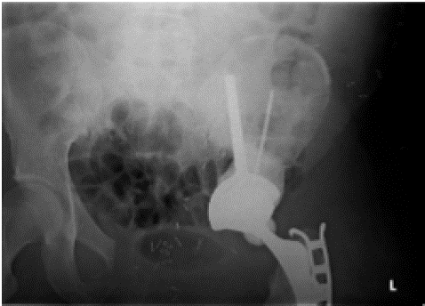
Figure 1: Initial post-operative x-ray following type 2+3 resection and reconstruction.
He underwent laparoscopic abdominal hernia repair 27 months post-op. He presented with mechanical pelvic pain with gradual worsening over a period of weeks around the 30th month post-op. Pelvic x-ray was unremarkable, Pelvic MRI and CT scan of the pelvis showed a stress response paralleling the stem of the coned implant and a small sacral stress fracture at the superior margin of the sacro-iliac joint. He was treated with analgesics and PWB with crutches.
He re-presented 8 weeks later with no improvement in the mechanical pain and x-ray showed fatigue fracture of the coned implant (Figure 2a). He was treated conservatively with analgesics, a period of rest and non-weight bearing. Serial x-rays demonstrate continued loosening around the distal part of the stem, whereas the proximal tip remained well fixed in the bone (Figure 2b).
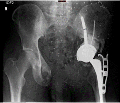
Figure 2a: Pelvic x-ray showing failure of the stem of the ice-cream cone implant.
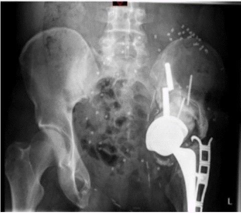
Figure 2b: Pelvic x-ray showing loosening around the distal part of prosthesis (18 months post-failure).
He managed to return to full time work with regular analgesia and using a pair of crutches for ambulation.
Patient 2: 30-year-old male with long-standing left hip pain, that was diagnosed as synovial chondromatosis based on the imaging and biopsy. He underwent a standard total hip replacement. Pathologic analysis of samples sent at that time revealed a secondary malignant transformation in keeping with chondrosarcoma. Baseline imaging demonstrated residual chondroid tumour in the pelvis and the patient was placed on follow-up with regular short interval scans as he was not willing to have pelvic resection at that time. Pelvic MRI done 22 months from total hip replacement, demonstrated an increase in the size of the chondroid lesion
After further work-up and staging, a type 2 extra-articular resection along with the proximal femur was done (Figure 3). The patient had no major complications in the immediate post-op period and returned to full-time work within a year of surgery. accompanied by leg swelling 20 months post op. X-ray demonstrated fatigue failure of the implant stem and the screws through the cement (Figure 4a).
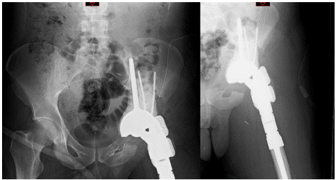
Figure 3: Initial post-op radiographs showing type-2 pelvic resection, proximal femur replacement, with appropriate placement of components.
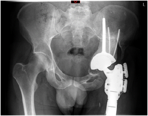
Figure 4a: Pelvic radiograph showing failure of the stem of the coned implant.
He was treated with analgesics, rest, activity modification and weight-bearing restrictions. He was placed on regular short interval follow-up with a serial x-ray which demonstrated no significant loosening or migration of the prosthesis over the following 12 months (Figure 4b). The leg swelling and pain improved gradually and he was able to walk pain-free without crutches for short distances. He returned to full-time work and was placed back on a regular sarcoma surveillance schedule.
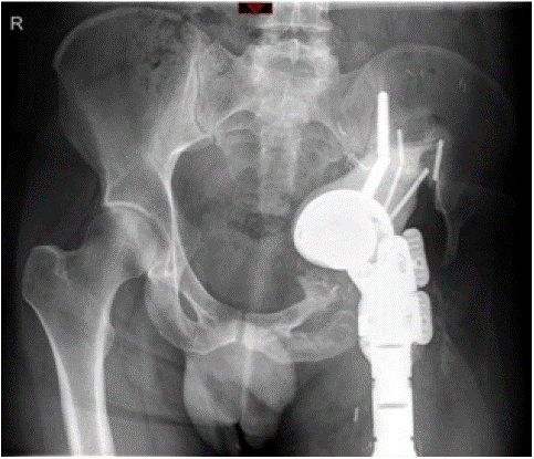
Figure 4b: Pelvic radiograph 18 months latter.
Discussion
Long term follow-up in our small series of 10 patients demonstrated a complication rate of 50 % (2 dislocations, 1 infection and 2 fatigue implant failures). This reflects the complex nature of the surgical management and in keeping with the complication rate observed in other studies of peri-acetabular reconstruction using the Stanmore conned hemi pelvic implant as well as other pedestal cup implants [3,9,16-19].
Despite the limitation of having only 10 patients, the mean follow-up of 67 months post operatively is longer than other studies of done with this implant. In addition, our description of the unique implant fatigue failures to our knowledge is not described elsewhere. The fact that these complications were seen in the medium follow up (20 and 30 months post-op) shows a combination of patient and biomechanical factors play a role in theses failures.
Use of navigation has been demonstrated to reduce complications and improve long term functional outcomes in other studies. All our surgeries were done under computer assisted/navigated surgery which allowed the stem to be implanted centred in the remaining ilium. This in conjunction with the unique design features of the implant have resulted in a stable fixation. This is further augmented by the partial Hydroxyapatite coating of the stem resulting in solid integration of the implant. This was evidenced by the fact that none of the patients had loosening around the stem in long run. This, we believe is due to the lack of micromotion at the interface between a well centred implant with adequate integration in to surrounding bone. However, this solid integration creates excess cantilever stress the part of the prosthesis not implanted into the bone resulting in fatigue failure at this point, as evidenced by the point of failure in our 2 cases. This risk is in part related to the degree of repetitive cantilever stress (cycles per million/lifetime) as well as the absolute stress applied in each cycle, both of which can be influenced by several biomechanics factors such as body habitus, alteration of gait biomechanics and level of activity.
The fact that both patients were young, active males who returned to an active lifestyle soon after surgery at least proves our theory, but needs to be studied further in biomechanics models/laboratory models. We hope our study serves as a basis for future studies.
Conclusion
Reconstruction after peri-acetabular tumour resection remains challenging with high rates of complications. Surgical techniques and implant options are evolving but the ideal implant remain to be established. Computer assisted surgical planning and use of navigation is improving results. The unique implant failure complications should be discussed with patients. Future studies should explore risk factors for implant failure and potential salvage options.
Author Statements
Conflict of Interest
On behalf of all authors, the corresponding author states that there is no conflict of interest.
References
- Enneking WF, Dunham WK. Resection and reconstruction for primary neoplasms involving the innominate bone. J Bone Joint Surg Am. 1978; 60: 731-46.
- Mayerson JL, Wooldridge AN, Scharschmidt TJ. Pelvic resection: current concepts. J Am Acad Orthop Surg. 2014; 22: 214-22.
- Ji T, Guo W. The evolution of pelvic endoprosthetic reconstruction after tumor resection. Ann Joint. 2019; 4: 29.
- Abudu A, Grimer RJ, Cannon SR, Carter SR, Sneath RS. Reconstruction of the hemipelvis after the excision of malignant tumours complications and functional outcome of prostheses. J Bone Joint Surg Br. 1997; 79: 773-9.
- Gupta S, Griffin AM, Gundle K, Kafchinski L, Zarnett O, Ferguson PC, et al. Long-term outcome of iliosacral resection without reconstruction for primary bone tumours. Bone Joint J. 2020; 102-B: 779-87.
- Ji T, Guo W. Periacetabular reconstruction. In: Surgery of the pelvic and sacral tumor. Springer Netherlands. 2020; 81-9.
- Ji T, Yang Y, Tang X, Liang H, Yan T, Yang R, et al. 3D-printed modular hemipelvic endoprosthetic reconstruction following periacetabular tumor resection: Early Results of 80 Consecutive Cases. J Bone Joint Surg Am. 2020; 102: 1530-41.
- METS Hemi-Pelvis Coned Surgical procedure Surgical Procedure Contents; n.d.
- Fisher NE, Patton JT, Grimer RJ, Porter D, Jeys L, Tillman RM, et al. Ice-cream cone reconstruction of the pelvis: A new type of pelvic replacement. Early results. J Bone Joint Surg Br. 2011; 93: 684-8.
- Lowe M, Jeys L, Grimer R, Parry M. Pelvic reconstruction using pedestal endoprosthesis—experience from Europe. Ann Joint. 2019; 4: 34.
- Fujiwara T, Sree DV, Stevenson J, Kaneuchi Y, Parry M, Tsuda Y, et al. Acetabular reconstruction with an ice-cream cone prosthesis following resection of pelvic tumors: does computer navigation improve surgical outcome?. John Wiley and Sons Inc. J Surg Oncol. 2020; 121: 1104-14.
- Aponte-Tinao L. CORR Insights®: reconstruction after hemipelvectomy with the ice-cream cone prosthesis: what are the short-term clinical results? Clin Orthop Relat Res. 2017; 475: 742-4.
- Matharu GS, Mehdian R, Deepu Sethi LJF. Severe pelvic bone loss treated using a coned acetabular prosthesis with conned pelvic implant. Acta Orthop Belg. 2013; 79: 680-8.
- McMahon SE, Diamond OJ, Cusick LA. Coned hemipelvis reconstruction for osteoporotic acetabular fractures in frail elderly patients. Bone Joint J. 2020; 102-B: 155-61.
- Bus MPA. Reconstructive techniques in musculoskeletal tumor surgery management of pelvic and extremity bone tumors reconstructive techniques in musculoskeletal tumor surgery. n.d.
- De Paolis M, Biazzo A, Romagnoli C, Alì N, Giannini S, Donati DM. The Use of Iliac Stem Prosthesis for acetabular Defects following Resections for periacetabular Tumors. ScientificWorldJournal. 2013; 2013: 717031.
- Bus MPA, Boerhout EJ, Bramer JA, Dijkstra PD. Clinical outcome of pedestal cup endoprosthetic reconstruction after resection of a peri-acetabular tumour. Bone Joint J. 2014; 96-B: 1706-12.
- Bus MPA, Szafranski A, Sellevold S, Goryn T, Jutte PC, Bramer JA, et al. LUMiC® endoprosthetic reconstruction after periacetabular tumor resection: short-term results. Clin Orthop Relat Res. 2017; 475: 686-95.
- Issa SP, Biau D, Babinet A, Dumaine V, Le Hanneur M, Anract P. Pelvic reconstructions following peri-acetabular bone tumour resections using a cementless ice-cream cone prosthesis with dual mobility cup. Int Orthop. 2018; 42: 1987-97.
- Henderson ER, Groundland JS, Pala E, Dennis JA, Wooten R, Cheong D, et al. Failure mode classification for tumor endoprostheses: retrospective review of five institutions and a literature review. J Bone Joint Surg Am. 2011; 93: 418-29.
- Young PS, Bell SW, Mahendra A. The evolving role of computer-assisted navigation in musculoskeletal oncology. The Bone & Joint Journal. 2015; 97-B: 258–264.