Research Article
Austin J Musculoskelet Disord. 2015;2(1): 1014.
Clarification of the Distribution Pattern of the Twig(s) of Radial Nerve Innervating Brachial Muscle in Human
Jun Yan1*, Kazuki Masu1, Karen Tokunaga2, Yoshie Nagasawa3 and Jiro Hitomi1
1Department of Biokinetics, Exercise and Leisure Sciences, University of Kwa Zulu-Natal, South Africa
2Department of Sport, Rehabilitation and Dental Sciences, Tshwane University of Technology, South Africa
*Corresponding author: Jun Yan, Department of Anatomy, Iwate Medical University, 2-1-1, Nishi-Tokuta, Yahaba-cho, Shiwa-Gun, Iwate, 028-3694, Japan
Received: January 01, 2015; Accepted: February 24, 2015; Published: February 24, 2015
Abstract
To clarify the distributional pattern of the radial nerve branches innervating the lateral lower part of the brachial muscle, 76 Japanese cadavers (152 sides) were examined, and the number and patterns of the branches were recorded and classified. The results showed that (1), the mean lengths from the lateral epicondyle to the point where the radial nerve pierces through the lateral intermuscular septum were 97.4 mm (range, 70-140 mm) and 96.7 mm (range, 65-126 mm) on the left and right sides, respectively (2), the mean lengths between the lateral epicondyle and the points of the first twig branching from the radial nerve were 64.7 mm (range, 25-130 mm) and 68.9 mm (range, 40-120 mm) on the left and right sides, respectively (3) ,the mean lengths of the branches were 16.6 mm (range, 3-37 mm) and 15.4 mm (range, from 3-80 mm) on the left and right sides, respectively, and (4) the distribution pattern on the brachialis muscle could be divided into 4 types. These data will be useful for clinical practice and helpful for medical students to understand double innervation of muscles.
Keywords: Brachial muscle; Musculocutaneous nerve; Radial nerve; Double innervation; Human
Introduction
In general, the brachial muscle is innervated by the musculocutaneous nerve, as the muscle originates from the ventral myogenic region [1,2]. However, it is also consented that a fine branch from the radial nerve innervates the lateral lower part of the muscle [2-7]. In these twigs, motor fibers also exist [8-13], and these fibers have been shown to belong to the ventral component of the brachial plexus, as demonstrated using the fiber analysis method [14]. Recently, the distribution patterns of the twigs innervating the muscle were indicated and divided into three types however, the number of cases in this previous report was very small [10]. We think, however, as a basic date of human anatomy a certain number of the cases is necessary and reality of the twig(s) can be grasped by the observation of significance number of cases. Moreover, the current distribution pattern of the twig(s) was simple, and it is impossible to explain the variety of the twig(s). Therefore it is necessary to clarify the twig(s) of radial nerve innervating brachial muscle. With this in mind, the purpose of the present observation was to clarify the distribution pattern of the twig(s) of the brachial muscle and to compare the results with those of previous studies.
Materials and Methods
Seventy-six Japanese cadavers (152 sides; age: 60-102 years) were used for this investigation. The cadavers were fixed with 15% formalin through the radial artery and were preserved in 50% alcohol for six months. The cadavers were handled in compliance with the ethical guidelines of Iwate Medical University. During the gross anatomy course at the School of Medicine (2013 and 2014 terms) in Iwate Medical University, the superficial tissues in the brachial and elbow joint region were removed, and the radial nerve and lateral cutaneous nerve of the forearm were dissected (Figure 2). The lengths from the lateral epicondyle to the point where the radial nerve pierces through the lateral intermuscular septum, between the lateral epicondyle and the points of the first twig branching from the radial nerve, and of the twig(s) were examined using a digital caliper (SHINWA, JAPAN). The number and branching patterns of the twig(s) were identified by photography (EOS X3, CANON, JAPAN) and were sketched. A surgical microscope (OME, Olympus, Japan) was used for examination of cases in which fine vessels existed on the side of the twig(s).
Results
Lengths of the twig(s)
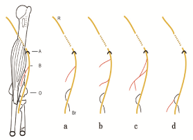
- As shown in Figure 1, the mean lengths from the lateral epicondyle to the point where the radial nerve pierces through the lateral intermuscular septum (OA) were 97.4 mm (range, 70-140 mm) and 96.7 mm (range, 65-126 mm) on the left and rights sides, respectively.
- The mean lengths between the lateral epicondyle and the points of the first twig branching from the radial nerve (Figure 1 OB) were 64.7 mm (range, 25-130 mm) and 68.9 mm (range, 40-120 mm) on the left and right sides, respectively.
- The mean lengths of the main twig were 16.6 mm (range, 3-40 mm) and 15.4 mm (range, 3-80 mm) on the left and right sides, respectively.

Figure 1: Sketch showing the measure points and the branching patterns of the twig(s) innervating the brachial muscle from the radial nerve. a: One twig is innervating the muscle, which shows no relation with the branch innervating the brachioradialis muscle, b: two separate twigs from the radial nerve; c: two or more twigs from the radial nerve distributing after anastomosis with each other, d: the twig(s) were from the brachioradialis muscle and not directly from the radial nerve. A: the point of the radial nerve exiting the lateral head of the triceps muscle; B: the point of the twig(s) branching from the radial nerve; O: the point of the lateral epicondyle, R: radial nerve.
The number and branching pattern of the twig(s)
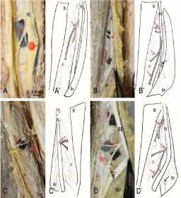
- On the left side, the incidence of twigs was 66/76 cases (86.8%), and 30/76 cases showed two or more branches (39.5%). The incidence on the right side was 69/76 cases (90.8%), and 23/76 cases showed two or more branches (47.4%) (Figure 2).
- As shown in Figure 1, the distribution pattern of the twigs could be divided into four types: one twig only (76/152 sides; 50.0%) (Figure 1a), two twigs from the radial nerve separately entering the muscle (35/152 sides; 23.0%) (Figure 1b), two twigs from the radial nerve merging and entering the muscle (10/152 sides; 6.6%) (Figure 1c), and a twig from the branch innervating the brachioradialis muscle (31/152 sides; 20.4%) (Figure 1d).

    Figure 2:  Photographs (X) and corresponding sketches (X') showing the
twig(s) from the radial nerve innervating the brachial muscle. A, A' and C,
C': one twig from the radial nerve is innervating the muscle (A: left; C: right);
B and B': there are some twigs from the radial nerve to the muscle, and no
relation with the branch innervating the brachioradialis muscle is observed,
D and D': the twigs innervating the muscle originate from the radial nerve
and the branch innervating the brachioradialis muscle. B: superior part of the
brachial muscle, red asterisk: lateral lower part of the brachial muscle, Br:
brachioradialis muscle, R: radial nerve; red arrows: the twig(s) innervating
the brachial muscle, black arrows: the branch innervating the brachioradialis
muscle.
Discussion
The presence of the nerve branch described herein is already recognized; however, there are varying reports on its incidence (between 62% and 100%) [10,15-18,]. In the present observation, the incidences of twig(s) were 86.8% on the left side and 90.8% on the right side. These incidences were similar to that described by Mahakkanukrauh (81.6%) [18]. We speculate that the cause of the different reported incidences in the literature is related to the relatively small number of cases in most previous studies on the topic [4,9,10]. In fact, the 100% incidence reported in one previous study may be incorrect, and may have resulted from fine vessels being mistakenly measured as nerve twigs. On the other hand, in the present report, we observed a relatively high incidence of cases with two twigs; to our knowledge, this has not been previously described in the literature. In terms of the distribution pattern of the twig(s), three types have been reported; however, this classification is not perfect, as the numbers of examined cases were only 6 [5] and 13 sides [10] in the previous studies. In the present observation, 152 sides were examined, and based on our findings, four types of distribution patterns were proposed. We believe that this classification is more appropriate and can adequately classify the nerve twigs. Regarding the nature of the nerve twig(s), it has been reported that sensory and/or motor fibers exist in the twig(s), as determined by clinical examination and electromyogram [2,10,16]. Herein, we focused on the relationship of the twig(s) with the branch innervating the brachioradialis muscle. The brachioradialis muscle is a flexor of the forearm [2], however, it is classified as a dorsal (extensor group), as it is innervated by the radial nerve [19]. In the present observation, the incidence of twigs originating from the branch innervating the brachioradialis muscle was 20.4% (31/152 sides). Because the fibers in the twigs belong to the ventral division of the brachial plexus [14], the nerve fibers in the branch of the brachioradialis muscle also belong to the ventral division of the brachial plexus, and a more appropriate classification of the brachioradialis may be a ventral muscle of the forearm.
When using a standard anterior approach to the humerus [20], the twigs have been reported to be important [4,5]. However, the advanced value of the mean lengths between the lateral epicondyle and pierce point of the twig(s) and of the first twig branching from the radial nerve has not been discussed in the literature, although very simple measurements have been provided [5,10]. In the present study, detailed data were offered, and we clarified that there were no sex differences or left/right differences; accordingly, we believe that the results will be useful for clinical practice and helpful for medical students to better understand double innervation of muscles.
Conclusion
- The incidences of twig(s) innervating the brachial muscle were 86.8% and 90.8% on the left and right sides, respectively.
- The mean lengths between the lateral epicondyle and piercing point of the twig(s) were 97.4 mm (left side) and 97.7 mm (right side). The mean lengths of the first twig branching from the radial nerve were 64.7 mm (left side) and 68.9 mm (right side).
- Four types of innervating patterns were observed.
- Lastly, our results suggest that the brachioradialis muscle could potentially be classified as a ventral (flexor group) muscle, according to the anastomosis with the twig(s).
Acknowledgment
We thank Mr. S. Takahashi and Mr. N. Sasaki (Iwate Medical University) for their technical advice. This work was supported financially by the Advanced Medical Science Center of Iwate Medical University (No. 9250020).
References
- Hollinshead WH. Anatomy for surgeons. Vol 3. London: Cassell and Co. 1958; 365.
- Sinclair DC. Muscles and fasciae, in Canningham's Textbook of Anatomy. Romanes GJ, editor. 12th edn. New York: Oxford University Press. 1981; 322-326.
- Abrams RA, Ziats RJ, Lieber RL, Botte MJ. Anatomy of the radial nerve motor branches in the forearm. J Hand Surg (Am).1997; 22: 232-237.
- Blackburn SC, Wood CPJ, Evans DJR, Watt DJ. Radial nerve contribution to brachialis in the UK Caucasian population: position is predictable based on surface landmarks. Clin Anat. 2007; 20: 64-67.
- Frazer EA, Hobson M, McDonald SW. The distribution of the radial and musculocutaneous nerves in the brachialis muscle. Clin Anat. 2007; 20: 785-789.
- Leonello DT, Galley IJ, Bain GI, Garter CD. Brachialis muscle anatomy. Astudy in cadavers. J Bone Joint Surg Am. 2007; 89: 1293-1297.
- Linell EA. The distribution of nerves in the upper limb, with reference to variabilities and their clinical significance. J Anat. 1921; 55: 79-112.
- Srimathi T, Sembian U. A study on the radial nerve supply to the human brachialis muscle and its clinical correlation. JClinDiagn Res. 2011; 5: 986-989.
- Oh CS, Won HS, Lee KS, Chung IH. Origin of the radial nerve branch innervating the brachialis muscle. Clin Anat. 2009; 22: 495-499.
- Bendersky M, Bianchi HF. Double innervation of the brachialis muscle: anatomic-physiological study.RurgRadiol Anat. 2012; 34: 865-870.
- Trojaborg W. Motor and sensory conduction in the musculocutaneousnerve. JNeurol Neurosurg Psychiatry. 1976; 39: 890-899.
- Braddom RL, Wolf C. Musculocutaneous nerve injury after heavy exercise. ArchPhys Med Rehabil. 1978; 59: 290-293.
- Dundore DE, DeLisa JA. Musculocutaneous nerve palsy: an isolated complication of surgery.ArchPhys Med Rehabil. 1979; 60: 130-133.
- Yan J, Aizawa Y, Honma S, Horiguchi M. Re-evaluation of the human brachialis muscle by fiber analysis of supply nerves.ActaAnat Nippon. 1998; 73: 247-258.
- Spinner RJ, Pichelmann MA, Birch R. Radial nerve innervation to the inferolateral segment of the brachialis muscle: from anatomy to clinical reality.Clin Anat. 2003; 16: 368-369.
- Jones FW. Voluntary muscular movements in cases of nerve lesions. JAnat London.1919; 54: 41-57.
- Ip MC, Chang KSF. A study on radial supply of the human brachialis muscle. AnatRec. 1968; 162: 363-371.
- Mahakkanukrauh P, Somsarp V. Dual innervation of the brachialis muscle.Clin Anat. 2002; 15: 206-209.
- Ouchi H, Murakami T. Myology, in BunTan Anatomy, Okawa T, editor, 11th edn. Tokyo: Kanehara. 1982; 342-345.
- Jordan C, Mirzabeigi E. Atlas of orthopaedic surgical exposures. New York: Thieme Medical Publishers Ltd. 2000.