Research Article
Austin J Musculoskelet Disord. 2015;2(2): 1021.
The Use of Motion Tracking Analysis Program in Detecting Linear Muscular Displacement of the Extensor Digitorum Communis in Patients with Lateral Epicondylalgia: A Cross-Sectional Study
Dones VC III¹*, Suarez CG², Rimando CD³, Yap M4, Seril4, Lopez JF4, Dizon LB4, Carbajal JO4, Francisco CD4, Macaranas JD4 and Paris JH4
¹Senior Researcher - Centre for Health Research and Movement Sciences, Assistant Professor - College of Rehabilitation Sciences, University of Santo Tomas, Philippines
²Director, Centre for Health Researches and Movement Science, College of Rehabilitation Sciences, University of Santo Tomas Professor, College of Rehabilitation Sciences, University of Santo Tomas
³Instructor/Physical Therapy Internship Coordinator - College of Rehabilitation Sciences, University of Santo Tomas, Philippines
4Physical Therapy Graduates - College of Rehabilitation Sciences, University of Santo Tomas, Philippines
*Corresponding author: Valentin C. Dones III, Senior Researcher - Centre for Health Research and Movement Sciences, Assistant Professor - College of Rehabilitation Sciences, University of Santo Tomas, Philippines
Received: May 06, 2015; Accepted: June 18, 2015; Published: June 23, 2015
Abstract
Background/Objectives: The pathology underlying Lateral Epicondylalgia (LE) remains debatable. Abnormality in the movements of the Extensor Digitorum Communis (EDC) was amongst the cited sources of pain in LE. Using the Motion Tracking Analysis Program (MTAP), this study compared the amount of linear displacement of the EDC in elbows with and without LE. Methodology: 54 participants with Lateral Epicondylalgia confirmed by replication of lateral elbow pain in either one of the Cozen, Mill or Maudsley’s test were enrolled in the study. A sonologist scanned both elbows of participants using Musculoskeletal Ultrasound (MSUS). The linear displacement of the EDC shown in the MSUS video was quantified using the Motion Tracking Analysis Program (MTAP). Results: A general trend towards significantly increased linear displacement of EDC in elbows with LE compared to elbows without LE was noted (p=0.06). Based on age, gender, Chronicity of pain, and handedness, no significant differences in the linear displacement of EDC using MTAP were found. Conclusion: Albeit bordering significance (p= 0.06), the increased linear displacement of the extensor digitorum communis reflected its decreased stiffness in elbows with lateral epicondylalgia.
Keywords: Tennis elbow; Motion tracking; Motion tracking analysis program; Extensor digitorum communis
Introduction
Lateral Epicondylalgia (LE) causes pain on the lateral aspect of the elbow attributed to forceful and repetitive hand, wrist and elbow movements. Lateral epicondylalgia is found in 1.1–4.0% in women and 1.0-1.3% in men [1] commonly affecting people in the age range of 35-60 years albeit may also be observed in age younger than 35 and older than 60 years old [1,2]. The higher incidence of LE in women was attributed to the usual activities they are engaged in requiring repetitive movements of their elbow, forearm, wrist and hand such as doing laundry and household keeping. Potential sources of LE include pathological findings in the Extensor Digitorum Communis (EDC) and Extensor Carpi Radialis Brevis (ECRB) [3]. Suggested pathomechanism involved pulling of the EDC on the lateral epicondyle during forceful wrist extension [3-5] and pulling of the ECRB on the lateral epicondyle during resisted wrist extension (Roles and Maudsley 1972). Overuse of these muscles will result to pain and difficulty in moving the wrist especially in to extension (Roles and Maudsley, 1972). The EDC and ECRB stabilize the wrist during gripping activities, counteracting the finger flexor moment force. Forceful handgrip increases stresses on the Common Extensor Origin (CEO) associated with lateral elbow pain [6]. Pathological findings in the EDC and ECRB in elbows with LE are investigated through use of Musculoskeletal Ultrasound (MSUS). According to Iagnocco et al [7]. MSUS is the reference standard in evaluating pathology found in joints involved in various musculoskeletal conditions yielding high resolution images [6]. In an attempt to characterize the linear displacements of the forearm extensor muscles and associate it with Lateral Epicondylalgia, Dones et al. And Liu et al. used MSUS to quantify the movement of these forearm extensor muscles. Additionally, the (ECRB) has small cross-sectional area that made it challenging to be scanned using MSUS [3]. The Motion Tracking Analysis Program (MTAP) by Nicoud et al. quantified the linear displacement of the Extensor Carpi Radialis Brevis in the video images of MSUS [8]. Dones et al. reported the reliability of quantifying the linear displacement of EDC in the proximal half of the forearm using MTAP (ICC=1, 0.99-1.00). Considering that the EDC shares a common origin with the ECRB, both enveloped by a common fascia, the researchers are hypothesizing that the EDC might be associated with LE. Due to the lack of evidence associating Lateral. Epicondylalgia with movement of EDC, this study aimed to compare the linear displacement of the EDC between elbows with LE and elbows without LE. This study determined the association of age, gender, chronicity of symptoms, and handedness with the linear displacement of EDC.
Methodology
Study design
This study was divided into two phased namely:
• Phase 1. This is a reliability study that established the intertester and intra-tester reliability of the three MTAP readers.
• Phase 2. This is a cross-sectional case-control study that compared the EDC movement in elbows with LE and without LE.
Ethics approval
This study was approved by the Human Research Ethics Committee of the University of South Australia (Ethics protocol number 21929) and the Institutional Review Board of the Santo Tomas University Hospital (IRB-AP210-D-LEPS).
Setting
Department of Rehabilitation Medicine of the Faculty of Medicine and Surgery of the University of Santo Tomas
Assessors
The physiotherapist who screened the participants had 5 years of experience in manual physiotherapy. The sonologist has 20 years of experience in rehabilitation medicine and 5 years of experience in the use of musculoskeletal ultrasound with a musculoskeletal case load of 5-7 patients a day for 5 days in a week. In a published intertester reliability study, the sonologist’s reliability in scanning elbows with LE for bony lesions, neovascularity and fluid was comparable to a sonographer who had 20 years of experience in the use of musculoskeletal ultrasound (Kappa=0.53-1.0) [9]. Three 5th year physical therapy students with 11 months of clinical physiotherapy experience quantified the linear displacement of the EDC using the Motion Tracking Analysis Program (MTAP).
Equipment Used
Musculoskeletal ultrasound
Ultrasound measurements were made with a Siemens Antares Sonoline Ultrasound machine [Siemens Medical Solutions, USA, Inc, Ultrasound Group Issaquah, and WA] with a 5-13 MHz linear array broadband transducer.
Motion tracking analysis program
The Motion Tracking Analysis Program (MTAP) by Nicoud et al. is a program that quantifies the linear displacement of anatomical structures in pixel as unit of measurement, such as the EDC which is the variable of interest in this study [8]. During EDC tracking, a muscle’s location is determined & given coordinate values in each frame of a video. MTAP uses normalized cross-correlation to track a rectangular area known as a template from frame-to-frame in the video. Dones et al. and Nicoud et al. reported the mechanism of quantifying the movements of muscles using MTAP [8,9].
Participant Selection
Participants were recruited from hospitals, clinics, sports centres and barangay health centres. Flyers and social networking sites were used to disseminate information. Potential participants with lateral elbow pain were screened by an experienced physiotherapist using the initial screening checklist (Appendix 1). Participants were given and signed consent forms. Participants whose lateral elbow pain was replicated by either one of the Cozen, Mill or Maudsley’s test were included in the study. Exclusion criteria were: current general body malaise, diagnosis of cancer, previous or current fractures in the upper limb, osteoarthritis of the elbow, recent blunt trauma to the elbow, and previous surgery to the elbow. Consequently, regardless of side of pain, both elbows of participants were scanned by a sonologist using a musculoskeletal ultrasound.
Musculoskeletal ultrasound protocol
Elbows of seated participants were placed in a mechanical contraption keeping the elbow extended while the wrist was flexing (Figure 1). The right elbow was scanned first before the left elbow. The wrist was flexed only once. During wrist flexion, the sonologist placed the transducer head on the proximal half of the dorsal side of the forearm (Figure 1).

    Figure 1:  Musculo skeletal ultrasound protocol.
(a) Mechanical contraption for isolating wrist movement
(b) Placement of US transducer head
Motion tracking analysis program protocol
An independent researcher coded the MSUS videos and classified the MSUS videos based on presence or absence of LE. The coded MSUS videos were randomly assigned to the three blinded assessors. The assessors used three laptops with installed MTAP and with uniform screen dimensions (14 inches) and resolutions (1366x768x59 Hz).
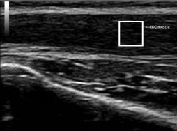
The MSUS video was uploaded on the MTAP software. The initial template was determined on the first image of the MSUS video. The initial template contained hypoechogenic variable of interest found inside the EDC (Figure 2) with dimension of 2cmx2cm. The initial template was cross-correlated with the second frame of the MSUS video and the region of best match generates the next version of the template which was tracked in the subsequent frame and so on until the last frame was processed. This adaptive template approach accommodates the small changes which occur in the feature being tracked over several frames and improves the tracking greatly. Horizontal displacements of the EDC (x- axis) were obtained and collated using Microsoft Excel. The greatest and lowest values were obtained after arranging the coordinates. The difference of these values is considered to be equivalent to the linear displacement of the EDC muscle. The values of vertical displacement (y-axis) were neglected because it is not within the scope of the study. This study was divided into two phases namely: a. preliminary phase that had established the reliability of the three physical therapy interns and; b. observational cross-sectional study that examined the linear displacement of the EDC through use of MSUS.

Figure 2: Initial template containing hypoechogenic portion of EDC.
Phase 1: Reliability Testing
Intra-tester reliability
From the collected MSUS videos of eligible participants, 10 MSUS videos were randomly assigned to three blinded assessors using the fishbowl technique. These MSUS videos contained the EDC movements of LE and non-LE elbows. Each video was run thrice by each of the assessors using MTAP.
Inter-tester reliability
From the collected MSUS videos of eligible participants, one MSUS video was randomly chosen and used to determine the intertester reliability of the three assessors. MTAP Readings were exported to Microsoft Excel to sort the linear displacement of EDC based on coordinates. The lowest coordinate value was subtracted from the highest coordinate value yielding the linear displacement of EDC.
Data analysis
The computed linear displacement values were analyzed for intra-tester and inter-tester reliability using Intraclass Correlation Coefficient by MedCalc. Intraclass correlation coefficient is a measure of the reliability of measurements or ratings. ICC using Absolute agreement determined the reliability of the three assessors based on the collected data. The Intra-Class Correlation Coefficient (ICC) was used to determine the intra-tester reliability of the junior investigators. ICC with same raters and of absolute agreement was used to determine the intra-tester reliability. Absolute agreement considered systematic differences involved in the process of quantifying SMHGT of included patients. Intra-class correlation coefficients were interpreted in this study as follows:
• 0-0.2: poor agreement
• 0.3-0.4: fair agreement
• 0.5-0.6: moderate agreement
• 0.7-0.8: strong agreement
• >0.8: almost perfect agreement
Stockehndahl et al. suggested that a Kappa score of =/>0.4 is clinically acceptable in establishing the intra-tester reliability of examiner in evaluating joints and muscles of individuals with musculoskeletal conditions [10].
Phase 1 established the reliability of the assessors. This was followed by Phase 2 that aimed to quantify the linear displacement of the EDC in LE and non-LE elbows.
Phase 2: Observational Cross-Sectional Study
Data analysis
Motion Tracking Analysis Program readings were exported to Microsoft Excel (2007) to sort the linear displacement of EDC based on coordinates. The lowest coordinate value was subtracted from the highest coordinate value yielding the linear displacement of EDC. The D’Agostino-Pearson Test for Normal Distribution was used to determine the homogeneity of the readings by the three assessors. Following the determination of the homogeneity of the distribution, the following non-parametric tests were used:
Wilcoxon test
Based on presence or absence of LE, and hand dominance, this test was used to determine the presence of significant differences in EDC movement between LE elbow and the corresponding non-LE elbow of the same participant. Wilcoxon test for paired samples is the non-parametric equivalent of the paired samples t-test [11].
Mann whitney U test
Based on gender and Chronicity of symptoms, this test was used to determine presence of significant differences in linear displacement of EDC between LE and non-LE elbows. It is a non-parametric equivalent of the independent samples-t test. The Mann-Whitney test is the non-parametric equivalent of the independent samples t-test [11].
Kruskal-wallis test
Based on age, this test was used to presence of significant differences in linear displacement of EDC between LE and non-LE elbows. The Kruskal-Wallis test is an extension of the Wilcoxon test and can be used to test the hypothesis that a number of unpaired samples originate from the same population [11].
Results
Baseline characteristics of participants
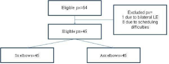
Fifty-four participants (39 female, 15 male) aged between 15 to 66 years (mean (SD):41(13)) were included in the study. Eight (8) participants could not participate in the MSUS scan because of scheduling difficulties. Of the 45 available participants (90 elbows, 31 females, 14 males), 41 were right handed. One participant was ambidextrous. Of the 45 symptomatic elbows, 36 elbows were on the dominant side (34 on the right, 1 on the left, and 1 participant was ambidextrous). Twenty-seven (27) elbows had LE for more than 6 weeks. Eighteen (18) elbows had LE for less than 6 weeks (Figure 3).

Figure 3: Flow chart of participants.
Phase 1: Reliability testing
All three junior investigators who read the MSUS video using MTAP had moderate to good intra-tester and inter-tester reliability (ICC=0.67 to 1.00) (Appendices 2 and 3) show results on intra-rater and inter-tester reliability of three blinded junior investigators.
Phase 2: Motion tracking of EDC
9.3.1. Normality test to determine homogeneity of data: Using D’Agostino-Pearson test for Normal Distribution, the results of the measurements on linear displacement (MTAP) of Extensor Digitorum Communis demonstrated a non-normal distribution (P<0.0001).
Linear muscular displacement of EDC based LE diagnosis
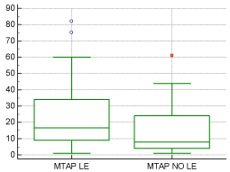
Using Wilcoxon test, there was a general trend towards presence of significant difference in the linear displacement of the Extensor Digitorum Communis between elbows with LE and elbows without LE with a p-value of 0.06 as reported in (Table 1). ( Figure 4) shows that elbows with LE had greater linear displacement of the EDC muscle as compared to elbows without LE approaching significant difference (p=0.06).
    N=elbows
    Median
    95% CI for Median
    p value


    With LE
    45
    16.50
    9.88-25.00
    0.06


    Without LE
    45
    8.00
    5.00-16.00


    Key:CI, confidence interval; LE, lateral    epicondylalgia.
Table 1: Comparison of MTAP Results of elbows with LE vs elbow without LE.

    Figure 4:  Box-and-Whiskers Plot comparing MTAP results of elbow with LE
vs elbow without LE
Key: LE, Lateral epicondylalgia; MTAP, Motion Tracking Analysis Program.
Linear displacement of EDC based on hand dominance
Using the Wilcoxon test, no significant difference in the linear displacement of the Extensor Digitorum Communis was found between dominant and non-dominant elbows (P=0.21), as shown in (Table 2).
    N=elbows
    Median
      (pixel/s)
    95% CI for Median
      (pixel/s)
    p value


    Dominant Hand
    36
    16.00
    9.00-23.00
    0.21


    Non-dominant Hand
    9
    8.00
    6.46-16.00


    Key: CI, confidence interval; LE, lateral    epicondylalgia; MTAP, motion tracking analysis program.
Table 2:  Comparison of MTAP Results of elbows with LE and elbows without LE
based on hand dominance.
Linear displacement of EDC based on chronicity of symptoms
Using the Mann-Whitney test, no statistical significant difference in the linear displacement of the EDC between elbows with LE and elbows without LE based on chronicity of symptoms was found (P=0.28). (Table 3) reports the comparison of MTAP results on EDC based on chronicity of symptoms.
    N=elbows
    Median
      (pixel/s)
    95% CI for Median
      (pixel/s)
    p value


    < 6 weeks
    18
    10.00
    9.00-16.98
    0.28


    > 6 weeks
    27
    20.00
    9.00-26.70


    Key: E, lateral epicondylalgia; MTAP, motion    tracking analysis program.
Table 3: Comparison of MTAP results for LE chronicity of <6 weeks vs >6 weeks.
Linear displacement of EDC based on age of participants
Kruskal-Wallis test resulted to no significant difference on linear displacement of EDC based on age groups (<40 y/o, 40-60 y/o, >60 y/o) (p = 0.34). (Table 4) reports the comparison of MTAP results based on different age groups.
    N=participants
    Median
    95 % CI for median
    p value


    <40 y/o
    18
    17.5
    12.34-31.33
    0.34


    40-60 y/o
    24
    15.5
    8.75-28.77


    >60 y/o
    3
    10
    (-)28.43-59.09


    Key: N, number of participants;    y/o, year old.
Table 4:  Comparison of MTAP results on different age groups as referenced by
the median.
Linear displacement of EDC based on gender
Using Mann-Whitney Test, no significant difference in MTAP values of EDC was found between males and females (p = 0.78). (Table 5) reports the comparison of MTAP results on EDC based on gender.
    N=participants
    Median
    95% CI for Median
    p value


    Male
    14
    15
    8.53-30.74
    0.78


    Female
    31
    14
    9-23


    Key: N, number of participants.
Table 5: Comparison of MTAP results of EDC based on gender.
Discussion
Using the Motion Tracking Analysis Program (MTAP) by Nicoud et al. this is the first study to compare the movement of the Extensor Digitorum Communis (EDC) in musculoskeletal video images between LE and non-LE elbows of patients with unilateral LE. The results of this study were as follows a. There was a general trend towards significantly greater linear displacement of the EDC in LE elbows compared to non-LE elbows and b. No significant differences in the linear displacement of EDC between elbows were found based on hand dominance, chronicity of symptoms, age of participants and gender. Albeit bordering significant difference, a greater displacement of EDC in LE elbows compared to non-LE elbows underpins decreased stiffness in the elbow region. Dones et al. reported on inherent tightness of human elbows due to the arrangement of the EDC in the proximal forearm. The EDC was tightly attached to the deep fascia on the lateral epicondyle and distal to the radiocapitellar joint. On the anterior aspect of the lateral epicondyle, the EDC together with the Extensor carpi radialis brevis (ECRB) were covered by tight deep fascia a.k.a regular deep connective tissue [3,12]. Distal to the radiocapitellar joint, the proximal bellies of the EDC appeared tightly attached to the deep fascia. Separation of the EDC on the deep fascia caused dismemberment of its muscle fibres. Repetitive and forceful handgrip activities require a level of tightness in the elbow [13]. The seemingly increased linear displacement of the EDC in LE elbows investigated in this study suggests decreased resistance of EDC muscle to wrist flexion movement underpinning decreased tightness. Decreased tightness suggests a lower capacity of elbow to oppose rapidly changing forces of handgrip activities. Chourasia et al. reported that increased risk for injury or recurrence of injury may be associated with reduction in ability to oppose changing forces in the elbow on side of repetitive and forceful grip [13]. Chronicity of symptoms, age and gender are reported to be associated with tightness of tissues [14,15]. Chronicity of symptoms were commonly associated with stiffness of tissues secondary to accumulation of adhesions and fibrosis brought about by repetitive micro-injury [16]. Tightness of the actin-myosin cross bridges were seen in older age groups secondary to increased tightness in the actin-myosin cross-bridges [14]. Males demonstrated less active & passive muscle flexibility as compared to females [15]. Despite that these factors were reported to be associated with tightness; our current study did not show any associations between chronicity of symptoms, age and gender and stiffness of EDC. Furthermore, hand dominance did not significantly alter the amount of linear displacement of EDC in LE elbows. The elbows scanned in this study were true representative of elbows with LE and without LE. Standard clinical provocation tests were used to determine elbows with LE and without LE. Participants, albeit recruited through convenience sampling, represented patients from inpatient and outpatient settings. Albeit only 45 out of 54 participants’ LE and non-elbows were compared, the data collected assumed
Limitations of the Study
Only 45 out of 54 participants’ LE and non-elbows were compared which may have contributed to the non-homogenous distribution of collected data on linear muscular displacement of EDC. A greater number of scanned elbows could have provided a better contrast on muscular displacement of EDC between LE and non-LE elbows. Albeit elbows included in this study were true representative of LE and non-LE elbows based on diagnostic examinations and inclusion of inpatient and outpatient sample population, a higher sample size is recommended in future studies. The quality of the MSUS videos might have affected the template used in quantifying the EDC displacement. Artefacts in MSUS video images may be secondary to the accessory movement of the wrist during flexion. These accessory movements may be constrained by using a mechanical contraption machine that fits the size of the wrist, preventing excess wrist movement. An automatic system where a video display tube signals and electronically starts wrist movement may decrease the time lag between verbal prompting of the sonologist and the initiation of the wrist flexion by the participant. This will eliminate unnecessary waiting time prior to start of wrist flexion. This time lag due to the manual process used in this study was not quantified. Due to unavoidable errors in scanning such as the transducer head slipping off the skin while the wrist was flexing, a number of patients had to repeat the wrist flexion movement more than once. This could have influenced the flexibility of the EDC affecting its linear displacement.
Conclusion
It appears that presence of LE may increase the linear displacement of EDC in LE elbows. The decreased tightness of the EDC may be secondary to injuries accumulated in the CEO due to repetitive and forceful handgrip activities. Hand dominance, chronicity of symptoms, and gender were not associated with the linear displacement of the EDC.
Acknowledgment
The authors would like to acknowledge Professor John Thomas and Dr Peter Lesniewski of the University of South Australia for consenting use of the Motion Tracking Analysis Program.
Funding
This study was funded by the Department of Science and Technology – Philippine Council for Health Research and Development.
References
- Murgia A, Harwin W, Prakoonwit S, Brownlow, H. Preliminary observations on the presence of sustained tendon strain and eccentric contractions of the wrist extensors during a common manual task: implications for lateral epicondylitis. Medical Engineering & Physics.2011; 33: 793-797.
- Herquelot E, Guéguen A, Roquelaure Y, Bodin J, Sérazin C, Ha C, et al. Work-related risk factors for incidence of lateral epicondylitis in a large working population. Scand J Work Envin Health: 2013; 39: 578-588.
- Dones VCIII, Milanese S, Worth D, Grimmer-Sommers K. The anatomy of the forearm extensor muscles and the fascia in the lateral aspect of the elbow joint complex.Anatomy & Physiology. 2013; 3: 1-7.
- Bunata RE, Brown D, Capelo R. Anatomic factors related to the cause of tennis elbow. The Journal of Bone & Joint Surgery. 2007; 89: 1955-1962.
- Takasaki H, Aoki M, Muraki T, Uchiyama E, Murakami G, Yamashita T. Muscle strain on radial wrist extensors during motion-stimulating stretching exercise for lateral epicondylitis: a cadaveric study. J Shoulder Elbow Surg. 2007; 16: 854-888.
- Campbell BJ. Wrist extension counter-moment force effects on muscle activity of the ECR with gripping: implications for lateral epicondylalgia [dissertation]. Auburn University, 2006.
- Iagnocco A, Naredo E, Bijlsma JWJ. Becoming a musculoskeletal ultrasonographer. Best Practice & Research Clinical Rheumatology. 2013; 27: 271-281.
- Nicoud F, Castellazzi G, Lesniewski P, Thomas JC. Velocity determination of nerve tissue from ultrasound video images.Australian Institute of Physics. 2008: 73-76.
- Dones VCIII, Grimmer-Sommers K, Thoirs K, Gonzalez-Suarez C. Inter-tester reliability of sonographers in detecting pathological lesions in elbows of individuals with lateral epicondylar pain. Journal of Musculoskeletal Research. 2011; 14: 1-14.
- Stochkendahl MJ. Christensen HW. Hartvigsen J. Vach W. Haas M. Hestbaek L. et al. Manual examination of the spine: a systematic critical literature review of reproducibility. Journal of Manipulative and Physiological Therapeutics. 2006: 29; 475-485.
- Wilcoxon Test.
- Van De Wal J. The architecture of the connective tissue in the musculoskeletal system: an often overlooked functional parameter as to proprioception in the locomotor apparatus. Int J Ther Massage Bodywork.2009; 2:9-23.
- Chourasia AO, Buhr KA, Rabago DP, Kijowski R, Sesto ME. The effect of lateral epicondylosis on upper limb mechanical parameters.Clin Biomech (Bristol, Avon). 2012; 27:124-130.
- Ochlala J, Frontera WR, Dorer DJ, Van hoecke J, Krivickas LS. Single skeletal muscle fiber elastic and contractile characteristics in young and older men. J Gerontol A Biol Sci Med Sci. 2007; 62: 375-381.
- Blackburn JT, Riemann BL, Padua DA, Guskiewicz KM. Sex comparison of extensibility, passive and active stiffness of the knee flexors. Clin Biomech (Bristol, Avon). 2004; 19: 36-43.
- Spina A. Treatment of proximan hamstring pain using active release technique applied to the myofascial meridian: a case report. Sports Performance Centres.
Citation: Dones VC III, Suarez CG, Rimando CD, Yap M, Seril, Lopez JF, Dizon LB, et al. The Use of Motion Tracking Analysis Program in Detecting Linear Muscular Displacement of the Extensor Digitorum Communis in Patients with Lateral Epicondylalgia: A Cross-Sectional Study. Austin J Musculoskelet Disord. 2015;2(2): 1021. ISSN : 2381-8948