Clinical Image
Austin J Musculoskelet Disord. 2016; 3(1): 1030.
Park-Harris Growth Arrest Lines Associated with Systemic-Onset Juvenile Idiopathic Arthritis
Sifuentes Giraldo WA*, Boteanu AL and Gámir Gámir ML
Pediatric Rheumatology Unit, Ramón y Cajal University Hospital, Spain
*Corresponding author: Sifuentes Giraldo WA, Pediatric Rheumatology Unit, Ramón y Cajal University Hospital, Madrid 28034, Spain
Received: May 18, 2016; Accepted: May 19, 2016; Published: May 20, 2016
Keywords
Park-Harris growth arrest lines; Systemic-onset juvenile idiopathic arthritis
Clinical Image
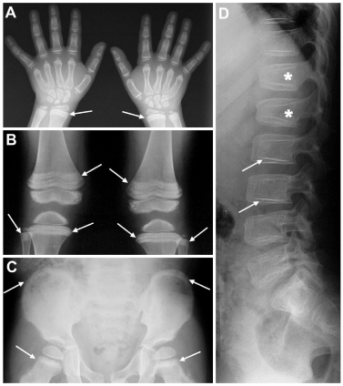
A 17-year-old boy was diagnosed with systemic-onset Juvenile Idiopathic Arthritis (JIA) at age 2 years based on spiking fever, evanescent macular rash and polyarthritis. The patient was treated with intra-articular and high-dose systemic corticosteroids, methotrexate, anakinra (discontinued due to injection site reaction) and tocilizumab, but he developed recurrent episodes of macrophage activation syndrome, growth retardation and osteoporosis with vertebral compression fractures (Figure 1D, asterisks) and was also treated with calcium plus vitamin D supplements, pamidronate and growth hormone. Radiographs at age 10 years revealed multiple radio-opaque horizontal lines in the distal radius, proximal and distal femur, proximal tibia and fibula, and iliac crest (Figure 1A, B and C, arrows) and parallel lines to endplates in both thoracic and lumbar spine (Figure 1D, arrows). These findings were consistent with Park-Harris growth arrest lines [1]. These lines were less evident in subsequent radiographs and disappeared at age 16 years. The Park-Harris growth arrest lines are due to thin transverse bone layers formed below zones of provisional calcification during the time when growth has recovered after a period of cessation. When growth is re-established, cartilaginous proliferation and increased osteoblastic activity contribute to the thickening and metaphyseal migration of the transverse lines [2]. The growth arrest lines are most common in regions associated with significant longitudinal growth such as the distal femur and proximal tibia and the lines become thinner as the distance from the physis increases [1]. These lines may occur as a result of repetitive trauma, infection, nutritional disorders (malnutrition, rickets), hypervitaminosis D, radiation, immobilization, hypoparathyroidism, alcohol consumption during growth, JIA, psychosocial short stature, endemic skeletal fluorosis, bisphosponate therapy, heavy metal poisoning or leukaemia [1- 3]. After normal growth resumes and longitudinal trabeculae are restored, the Park-Harris lines may eventually disappear [1].

    Figure 1: (A) Plain radiograph, Posterior Anterior (PA) view of the hands, (B)
Plain radiograph, PA view of the knees, (C) Plain radiograph, PA view of the
pelvis, (D) Plain radiograph, and lateral view of the lumbar spine.
References
- Lee TM, Mehlman CT. Hyphenated history: Park-Harris growth arrest lines. Am J Orthop (Belle Mead NJ). 2003; 32: 408-411.
- Sajko S, Stuber K, Wessely M. Growth Restart/Recovery Lines involving the vertebral body: a rare, incidental finding and diagnostic challenge in two patients. J Can Chiropr Assoc. 2011; 55: 313-317.
- Sifuentes Giraldo WA, Vázquez Díaz M, Barrios Castellanos R. The “bone within a bone” signs in idiopathic hypoparathyroidism. Reumatol Clin. 2012; 8: 372-374.