Clinical image
Austin J Nephrol Hypertens. 2014;1(2): 1008.
Pericardial Effusion and Dilated Cardiomyopathy in Acute Kidney Failure Patients: Role of Nephrologists’ Performed Echocardiography
Di Lullo L1*, Rivera R2 and Santoboni A1
1Department of Nephrology and Dialysis, L. Parodi Delfino Hospital, Italy
2Department of Nephrology, S. Gerardo Hospital, Italy
*Corresponding author: Di Lullo L, Department of Nephrology and Dialysis, “L. Parodi Delfino Hospital”, Colleferro (Roma), Italy
Received: June 30, 2014; Accepted: July 21, 2014; Published: July 23, 2014
Introduction
Cardiovascular diseases such as coronary artery disease, congestive heart failure, arrhythmias and sudden cardiac death represent main causes of morbidity and mortality in patients with acute (AKF) and chronic kidney disease (CKD). Pathogenesis includes close linkage between heart and kidneys and involves traditional and non-traditional risk factors.
Patients with acute kidney failure often present to emergency units with dyspnoea and anuria. Echocardiography usually shows the presence of dilated cardiomyopathy with left atrium enlargement also accompanied by mild to severe pericardial effusion.
Here we present two imagine case reports concerning two patients referred to our nephrology unit for acute kidney failure with oligo – anuria, dyspnoea and massive low limbs edema.
Case Presentation N° 1
66 years old male referred to emergency unit for growing dyspnoea, affected by chronic kidney disease (eGFR EPI 15 ml/ min/1.73 m2) due to diabetic nephropathy. He was a hypertensive patient on calcium channel blockers therapy (manidipine 20 mg/die) with chronic heart failure followed by nephrology ambulatory unit.
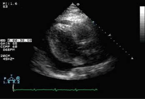
When he was admitted to nephrology unit, an echocardiography was performed by certified nephrologists and massive pericardic effusion (Figure 1) with left atrium enlargement (Figure 2) and EF (ejection fraction) decline were reported. Serologic tests showed an increase in creatinine (9.2 mg/dl) and BUN levels (98 mg/dl) with normokaliemia (4.4 mEq/L) and preserved bicarbonate levels (21mEq/L).
Figure 1: Massive pericardial effusion.
Figure 2: Left atrium enlargement.
CVVHD renal replacement therapy was started with 12 hours treatment. After one week hospitalization and daily renal replacement therapy schedule, patient recovered from heavy dyspnoea and echocardiography showed regular left atrium area with no pericardial effusion and EF recovery ‘till 55% (Figure 3).
Figure 3: Echocardiographic pattern after one week daily hemodialysis.
Case Presentation N° 2
62 years old female with diagnosis of type 2 diabetes mellitus on metformin treatment, chronic kidney disease (eGFR EPI: 36 ml/min) and arterial hypertension on alpha blockers and calcium channel blockers therapy.
Patient referred to emergency unit for oligo–anuria and dyspnoea, hypotension and tachycardia. Blood tests revealed impaired renal function (creatinine 10.3 mg/dl) with hyperkaliemia (7.3mEq/L) and metabolic acidosis (HCO3- 14 mEq/L). An echocardiography was performed by certified nephrologist showing large pericardial effusion (Figure 4) with preserved systolic function (EF: 58%).
Figure 4: Severe pericardial effusion in acute kidney failure.
Patient underwent CVVHD and recovered from pericardial involvement (Figure 5) into two weeks with dramatic improvement on clinical picture.
Figure 5: Echocardiographic picture after four CVVHD sessions.
Conclusion
Above cited case reports underline crucial importance of nephrologist performing echocardiography helpful to assess fluid status in AKF patients.
Echocardiography, together with clinical picture and serological assays, allows obtaining wide assessment of patient asset leading to correct diagnosis and treatment.
Nephrologists have to close interact with cardiologists to take care of patients often presenting an overlap of cardio renal symptoms and complications.
Ultrasound imaging is cheap, non invasive and quotable but it needs of certified and skilled physicians; nephrologists, already confident with ultrasound devices, could enlarge their “know how” to perform basic echocardiography and provide more complete diagnosis in cardio – renal disease, especially in patients with acute kidney failure.


