
Research Article
Austin J Nephrol Hypertens. 2025; 11(1): 1113.
Fractures in Chronic Hemodialysis Patients
Boumaiz Firdaous*
Department of Nephrology, 4 Rue Du Professeur Robert Debre, 30900 Nimes, Morocco
*Corresponding author: Boumaiz Firdaous, Department of Nephrology, 4 Rue Du Professeur Robert Debre, 30900 Nimes, Morocco Tel : +212661243455; Email: dr.firdaousboumaiz@gmail.com
Received: July 01, 2025 Accepted: July 23, 2025 Published: July 25, 2025
Abstract
Background: Chronic kidney disease (CKD) patients undergoing hemodialysis face significant risks of mineral and bone metabolism disorders (MBD), which are associated with an increased incidence of fractures. This study aims to evaluate the prevalence of pathological fractures in chronic hemodialysis patients, analyze associated clinical characteristics, and identify risk factors.
Methods: A retrospective, descriptive study was conducted involving 57 chronic hemodialysis patients with documented fractures at Ibn Sina Hospital in Rabat. Data were collected from medical records, including demographics, comorbidities, fracture details, and biological parameters. Statistical analyses were performed using Chi-squared and Student’s t-tests.
Results: The cohort consisted of 56% males and 44% females, with a mean age of 62 ± 13 years and an average dialysis duration of 8.2 ± 4.1 years. The most common comorbidities included diabetes (45%) and secondary hyperparathyroidism (38%). Fractures predominantly occurred in the femoral neck (53%), with 60% attributed to minor trauma. Significant risk factors identified included advanced age, female sex, low body mass index, prolonged dialysis duration, elevated parathyroid hormone levels, and vitamin D deficiency.
Conclusion: The high prevalence of fractures among chronic hemodialysis patients underscores the urgent need for proactive management of MBD and targeted prevention strategies. Future research should focus on personalized interventions to mitigate fracture risk and improve patient outcomes.
Introduction
Hemodialysis serves as a critical life-sustaining therapy for individuals facing end-stage chronic kidney disease (CKD), complementing other modalities such as peritoneal dialysis and kidney transplantation. Among the myriad complications associated with CKD, mineral and bone metabolism disorders (MBD) are particularly prevalent. These disorders significantly elevate the risks of fractures, cardiovascular events, and overall mortality. Numerous studies have established a clear link between MBD, especially secondary hyperparathyroidism, and the occurrence of extra-skeletal calcifications, particularly in vascular and valvular tissues [1-3].
Understanding the dynamics of fractures in hemodialysis patients is of paramount clinical importance. It not only enhances our comprehension of the complications linked to renal failure but also aids in refining prevention strategies. The Dialysis Outcomes and Practice Patterns Study (DOPPS) provides a robust framework for evaluating fracture incidence through meticulously collected clinical and radiological data, allowing for reliable comparisons across different patient cohorts [4].
In the context of chronic hemodialysis (CHD), MBD often manifests early in the disease course, underscoring the necessity for timely prevention and treatment aligned with contemporary clinical guidelines [5]. The Kidney Disease Improving Global Outcomes (KDIGO) has issued updated recommendations aimed at refining the management of MBD, building upon the earlier Kidney Disease Outcomes Quality Initiative (K/DOQI) guidelines established in 2003 [6-8].
The term Chronic Kidney Disease-Mineral and Bone Disorder (CKD-MBD) encompasses a spectrum of abnormalities related to calcium, phosphorus, intact parathyroid hormone (iPTH), and vitamin D metabolism. These disturbances are associated with significant morbidity and mortality, particularly in the form of increased fracture risk [9,10,11].
A pathological fracture is defined as one occurring spontaneously or as a result of minimal trauma [12,13]. Notably, the DOPPS study from 2014 indicated that the incidence of hip fractures in hemodialysis patients is fourfold greater than that observed in the general population [14]. Prevalence rates vary globally; for instance, Brunerov et al. reported a prevalence of 11.9%. Key risk factors for fractures have been identified, including advanced age, female sex, low body mass index (BMI), prolonged dialysis duration, opioid use, and hypophosphatemia [12].
This study aims to evaluate the prevalence of pathological fractures among our chronic hemodialysis patients, analyze their clinical characteristics, and identify associated risk factors while assessing phosphocalcic status and the prevalence of MBD.
Materials and Methods
This retrospective, descriptive, and analytical study was conducted in the nephrology, dialysis, and kidney transplantation department at Ibn Sina Hospital in Rabat. The study included 57 chronic hemodialysis patients who presented with documented bone fractures. Data were gathered from patient medical records between January 2019 and May 2024.
Inclusion and Exclusion Criteria
Inclusion criteria were as follows:
• Patients aged over 18 years,
• Undergoing hemodialysis for at least one year,
• Documented fractures confirmed via radiographs. Exclusion criteria included:
• Patients with congenital bone diseases,
• History of major orthopedic surgery.
Data Collection Methods
Data collection involved thorough reviews of medical records, encompassing fracture history, comorbidities, and relevant biological parameters. Ethical approval was obtained to conduct this study, adhering to institutional guidelines.
Studied Variables
The variables examined included age, sex, duration of dialysis, fracture history, comorbidities (notably diabetes, hypertension, heart disease, and secondary hyperparathyroidism), and biological parameters (calcium, phosphorus, parathyroid hormone (PTH), and 25-hydroxyvitamin D). Fractures were classified by anatomical location (femoral neck, pertrochanteric, tibia, humerus) and mechanism (traumatic or spontaneous).
Statistical Analysis
Results were presented in tables and graphs, illustrating the distribution of fractures and associated risk factors. Statistical analyses were performed using JAMOVI software, employing the Chi-squared test for percentage comparisons and the Student's t-test for mean comparisons.
Results
Demographic Characteristics of Patients
Among the 57 patients included in the study, 56% were male and 44% were female, with a mean age of 62 ± 13 years. The average duration of dialysis was 8.2 ± 4.1 years. The most prevalent comorbidities were diabetes (45%) and secondary hyperparathyroidism (38%) (Table 1).

    Table 1: Demographic and Clinical Characteristics of Chronic Hemodialysis
Patients.
Distribution of Fractures
Fracture Location: Fractures were predominantly located in the femoral neck (53%), with pertrochanteric fractures accounting for 25%, and tibial fractures comprising 15%. The remaining fractures (7%) involved the wrist and humerus (figure 1)
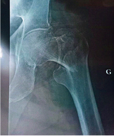
Figure 1: Pertrochanteric fracture.
Mechanism of Fractures: 60% of fractures were attributed to minor trauma, while 40% were classified as atraumatic (spontaneous) (Table 2, Figure 1).
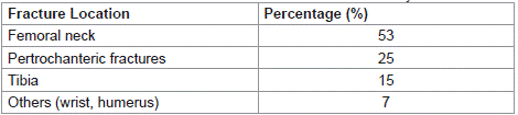
Table 2: Distribution of Fracture Locations in Chronic Hemodialysis Patients.
Associated Risk Factors
The following risk factors were identified in our study:
Advanced Age: Patients over 65 years exhibited a heightened fracture risk.
Female Sex: Women, particularly those who are postmenopausal, are more susceptible to fractures due to decreased estrogen levels.
Low Body Mass Index (BMI): A BMI below 22 kg/m² was associated with an increased fracture risk.
Prolonged Duration of Dialysis: Patients undergoing dialysis for more than 10 years demonstrated a higher fracture incidence.
Secondary Hyperparathyroidism: Elevated PTH levels (> 300 pg/mL) were significantly correlated with fracture risk.
Vitamin D Deficiency: Levels of 25-hydroxyvitamin D < 20 ng/ mL increased fracture risk (Table 3).

table 3: Risk factors.
Contingency Table Analysis
Contingency table analysis revealed the relationship between PTH levels (normal vs. hyperparathyroid) and the occurrence of fractures based on trauma mechanism. Of the 39 patients analyzed, 25 had normal PTH levels; 11 experienced trauma (44%) and 14 did not (56%). Conversely, among the 14 patients with hyperparathyroidism, 4 experienced trauma (28.6%) and 10 did not (71.4%).
Statistical analysis via the Chi-squared test yielded a Χ² value of 0.903 with a p-value of 0.342, indicating no statistically significant relationship between PTH levels and trauma mechanism in our sample. This suggests that while PTH levels are associated with fractures, the type of trauma (traumatic vs. non-traumatic) does not appear to be influenced by PTH levels within this cohort.
Surgical and Orthopedic Treatment
Surgical Intervention: 40% (n = 23) of patients required surgical intervention. Indications for surgery included displaced, unstable, or complex fractures that could not be adequately managed through conservative means.
• Types of Surgeries:Plate and screw osteosynthesis: 22% (n = 12)
• Intramedullary nail: 10% (n = 5)
• External fixator: 8% (n = 4)
Casting: 60% (n = 34) of patients were treated with orthopedic immobilization using casts, preferred for non-displaced, stable fractures or those situated in areas conducive to healing under immobilization.
Types of Casts Used:
• Circular cast: 35% (n = 19)
• Splint cast: 25% (n = 15)
Discussion
Fractures and Chronic Kidney Disease
Bone fractures represent a common and serious complication among patients on chronic hemodialysis, contributing to increased morbidity and mortality [1]. The high incidence of fractures observed in our study aligns with existing literature, which consistently indicates that hemodialysis patients are at a significantly elevated risk for fractures, particularly at the femoral neck [2]. Previous studies have demonstrated that fracture incidence in this population can be four to five times greater than that seen in the general population, highlighting the critical need for vigilant monitoring [3].
Role of Risk Factors
Identifying the risk factors associated with fractures in hemodialysis patients is essential for implementing targeted prevention strategies. Our findings indicate that elevated PTH levels and prolonged dialysis duration are significant risk factors, corroborating conclusions from other studies [4,5]. For example, research by Ketteler et al. has shown that PTH levels exceeding 300 pg/mL considerably heighten fracture risk, emphasizing the necessity of effective management of MBD [6]. Additionally, our observation that women and older patients are more likely to suffer fractures is further supported by the literature [7].
Functional and Psychological Consequences
Fractures in hemodialysis patients lead not only to physical complications but also to significant psychological ramifications. The results of our study suggest that patients may experience a loss of autonomy and a diminished quality of life, consistent with findings from other studies that highlight the association between fractures and depression in this population [8]. Functional rehabilitation is crucial for restoring limb function, yet it may be complicated by prevalent comorbidities such as cardiovascular disease and diabetes [9].
Prevention and Treatment Approaches
Preventing fractures in hemodialysis patients necessitates focused attention. Our findings underscore the importance of proactive management of mineral imbalances, particularly through the control of calcium, phosphorus, and PTH levels, as recommended by KDIGO guidelines. These guidelines advocate for target levels of calcium between 8.4 and 9.5 mg/dL, phosphorus between 3.5 and 5.5 mg/ dL, and PTH between 2 and 9 times the upper limit of normal [10]. Furthermore, KDIGO recommends vitamin D supplementation to achieve levels of 25-hydroxyvitamin D between 30 and 60 ng/mL, aimed at optimizing bone health and mitigating fracture risk. This approach is supported by our observations and aligns with existing literature [11].
Additionally, it is vital to acknowledge that the interplay between these minerals and hormones plays a pivotal role in bone remodeling. Recent studies have indicated that dysregulation of phosphocalcic metabolism, commonly observed in hemodialysis patients, can lead to structural changes in bone, thereby increasing the risk of pathological fractures [12]. Integrating tailored exercise programs and targeted nutritional strategies can also enhance clinical outcomes by strengthening musculature and reducing fall risk, which is critically important for this vulnerable population [13].
Conclusion and Future Perspectives
The high prevalence of fractures among chronic hemodialysis patients necessitates ongoing vigilance from clinicians in the prevention and management of MBD. Future research should focus on identifying novel biomarkers and evaluating the efficacy of various treatment and prevention strategies. Longitudinal studies are essential for elucidating the underlying mechanisms of fractures in this population and for developing personalized interventions that consider individual patient characteristics [14]. By incorporating these elements into clinical practice, we can aspire to reduce fracture incidence and enhance the quality of life for hemodialysis patients.
References
- Coen G, Ballanti P, Bonucci E, et al. Bone turnover, osteopenia and vascular calcifications in haemodialysis patients. Nephrol Dial Transplant. 1996; 11: 479-484.
- London GM, Marty C, Marchais SJ, et al. Arterial calcifications and bone histomorphometry in end-stage renal disease. J Am Soc Nephrol. 2004; 15: 1943-1951.
- Tentori F, Blayney MJ, Albert JM, et al. Mortality risk for dialysis patients with different levels of serum calcium, phosphorus, and PTH: the Dialysis Outcomes and Practice Patterns Study (DOPPS). Am J Kidney Dis. 2008; 52: 519-530.
- Jadoul M, et al. Fracture risk in dialysis patients: a meta-analysis of cohort studies. Bone. 2020; XX(X).
- Ganesh SK, Stack AG, Levin NW, et al. Association of elevated serum PO4, Ca×PO4 product, and parathyroid hormone with cardiac mortality risk in chronic hemodialysis patients. J Am Soc Nephrol. 2001; 12: 2131-2138.
- Ketteler M, Evenepoel P, et al. Lower serum magnesium levels are associated with a higher risk of fractures and vascular calcifications in chronic kidney disease. Clin Kidney J. 2025; 18: sfae381.
- Mizobuchi M, Towler D, Slatopolsky E. Vascular calcification: the killer of patients with chronic kidney disease. J Am Soc Nephrol. 2009; 20: 1453- 1464.
- Ketteler M, Block GA, Evenepoel P, et al. Executive summary of the 2017 KDIGO Chronic Kidney Disease-Mineral and Bone Disorder (CKD-MBD) Guideline Update: what’s changed and why it matters. Kidney Int. 2017; 92: 26-36.
- Sprague SM, et al. Diagnostic accuracy of bone turnover markers and bone histology in patients with CKD treated by dialysis. Am J Kidney Dis. 2016; 67: 559-566.
- Kidney Disease: Improving Global Outcomes (KDIGO) CKD-MBD Work Group. KDIGO clinical practice guideline for the diagnosis, evaluation, prevention, and treatment of chronic kidney disease-Mineral and Bone Disorder (CKD-MBD). Kidney Int Suppl. 2009; 113: 51-130.
- London GM, et al. Vascular calcifications and bone disease in chronic kidney disease: a review. Nephrol Dial Transplant. 2010; 25: 2204-2210.
- Tentori F, et al. Mortality risk for dialysis patients with different levels of serum calcium, phosphorus, and PTH: the Dialysis Outcomes and Practice Patterns Study (DOPPS). Am J Kidney Dis. 2008; 52: 519-530.
- Hansen D, Jørgensen HS, et al. Multidisciplinary team approach for CKDassociated osteoporosis. Nephrol Dial Transplant. 2025; 40: 48-59.
- Ketteler M, et al. Lower serum magnesium levels are associated with a higher risk of fractures and vascular calcifications in chronic kidney disease. Clin Kidney J. 2025; 18: sfae381.