
Case Series
Austin J Nephrol Hypertens. 2015;2(1): 1030.
Single Photon Emission Tomography (Spect) Imaging is Essential to Show Hernia of Peritoneum in Peritoneal Scintigraphy in Peritoneal Dialysis Patients
Koc ZP1*, Balci TA1 and Dogukan A2
1Firat University Medical Faculty, Nuclear Medicine Department, Turkey
2Firat University Medical Faculty, Nephrology Department, Turkey
*Corresponding author: Zehra Pinar Koç, Firat University Medical Faculty, Department of Nuclear Medicine 23119 / Elazig/Turkey
Received: October 30, 2014; Accepted: January 06, 2015; Published: January 08, 2015
Abstract
Imaging of the complications of peritoneal dialysis is possible by means of morphologic imaging methods and peritoneal scintigraphy. Peritoneal scintigraphy is a new entity and improvement of this modality might result in better results. Aim of this study is to investigate diagnostic performance of additional SPECT imaging in peritoneal scintigraphy in chronic peritoneal dialysis patients.
Four peritoneal dialysis patients (3 F, 1M; mean: 46±5,4 years old) with suspicion of mechanical peritoneal complications (leak, hernia or fistula) were included to the study. Peritoneal scintigraphy with Tc-99m Macroagregated Albümin (MAA) and additional SPECT imaging were performed to the patients. Dynamic and planar images and SPECT images were compared according to diagnostic considerations. In estimation of SPECT imaging, inguinal hernia was observed in one patient and hernias in the Douglas space were observed in two patients additional to planar peritoneal scintigraphy. Additional SPECT imaging might improve the diagnostic performance of peritoneal scintigraphy significantly according to our results in peritoneal dialysis patients.
Keywords: Peritoneal scintigraphy; Peritoneal dialysis; Hernia
Case Presentations
Four peritoneal dialysis patients (3 F, 1M; mean: 46±5,4 years old) were involved to this study. Mean peritoneal dialysis duration of the patients was 6±3 years. All the patients had suspicion of a mechanical complication (leak, hernia, and fistula) prior to the study. After routine physical examination ultrasonography of abdomen was performed to all patients.
Procedure
The peritoneal scintigraphy was performed by intraperitoneal administration of 1 mCi Tc-99m Macroagregated Albumin (MAA) and immediately after administration of the dialysate fluid, dynamic imaging was started. Dynamic imaging was acquired in supine position 30sn per image and lasting for 15 minutes totally. Anterioposterior and lateral spot images at least 500.000 counts were also obtained. Additionally sequential SPECT imaging from abdominal region were performed by double head SPECT gama camera equipped with low energy all-purpose collimator (GE, infina). In case of suspicion of leakage of fluid to thorax additional images from thoracic region in 24 hour were also obtained. The images were analyzed and compared by an experienced Nuclear Medicine physician.
Case 1
First patient had history of hypertension and receive peritoneal dialysis for two years. Additionally there was suspicion of the intraabdominal hernia associated with peritoneal dialysis because of outflow failure. Dynamic imaging revealed no information regarding the hernia (Figure 1a) however SPECT imaging clearly demonstrated the hernia in right inguinal canal (Figure 1b). Surgery was performed to the patient for displacement of the catheter since it was found to be infected. Hemodialysis was started later on. The patient died two weeks after the surgery.
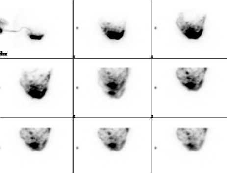
    Figure 1a:  Dynamic phase of planar Tc-99m MAA peritoneal scintigraphy in
anterior projection.
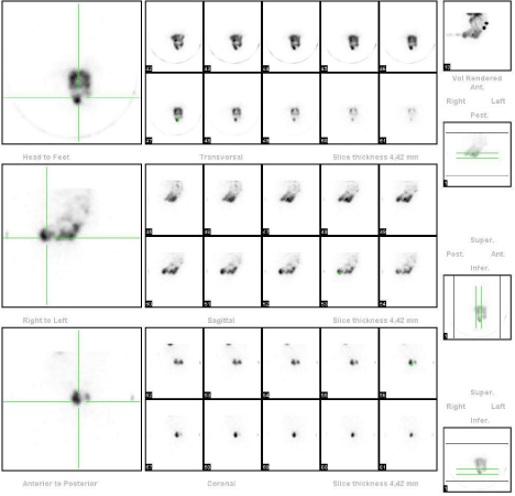
    Figure 1b:  Transaxial, coronal and sagittal slices of SPECT imaging showed
the inguinal hernia
Case 2
Second patient performed CAPD for three years and had resistant anemia and abdominal pain. Her planar scintigraphy revealed perisplenic accumulation of dialysate fluid and SPECT imaging showed additional hernia in Douglas pouch (Figure 2a, 2b).
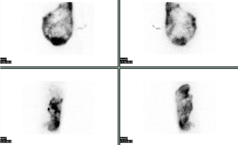
    Figure 2a:  Anteroposterior and lateral projection peritoneal scintigraphy
showed perisplenic accumulation.

Figure 2b: SPECT imaging revealed additional hernia in Douglas space.
Case 3
Third patient had history of sudden unset of fever, abdominal pain and nausea after CAPD for one year. Physical examination revealed a gross umbilical hernia which is in follow up. Also the patient had peritonitis. Planar peritoneal scintigrapy showed umbilical and bilateral inguinal hernia and SPECT imaging revealed additional hernia in Douglas space (Figure 3a, 3b).
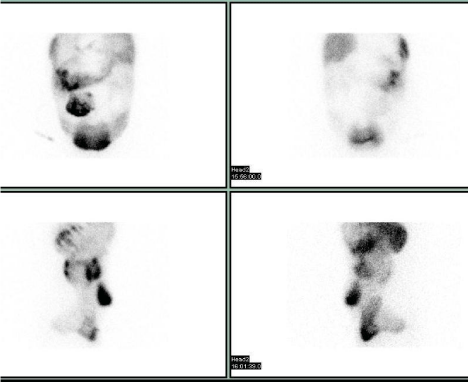
Figure 3a: Planar imaging showed umbilical and inguinal hernia.
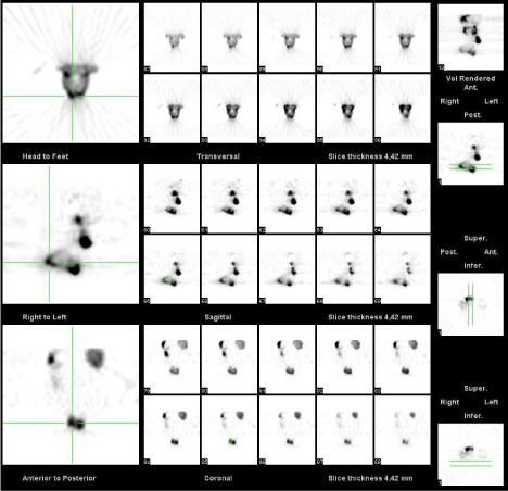
Figure 3b: Additional SPECT imaging pointed hernia in Douglas pouch.
Case 4
Fourth patient performed peritoneal dialysis for seven years and suspicion of peritoneal leak exists since the peritoneal fluid outcome was less than the income (outflow failure). Planar and SPECT peritoneal scintigraphy showed the fluid accumulation in the perihepatic area (Figure 4).
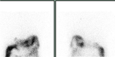
    Figure 4:  Anterioposterior imaging of thorax showed perihepatic accumulation
of the fluid.
Abdominal ultrasonography of the patients revealed no specialty and three patients were programmed for Hemodialysis.
Discussion
Peritoneal dialysis is a favorable renal replacement method however sometimes complication associated to peritoneal dialysis occurs. Besides the infectious complications like peritonitis non-infectious or mechanic complications might happen like catheter dysfunction, leakage, hernia [1]. Although the infectious complications are more common than the non-infectious complications these complications can also cause mortality or morbidity [2]. The mechanism of the non-infectious complications is the increase in abdominal pressure which results in the herniation or leakage [3]. Peritoneal scintigraphy is introduced as an alternative method to show the mechanic complications of Continuous Ambulatory Peritoneal Dialysis (CAPD) and aim of this study is to show if additional SPECT imaging might help in diagnosis of peritoneal complications.
Although peritoneal dialysis is a safe and easier procedure, unfortunately there are some complications associated with this method. Important percentages of the complications are infectious complications therefore it is important to perform the dialysis considering infection. Additionally there are non-infectious complications related to the peritoneal dialysis. Peritoneal scintigraphy is a relatively new modality to image the non-infectious complications of peritoneal dialysis. Since the peritoneal dialysis causes an increase in intra-abdominal pressure leak or hernias can appear in the defects of the abdominal muscles especially in the presence of a predisposing event like obesity or pregnancy [1]. Mechanical complications are sometimes confusing and morphologic imaging methods are not sufficient to demonstrate these complications thus functional imaging modality serves as an alternative diagnostic method. Computed Tomography (CT) is another peritoneal imaging option however the adverse effects of contrast media and limitation of additional delayed imaging due to radiation exposure limits the application of the method [4]. In a recent case report CT peritoneography and peritoneal scintigraphy successfully have demonstrated transdiaphragmatic peritoneal hernia by providing complimentary information [5].
Peritoneal scintigraphy can be performed by different tracers like Tc-99m sulphur colloid, Tc-99m DTPA and Tc-99m MAA [4,6,7]. Additional maneuvers might be helpful in determination of special circumstances like an obscure hernia in scrotal localization as reported previously [8]. Additional anatomic localization is a necessity due to lack of anatomic detail in peritoneal scintigraphy. Injection of Tc- 99m pertechnetate and body contouring is an option or the most ideal way is the SPECT/CT imaging [4,6,9]. However the SPECT/CT is a novel imaging modality is not available in every department. Thus we need to improve diagnostic efficiency of peritoneal scintigraphy by another method like SPECT imaging.
Our results showed that another possible cause of discomfort related to peritoneal dialysis is herniation in Douglas space which was not documented before. The clinical importance of this new entity has to be evaluated by prospective studies. Probably the abdominal pain, discomfort and/or insufficient dialysis were the consequences of the accumulation of the dialysate fluid in Douglas space and/ or in perihepatic or perisplenic spaces which we observed in peritoneal scintigraphies of the patients. The patients were referred to Hemodialysis and were free of the previous complaints in the follow up.
In one patient we showed that hernias in inguinal canal may be obscure and planar scintigraphy can underestimate these hernias. Juergensen et al. have showed in a large study population that in planar imaging by peritoneal scintigraphy there might be equivocal results [10]. Same researchers divided peritoneal dialysis complications into four groups; abdominal wall swelling, inguinal or genital swelling, pleuroperitoneal leak and poor catheter drainage or ultra filtration. Pleuroperitoneal leak is an infrequent complication (1.6- 10%) however hernia is more frequent (9-24%) [11,12]. Peritoneal scintigraphy is an accurate method in diagnosis of Pleuroperitoneal leak (5, 7). Genital swelling and genital hernia is one of the most reported complications with scintigraphic images [13,14]. Peritoneal leakage is another pathology that peritoneal scintigraphy can easily demonstrate [15,16]. In fact the diagnostic power of peritoneal scintigraphy is so high that there are many reports that peritoneal scintigraphy might show subclinical hernia and also improvement of this hernia [17,8]. Tokmak et al. have underlined the importance of the peritoneal scintigraphy in the peritoneal dialysis complications in their report [18].
Additional SPECT imaging in peritoneal scintigraphy provided new information that planar imaging was insufficient to show thus SPECT imaging should be a conjunct imaging modality in patients with peritoneal dialysis complications. Especially SPECT might help in patients with outflow failure by showing the distribution of the fluid to unexpected spaces in the abdomen. Since SPECT imaging do not require additional radiation exposure, provides more information and easy to perform every department should add this modality in their peritoneal scintigraphy procedure.
References
- Stuart S, Booth TC, Cash CJ, Hameeduddin A, Goode JA, Harvey C, et al. Complications of continuous ambulatory peritoneal dialysis. Radiographics. 2009; 29: 441-460.
- Thodis E, Passadakis P, Lyrantzopooulos N, Panagoutsos S, Vargemezis V, Oreopoulos D. Peritoneal catheters and related infections. Int Urol Nephrol. 2005; 37: 379-393.
- Leblanc M, Ouimet D, Pichette V. Dialysate leaks in peritoneal dialysis. Semin Dial. 2001; 14: 50-54.
- Bhattacharya A, Mittal BR. Peritoneo-scrotal communication: demonstration by 99mtechnetium sulphur colloid scintigraphy. Australas Radiol. 2005; 49: 335-337.
- Coche E, Lonneux M, Goffin E. Transdiaphragmatic peritoneal hernia complicating peritoneal dialysis: demonstration with spiral computed tomography peritoneography and peritoneal scintigraphy. Eur Radiol. 2005; 15: 1667-1670.
- Chen YC, Su YC, Chiu JS, Wei CK, Wang YF. Peritoneo-scrotal shunting diagnosed by Tc-99m DTPA SPECT/CT imaging. Kidney Int. 2010; 78: 523.
- Hernandez Martinez AC, Marin Ferrer MD, Coronado Poggio M, Escabias Del Pozo C, Coya Viña J, Martín Curto L. [99mTc-MAA peritoneal scintigraphy in pleuroperitoneal comunication in peritoneal dialysis patients]. Rev Esp Med Nucl. 2010; 29: 84-86.
- Gupta SM, Bagga S, Gelfman N, Margules R. Demonstration of subclinical inguinal hernia by peritoneal scintigraphy. Clin Nucl Med. 1997; 22: 409-410.
- Farid K, Guyot M, Jeandot R, Durieux M, Allard M, Fernandez P. SPECT-CT improves detection of small peritoneal fistula. Clin Nucl Med. 2009; 34: 634-635.
- Juergensen PH, Rizvi H, Caride VJ, Kliger AS, Finkelstein FO. Value of scintigraphy in chronic peritoneal dialysis patients. Kidney Int. 1999; 55: 1111-1119.
- Canivet E, Lavaud S, Wampach H, Wuillai A, Randoux C, Liehn JC,et al. Detection of subclinical abdominal hernia by peritoneal scintigraphy. Adv Perit Dial. 2000; 16: 104-107.
- Ramon RG, Wallner M, Kalchmair H, Povacz F, Kramar R. Initial subcutaneous embedding of the peritoneal dialysis catheter: A critical appraisal of this new implantation technique. Nephrol Dial Transplant. 1997; 12: 1661-1667.
- Berman C, Velchik MG, Shusterman N, Alavi A. The clinical utility of the Tc-99m SC intraperitoneal scan in CAPD patients. Clin Nucl Med. 1989; 14: 405-409.
- Gupta SM, Bagga S, Gelfman N, Margules R . Demonstration of subclinical inguinal hernia by peritoneal scintigraphy. Clin Nucl Med. 1997; 22: 409-410.
- Wu PS, Lee BF, Chiu NT, Tu DG, Yao WJ, Wang MC. Peritoneal scintigraphy for diagnosing periumbilical leakage in a patient on continuous ambulatory peritoneal dialysis. Kaohsiung J Med Sci. 2000; 16: 432-436.
- Thimister PW, Van Kuijk WH, Halders SG, Heidendal GA. Scintigraphic detection of peritoneal leakage. Clin Nucl Med. 2002; 27: 829.
- Davidson PG, Usal H, Fiorillo MA, Maniscalco A. The importance of peritoneal imaging in the workup of genital edema in patients on continuous ambulatory peritoneal dialysis. Mt Sinai J Med. 1999; 66: 125-127.
- Tokmak H, Mudun A, Turkmen C, Sanli Y, Cantez S, Bozfakioqlu S. The role of peritoneal scintigraphy in the detection of continuous ambulatory peritoneal dialysis complications. Ren Fail. 2006; 28: 709-713.
Citation: Koc ZP, Balci TA and Dogukan A. Single Photon Emission Tomography (Spect) Imaging is Essential to Show Hernia of Peritoneum in Peritoneal Scintigraphy in Peritoneal Dialysis Patients. Austin J Nephrol Hypertens. 2015;2(1): 1030.
Citation: Bucci J and Hansen KE. Should we treat Secondary Hyperparathyroidism in Patients with Pre-Dialysis Chronic Kidney Disease?. Austin J Nephrol Hypertens. 2015; 2(4): 1046. ISSN : 2381-8964