
Special Article - Chronic Kidney Disease
Austin J Nephrol Hypertens. 2015;2(1): 1033.
Social Marginalization, A Problem in Controlling Childhood Chronic Kidney Disease
Medina-Escobedo Martha* and Martín-Soberanis Gloria
Research Unit Kidney Diseases, General Hospital “ Dr Augustine O’Horán “ Health Services Yucatán
*Corresponding author: Martha Medina- Escobedo, Hospital General O’Horán, Av. Itzáez por Jacinto Canek S/N, Col. Centro, C.P. 97000, Mérida, Yucatán, México
Received: December 12, 2014; Accepted: February 02, 2015; Published: February 04, 2015
Abstract
We report here the case of a 14-year-old male patient previously treated for a bilateral obstructive uropathy secondary to a urinary lithiasis and the associated urinary infection. Upon hospital admission, he was diagnosed with undernourishment, chronic renal failure, urinary infection, an endoparasitic disease (ascariasis) and sepsis. After clinical and surgical treatment the patient´s condition improved. The patient has been monitored over the course of seven years and this revealed a gradual deterioration due to a combination of factors: social marginalization, family illiteracy, irregular adherence to treatment, poor attendance of medical appointments, and incomplete health coverage (Seguro Popular).
Keywords: Chronic kidney disease; Urinary lithiasis; Social marginalization; Children
Introduction
Chronic kidney disease (CKD) is a serious problem worldwide, with an increasing incidence. Whereas diabetes mellitus is the main driver of CKD in adults [1], at pediatric ages it is usually derived by obstructions of the urinary system due to malformations, although urinary lithiasis may also cause of this condition, albeit with a lower frequency. Other factors such as glomerulopathies, urinary infections (although not as a main driver) and social marginalization (SM) can also contribute to the progression of kidney damage [2-5]. This report aims to present the clinical case of a patient in whom several risk factors, and most notably social marginalization, converge in the development of CKD.
Case Study
The subject of this study is 14-year-old male patient (ACU) who is original from Tepakan (Yucatán, Mexico). He comes from a large and illiterate family, with a low income and poor socio-economic background. They live in a humble home in sub-standard conditions due to the lack of basic sewage and sanitary services. Communication with the mother is difficult, even with the aid of a Mayan translator.
ACU has a history of urinary infection (UI) in pre-school years and he was admitted to hospital when he was 6 years and 9 months old with a rapidly evolving clinical condition characterized by anorexia, nausea, vomiting, fever, pyuria and generally poor conditions: dirty, with lice, a mycosis in the genital region and both feet, as well as symptoms of sepsis. Upon admission, he weighed 10 Kg and measured 82 cm in height, and he was registered with the Social Security (Seguro Popular), a social security scheme for a population without rights to other types of medical insurance.
Laboratory tests and medical examination identified showed retention of nitrogenous components in the blood (urea 445 mg/ dl and creatinine 7.3 mg/dl), hyperuricemia (8.7 mg/dl), anemia (Hb 6.77 gr/dl) and leukocytosis (29,400/mm3)). A coproparasite study identified Ascaris lumbricoides (roundworm) and the blood culture was positive for Pseudomona aeruginosa. Ultrasound studies identified urinary lithiasis (left ureter 5 mm and bladder lithiasis 6 x 4 mm) with pyelocalyceal dilation and bilateral ureteral dilation. A simple x-ray of the abdomen showed abdominal distention and dilated bowel with image of radiodense calculus at the level of the bladder (Figure 1). He was subsequently administered antibiotics, anti-fungal and anti-parasite medications, alkalizing agents, diuretics, and treated with peritoneal dialysis with a Tenckhoff catheter.
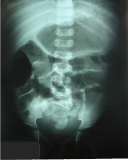
    Figure 1:  A simple x-ray of the abdomen showing abdominal distention and
dilated bowel loops with a radiodense calculus evident at the level of the
bladder.
Upon improvement of his medical condition, ureterolithotomy and cystolithotomy were performed and in a different surgical intervention (cystolithotomy), a sub-mucosal stone was removed from the proximal urethra. The patient left the hospital two months later and remained on dialysis for another four months, resulting in a decrease in the retention of nitrogenous components in the blood (urea 67 mg/dl and creatinine 2.1 mg/dl). The patient requested that the Tenckhoff catheter was removed due to his clinical improvement and his difficulties in attending the dialysis treatment. For 5 months, he suffered from recurring urinary infection, confirmed through urinary culture (Pseudomona aeruginosa, Morganella morganii and Enterobacter cloacae), and he was treated with a specific antibiotic based on the urinary culture and an antibiogram.
A renal gammagraphy showed good radionuclide captation in the right kidney and a delay in radionuclide release in the left kidney. A voiding cystogram was also performed (Figure 2). One month later, cystoscopy showed the dilation of the urethra and two months later, left ureteral reimplantation surgery was performed, after which the urine cultures were negative. However, gasometry revealed unbalanced metabolic acidosis after which, treatment with alkalizing agents begins.
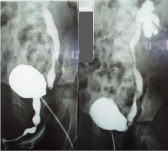
    Figure 2:  Voiding cystourethrography: left vesicoureteral reflux Grade IV,
piriform bladder and urethral stenosis.
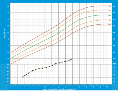
    Graph 1:  Evolution of height with age from the first admission in the hospital
until the last follow-up visit.
Due to family loss (maternal grandmother and aunt) and by separation from her partner, ACU´s mother fell into a depression that aggravated the family’s economic situation. This eventually led to the discontinuation of medical treatment and in poor attendance of the Pediatric Nephrology consultations. Overall, ACU has been monitored for seven years. He does not attend school, suffers from polyuria and he lacks control of his urinary sphincter. A delay in his physical growth has been observed (Figure 3) and clinical parameters indicate a renal osteodystrophy (Figure 4).
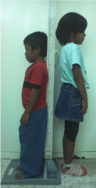
Figure 3: ACU (10-years-old) next to his five-year-old younger sister

    Figure 4:  Genu valgum derived from chronic kidney disease is observed in
ACU at 11 years of age.
At the last medical check (13 years and 9 months of age) he weighed 21.8 Kg and was 108.7 cm in height (Charts 1 and 2). His body mass index (BMI) was within normal ranges (Chart 3) and hypertension was not evident (80/60 mmHg). The blood test results were: hemoglobin 10.8 gr/dl, urea 74.9 mg/dl, creatinine 3.4 mg/dl, uric acid 5.7mg/dl, calcium 8.7 mg/dl, phosphorus 4.5 mg/dl and magnesium 2.3 mg/ dl. Gasometry showed a pH of 7.21 and bicarbonate levels of 14.0 mmol/l. Urine examination demonstrated leukocyturia and protein levels at 20mg/dl, although a urine culture was negative. Ultrasound tests showed a moderate left ureterohydronephrosis, discrete ectasia in the calices of the right kidney with an increase in renal echogenicity and evidence of chronic cystitis.
Discussion
The rise of chronic non-communicable diseases, which can predispose individuals to suffer from CKD, situates this medical condition as a major health problem worldwide. In addition, SM is an important risk factor for CKD in adults, although this has yet to be demonstrated specifically in pediatric populations [6, 7]
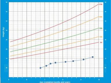
    Graph 2:  Evolution of weight with age, from the first admission to hospital
until an age of 9 years and 8 months.
In this case, ACU received healthcare and surgical assistance, leading to an improvement in this patient´s condition. However, since ACU´s mother was in charge of the treatment and of his attendance at the medical check-ups, the kidney damage has progressed to a predialysis stage with poor prospects for the near future, given the need to enter a program for functional kidney replacement [8]. It may not be appropriate to generalize from this case, because have been implemented dialysis and transplant programs for CKD patients of low income in poor countries [9].
It is noteworthy that several factors influenced the discontinuation of ACU´s treatment. Not only was it necessary for ACU to travel two hours by public transport to reach the medical centre but also, the lack of economic resources impeded regular attendance for followup appointments. A series of unfortunate events within the family and the mother’s depression aggravated this situation, contributing to the slow increased of blood nitrogenous components and severely affecting the patient´s physical growth.
The assessment of kidney function in this context is not an easy task. Whereas creatinine levels in an adult with normal body mass and optimal glomerular filtration may be 1.0 mg/dl, a child with low muscle mass may experience significant kidney damage even with 0.7 mg/dl creatinine [2]. Creatinine clearance (CCr) was initially estimated at 25.2 ml/min/m2 body surface area (BSA), according to Schwartz´s formula; but if adjusted for age (it should be borne in mind that the last height and weight data correspond to a child of 5 years of age) the CCr is lower (19.8 ml/min/m2BSA). In any case, given that normal values for males are 70 ± 14 ml/min/m2 BSA [10], the compromise in kidney function is clear. Hyperuricemia and metabolic acidosis are important risk factors for the development of kidney disease. Indeed, the patient´s average levels of uric acid over the last few years (5.6mg/dL) are indicative of hyperuricemia, which together with persistent metabolic acidosis, has contributed to the progression of the kidney damage [11].
A lack of physical growth, as recurrently noted in the ACU’s medical records, has been previously reported in patients with kidney problems [12]. Growth curves suggest that ACU´s condition started at one year of age. The etiopathogenesis of CKD patients is multifactorial and its severity depends on the age at which the disease appears, as well as on the degree to which kidney function is impaired. The risk factors are low caloric intake, osteodystrophy, electrolyte imbalance, polyuria, acidosis, anemia, infections and lack of sensitivity to growth factors [13]. Many of these factors usually become chronic in CKD patients.
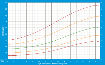
    Graph 3:  Evolution of BMI from the 2007 until the last Pediatric Nephrology
examination.
The socioeconomic context of the patient has not only affected his medical follow-up and attention but also, it has hampered the performance of specific tests (parathyroid hormone, vitamin D and other studies to determine the etiology of the polyuria) that are essential for a correct medical care. It is noteworthy that the patient is only covered by National Health Insurance, which does not cover the tests and treatments required by a patient diagnosed with CKD [14].
In Mexico the National Development Plan 2013-2018; [15] mentions that the conditions of life and life expectancy of the Mexican population has improved; likewise, it refers a plan to create and implement strategies to improve health services and therefore the health conditions of the population. At the same time, there have been studies in which inequality is evident in health systems, where coverage for Seguro Popular affiliates is not desirable; added to this, the problem of CKD in Mexico is emphasized, as a disease in progressive increase with higher prevalences, which affect more states with the highest marginalization and where the possibility of access to renal replacement program is low [5, 7]. Indeed, Yucatán and in particular Tepakán are classified among states and communities with high poverty index [16].
In conclusion, socioeconomic and educational factors are undoubtedly crucial in both the diagnosis and the appropriate treatment of kidney diseases, strongly affecting the medical followup of patients with CKD and their possible inclusion on a renal replacement therapeutic program. This is further aggravated by the therapeutic and technical limitations in the healthcare centres that are responsible for treating patients reliant on National Insurance.
References
- Centers for Disease Control and Prevention. National Diabetes Statistics Report: Estimates of Diabetes and Its Burden in the United States, 2014. Atlanta, GA: US Department of Health and Human Services; 2014.
- Warady BA, Chadha V . Chronic kidney disease in children: the global perspective. Pediatr Nephrol. 2007; 22: 1999-2009.
- Khan AM, Hussain MS, Moorani KN, Khan KM. Urolithiasis Associated Morbidity in Children. Journal of Rawalpindi Medical College (JRMC). 2014;18: 73-74. https://www.ncbi.nlm.nih.gov/pubmed/15831830
- Zorc JJ, Kiddoo DA, Shaw KN . Diagnosis and management of pediatric urinary tract infections. Clin Microbiol Rev. 2005; 18: 417-422.
- Franco-Marina F, Tirado-Gómez LL, Venado-Estrada AA, Moreno-López J, Pacheco-Domínguez RL, Luis Durán-Arenas L, et al. Una estimación indirecta de las desigualdades actuales y futuras en la frecuencia de la enfermedad renal crónica terminal en México. Salud Pública Méx 2011; 53: 506-515.
- Medina-Escobedo M, Sansores-España D, Villanueva-Jorge S . Kidney function in marginalized population: a pilot study. Rev Med Inst Mex Seguro Soc. 2014; 52: 156-161.
- Hoy W . Renal disease in Australian Aborigines. Nephrol Dial Transplant. 2000; 15: 1293-1297.
- Mercado-Martínez FJ, Hernández-Ibarra E, Ascencio-Mera CD, Díaz-Medina BA, Padilla-Altamira C, Kierans C . [Kidney transplant patients without social protection in health: what do patients say about the economic hardships and impact?]. Cad Saude Publica. 2014; 30: 2092-2100.
- Rizvi SA, Naqvi SA, Zafar MN, Akhtar SF . A kidney transplantation model in a low-resource country: an experience from Pakistan. Kidney Int Suppl (2011). 2013; 3: 236-240.
- Schwartz GJ, Furth SL . Glomerular filtration rate measurement and estimation in chronic kidney disease. Pediatr Nephrol. 2007; 22: 1839-1848.
- Staples A, Wong C . Risk factors for progression of chronic kidney disease. Curr Opin Pediatr. 2010; 22: 161-169.
- Sozeri B, Mir S, Kara OD, Dincel N . Growth impairment and nutritional status in children with chronic kidney disease. Iran J Pediatr. 2011; 21: 271-277.
- Salas P, Pinto V, Rodriguez J, Zambrano MJ, Mericq V. Growth Retardation in Children with Kidney Disease. Int J Endocrinol; 2013; ID970946.
- Comisión Nacional de Protección Social en Salud. Catálogo Universal de Servicios de Salud CAUSES 2014.
- Gobierno de la República. Programa Sectorial de Salud. Plan Nacional de Desarrollo 2013-2018.
- Índice de Marginación por Localidad.
Citation: Medina-Escobedo Martha and Martín-Soberanis Gloria. Social Marginalization, A Problem in Controlling Childhood Chronic Kidney Disease. Austin J Nephrol Hypertens. 2015;2(1): 1033.
Citation: Bucci J and Hansen KE. Should we treat Secondary Hyperparathyroidism in Patients with Pre-Dialysis Chronic Kidney Disease?. Austin J Nephrol Hypertens. 2015; 2(4): 1046. ISSN : 2381-8964