
Special Article - Dialysis Case Reports
Austin J Nephrol Hypertens. 2016; 3(1): 1057.
A New Technique using Balloon Assisted Placement of Tunnel Dialysis Catheter in the Internal Jugular Vein for Patients with End-Stage Renal Disease
Magbri A*, Brandt MCV, Mock L, Colosimo V, Zurawsky R and McCartney P
Dialysis Access Center of Pittsburgh, USA
*Corresponding author: Awad El-Magbri, Dialysis Access Center of Pittsburgh, 2030 Ardmore Blvd, Suite 125, Pittsburgh, USA
Received: July 09, 2016; Accepted: August 05, 2016; Published: August 11, 2016
Abstract
Two patients with end stage renal disease (ESRD) on hemodialysis (HD) were referred to the Dialysis Access Center of Pittsburgh for placement of tunneled hemodialysis catheter (TDC) while their fistula is maturing. A new approach for placement of TDC was discussed with the patients. Written consent was obtained from the patients and the procedure is explained to the patients. The procedures of placement of TDC were accomplished without problems in reasonable time. This approach of placement of TDC in the internal jugular vein has many advantages and may be adopted as the new way of catheter placement for ESRD on HD.
Keywords: Hemodialysis; Central veins; End stage renal disease
Case 1
A 60 year old (DN) male patient had end stage renal disease secondary to hypertension. The patient is referred to the Dialysis Access Center (DAC) of Pittsburgh for placement of central venous [1] catheter to be able to continue dialysis. After a written consent and explanation of the procedure to the patient is obtained, a tunneled hemodialysis catheter is placed in the right internal jugular vein using the new technique explained below. The procedure was accomplished without complication and the patient tolerated the procedure well.
Case 2
A 50 years (LJ) male patient had end stage renal disease secondary to diabetes and hypertension. The patient is referred to the DAC for placement of tunneled hemodialysis catheter to be able to continue dialysis till his arterial-venous fistula is maturing. A written consent with explanation of the procedure to the patient was done. The balloon assisted tunnel dialysis placement was adopted and the procedure is accomplished without complications. The patient is sent to the dialysis unit to complete his dialysis.
X ray films of the 2 cases are attached (Figure 1-9).
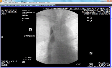
Figure 1:
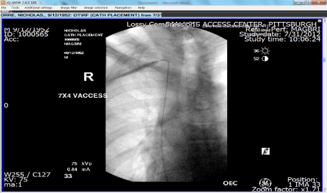
Figure 2:
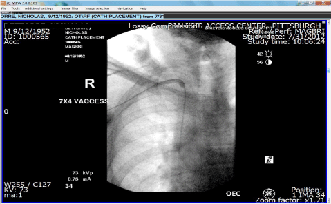
Figure 3:
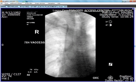
Figure 4:
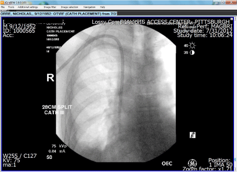
Figure 5:
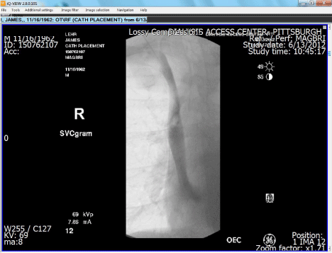
Figure 6:
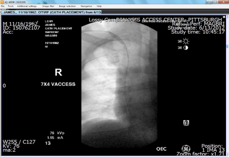
Figure 7:
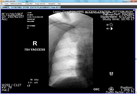
Figure 8:
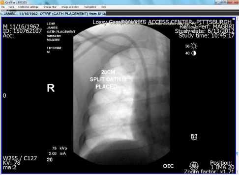
Figure 9:
Balloon assisted placement of tunneled hemodialysis catheter in the internal jugular vein
Sedation: The patient was sedated using versed and fentanyl injected into the central veins [2]. Vital signs were monitored by the nurse for the entire period of the procedure.
Cannulation of the vein: The skin at the cannulation site is infiltrated with local anesthesia (1% lidocaine), then using real time ultra-sound guidance, the right internal jugular vein was cannulated using a micro-puncture needle (0.018 inch guide wire, co-axial 3 and 5 Fr dilators). The puncture site to the internal jugular vein is between the medial and lateral heads of the sternocleidomastoid muscle about 1 cm above the clavicle. The 0.018 wire is advance into the vein and the needle is exchanged for the co-axial dilators. A 0.035 inch Bentson (Merit-Medical system Inc Jordan, Utah, USA) guide wire is placed with its distal tip in the inferior vena cava.
Balloon assisted catheter insertion and angioplasty of the venotomy site and the tunnel
1. After the vein was cannulated, the 0.018 wire is advance into the vein and the needle is exchanged for the co-axial 5 Fr sheath dilator which was placed over the micro-access wire. A 0.035 inch Bentson guide wire is exchanged and advance in the 5 Fr dilator with its distal tip in the inferior vena cava (IVC) under fluoroscopy.
2. A 1 cm incision was made at the venotomy site and the area was bluntly dissected with a pair of hemostat. A tunnel was created following a subcutaneous infiltration with 1% lidocaine along the expected course of the tunnel. The tunnel should be at least 8-10 cm in length. A # 11 blade is used to create a 5 mm incision on the chest wall at the desired entry site on the chest.
3. The tunnel was then dilated with a pair of hemostat from the exit site towards the venotomy site. The free tip of the guide wire (0.035 Bentson) was pulled back from the venotomy site with the hemostat to the exit site.
4. A 7x40 mm vaccess balloon (Bard peripheral vascular, Inc, Tempe, AZ) was then advanced over the guide wire from the exit site towards the venotomy site and eventually into the SVC.
5. The entrance to the central veins, the venotomy site and the tunnel to the exit site were then angioplastied and dilated with the balloon catheter.
6. The desired catheter length was advanced over the guide wire from the exit site in the anterior chest wall to the venotomy site and the SVC under direct fluoroscopy. The last step resembles exchanging a tunneled dialysis catheter.
7. The tip of the catheter is placed between the junctions of the SCV with the right atrium. Making sure there is no evidence of malposition, kinks of the catheter or complication.
8. The catheter ports are then tested by flushing them with 10cc syringe. The ports of the catheter are flushed with heparin and the catheter was sutured in place with 2-0 non-absorbable suture.
Potential advantages of the balloon assisted tunnel hemodialysis catheter insertion
1. The rate of all procedure- related complications has dropped significantly with image-guided insertion. Arterial puncture, pneumothoraces, and catheter malpositioning are virtually nonexisting with image guided insertion [3].
2. Venous laceration that often occurs with the traditional method of peel-away sheath dilator combination is avoided in the new method. The trauma and the pain caused by dilation of the track from the venotomy site to the central veins can be avoided also with balloon-assisted insertion [4,5]. Many times the rigid wire may not be in the IVC and the wire-dilator tandem punctures the wall of the vein in the traditional method, this is avoided completely with this method. By using the Bentson wire instead of rigid and stiff wire avoids the aforementioned complications.
3. Difficulty advancing the catheter through the peel-away sheath especially when the left-side approach is chosen. This challenge is totally avoided in the new method.
References
- Ray CE. Central venous access. Philadelphia: Lippincott Williams and Wilkins. 2001.
- Funaki B. Central venous access: a primer for the diagnostic radiologist. AJR. 2002; 179: 309-318.
- National Kidney foundation. Update on hemodialysis adequacy. 2006.
- Mandolfo S, Acconcia P, Bucci R, Corradi B, Farina M, Rizzo MA, et al. Hemodialysis tunneled central venous catheters: five-year outcome analysis. J Vasc Access. 2014; 15: 461-465.
- Engstrom BI, Horvath JJ, Stewart JK, Snyder RH, Miller MJ, Smith TP, et al. Tunneled internal jugular hemodialysis catheters: impact of laterality and tip position on catheter dysfunction and infection rates. J Vasc Interv Radiol. 2013; 24: 1295-1302.