
Case Report
Austin J Nephrol Hypertens. 2019; 6(2): 1084.
Pyoderma Gangrenosa in Post Donor Nephrectomy Patient
Singh G1*, Sharma VK2 and Patil N3
¹Department of Pathology, HBCH Varanasi, India
²Department of Urology, GB Pant Hospital, India
³Department of Pathology, ILBS New Delhi, India
*Corresponding author: Gyanendra Singh, Fellow in Onco-pathology, HBCH Varanasi, Department of Pathology, HBCH Varanasi, India
Received: November 04, 2019; Accepted: December 06, 2019; Published: December 13, 2019
Abstract
Pyoderma gangrenosa is an ulcerative disorder of unknown etiology, which is characterised by neutrophilic infiltration of deep dermis along with necrosis and ulceration of overlying epithelium. Association of pyoderma has been described with many condition like, inflammatory bowel disease, lymphoma, autoimmune disorder as well drug associated. Although previously described due to bacterial infection but recent study shows that immune dysregulation and abnormal neutrophilic function is main pathogenesis of pyoderma gangrenosa.
Pyoderma gangrenosa is very rare in renal transplant patient; here we describe a case report who presentation is typical of pyoderma gangrenosa both clinically and histopathologically.
Case Report
A 33 years old woman underwent a donor nephrectomy in March 2019. Three to four days after the surgical site closure she developed a small pustular lesion over the surgical site. General condition of the patient was stable, oriented to time, place and person.
Her vitals, pulse and temperature was within normal limits.
Investigation show increased in total leukocytes counts (24000/ microliter) with differential count within normal distribution. Platelets count was also within normal range. Her renal graft function was appearing to stable with urea and creatinine level of 22 mg/dl and 0.8 mg/dl respectively.
On muco-cutaneous examination, there was involvement of the left side of abdomen just over the surgical site mark. The lesion was in the form of well-defined deep ulcer with a necrotic slough and bluish undermined edge and violaceous margin (Figure 1). There was surrounding induration Significant tenderness was also present. No satellite lesion or pathergy noted.
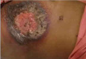
    Figure 1: Shows the lesion of the patient at the post nephrectomy site (Initial
lesion).
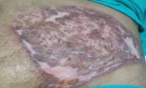
    Figure 2: Shows the lesion of the patient at the post nephrectomy site (after
the steroid treatment).
Histopathological examination shows stratified squamous epithelial lining with areas of ulceration and necrosis and neutrophilic exocytosis. The underlying dermis shows dense inflammatory cell infiltrate comprises of predominantly neutrophils. There is peri appandigeal neutrophilic infiltration (Figures 3-5).
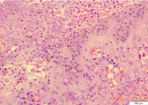
    Figure 3: Histopathology: Shows stratified squamous epithelium with focal
ulceration and necrosis and neutrophilic exudate.
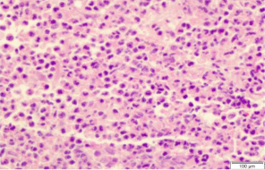
Figure 4: Histopathology shows dermis with dense neutrophilic infiltration.
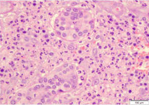
    Figure 5: Histopathology shows dermal appendage with dense neutrophilic
infiltrate.
For this lesion, she received oral and IV antibiotic but did not respond. After that, she also underwent surgical debridement twice with a diagnosis of surgical site infection. After skin biopsy, she started with steroid and lesion improve with steroid treatment (Figure 2).
Discussion
First described by Brunsting et al in 1930 [1]. Pyoderma gangrenosa is disorder of unknown etiology. Many factors are associated with pyoderma gangrenosa like myeloproliferative disorder, leukaemia, inflammatory bowel disease, autoimmune disorder like collagen vascular disorder, arthritis as well as malignancy. Association with certain drug has also been associated [1,2].
Previously pyoderma gangrenosa was considered due to bacterial infection but recent studies have shown no or very little association with bacterial infection. Recent studied shows various pathogenesis for pyoderma gangrenosa including dysregulation of immune system and abnormal neutrophil trafficking [3-6]. Few cases of pyoderma gangrenosa with positive family history also been reported suggesting that genetic susceptibility is also a contributing factor for it.
Pyoderma gangrenosa can occur at any age group from young age to adult age group. No sex prediction has been found.
As also celled as neutrophilic dermatosis, histologically it is characterized by ulceration and necrosis of epidermis, neutrophilic infiltrate in the deep dermis, perifollicular inflammation. Sometime leukocytoclastic vasculitis is also present.
Clinically four subtypes of pyoderma gangrenosa has been described [4]: (1) Classical or ulcerative, (2) Bullous (3) Pustular and (4) Vegetative.
Since the histopathological finding of pyoderma gangrenosa is non-specific [3]. Described the diagnostic criteria for pyoderma gangrenosa, which include both histopathological and clinical consideration. The describe it into major and minor criteria:
Major criterion: Biopsy of ulcer edge demonstrating a neutrophilic infiltrate.
Minor criteria:
1. Exclusion of infection
2. Pathergy
3. Personal history of inflammatory bowel disease or inflammatory arthritis
4. History of papule, pustule, or vesicle that rapidly ulcerated
5. Peripheral erythema, undermining border, and tenderness at site of ulceration
6. Multiple ulcerations (at least one occurring on an anterior lower leg)
7. Cribriform or “wrinkled paper” scar at sites of healed ulcers
8. Decrease in ulcer size within one month of initiating immunosuppressive medications
At least the major criterion and four minor criteria are necessary for diagnosis.
There is various disorder, which simulate pyoderma gangrenosa both histopathologically and clinically. Following should be kept in differential diagnosis [5] - cutaneous Crohn’s disease, ulcerative necrobiosis lipoidica, various cutaneous infection, venous disease, vasculitis, malignancy, drug reaction and exogenous tissue injury.
To conclude, pyoderma gangrenosa in post donor nephrectomy patient is very rare, with diagnosis confirmed by clinically and histopathologically and exclusion of other factor.
References
- Brunsting LA, Goeckerman WH, O’Leary PA. Pyoderma gangenosum: clinical and experimental observations in five cases occurring in adults. Arch Dermatol. 1930; 22: 655.
- Crowson AN, Mihm MC Jr, Magro C. Pyoderma gangrenosum: a review. J Cutan Pathol. 2003; 30: 97-107.
- Su WP, Schroeder AL, Perry HO, Powell FC. Histopathologic and immunopathologic study of pyoderma gangrenosum. J Cutan Pathol. 1986; 13: 323-330.
- Powell FC, Su WP, Perry HO. Pyoderma gangrenosum: Classification and management. J Am Acad Dermatol. 1996; 34: 395-409.
- Weenig RH, Davis MD, Dahl PR, Su WP. Skin ulcers misdiagnosed as pyoderma gangrenosum. N Engl J Med. 2002; 347: 1412.
- Al-Hwiesh AK. Pyoderma gangrenosum in a renal transplant recipient: a case report. Saudi J Kidney Dis Transpl. 2006; 17: 559-563.