Research Article
Austin J Neurol Disord Epilepsy. 2024; 10(1): 1055.
Risk Factor Assessment and Clinical Profile of Patients with Intracerebral Haemorrhage
Sanjay Bhat*; Supinder Singh; Sandeep Sadhu; Prabhjeet Kour
1Department of Medicine, ASCOMS & Hospital, Jammu, J&K, India
2Department of Pathology, ASCOMS & Hospital, Jammu, J&K, India
*Corresponding author: Sanjay Bhat, Department of Medicine, ASCOMS & Hospital, Jammu, J&K 180017, India. Tel: 9419127816 Email: drbhatsanjay@gmail.com
Received: October 14, 2024; Accepted: November 01, 2024 Published: November 08, 2024
Abstract
Title: Risk factor Assessment and clinical profile of patients with Intracerebral Haemorrhage.
Background: Incidence and prevalence of Cerebrovascular Diseases (CVD) are rising among Indians. Intracerebral Haemorrhage (ICH), a subtype of stroke, is a devastating condition whereby a hematoma is formed within the brain parenchyma with or without blood extension into the ventricles.
Aim: To determine the clinical profile of Intracerebral Haemorrhage Patients with assessment of risk factors.
Material and Methods: The present observational study was conducted for a period of one-year on the patients admitted in a tertiary care hospital. Total 60 patients presented with acute focal neurological deficit consistent with stroke and proved to be ICH on neuro imaging were included. Neurological assessment was done by taking the history including the demographic, various risk factors, comorbid etiological diseases and treatment history regarding use of any anti-coagulation or hematological disease. The onset stroke severity was assessed by Glasgow coma scale and neurological deficit on admission by NIH stroke Scale based on detailed neurological evaluation. The final outcome at discharge was evaluated by Glasgow Outcome Score (GOS) as well as by the Modified Rankin Scale (MRS).
Results: It was observed that 55% of the patients were in the age- group 61-70 years and 60% of the patients were males. 78% patients had systemic hypertension alone or with associated co-morbidities. It was observed that 26.67% presented with motor weakness in the form of hemiplegia and 20% presented with LOC. 50% patients were capsuloganglionic. 28.3% had lethal outcome with GOS-1 and 28.33% had lethal outcome with MRS 6.
Conclusion: The study concluded that poor outcome of ICH patients was significantly associated with location of Hematoma, Arterial Blood Pressure, NIHSS at baseline, age and comorbid conditions.
Keywords: Cerebrovascular disease; Intracerebral haemorrhage; Brain parenchyma.
Introduction
Due to the fast-rising risk factors for Cardiovascular Disease (CVD), which affect a significant section of the adult population, such as obesity, smoking, diabetes mellitus, and hypertension, the prevalence and incidence of CVD are rising in India.
Intracerebral Haemorrhage (ICH), a subtype of stroke, is a devastating condition whereby a hematoma is formed within the brain parenchyma with or without blood extension into the ventricles. Non-traumatic ICH comprises 10-15% of all strokes and is associated with high morbidity and mortality [1].
The current World Health Organization definition of stroke (introduced in1970 and still used) is “rapidly developing clinical signs of focal (or global) disturbance of cerebral function, lasting more than 24 hours or leading to death, with no apparent cause other than that of origin”. Intracerebral Hemorrhage (ICH) is usually caused by rupture of small penetrating arteries secondary to hypertensive changes or other vascular abnormalities [2-4].
Spontaneous Intracerebral Haemorrhage (SICH) is the second most common cause of stroke and accounts for 7.5-30% of all strokes [3,5]. Hemorrhagic stroke is generally associated with higher morbidity and mortality rates than ischemic stroke [6-8].
National Commission on Macroeconomics and Health, estimated 1.67 million stroke cases in India by the year 2015, suggesting that stroke will be arising epidemic in India in the near future. This may be due to the high prevalence of hypertension, diabetes, dyslipidemia, the fast-changing lifestyles and restructuring of the population [9].
Accordingly, it is important to analyse the factors causing significant decline in neurological status as well as affecting the Glasgow outcome score. Therefore, the present study is conducted to determine the clinical profile of Intracerebral Haemorrhage Patients with assessment of risk factors.
Material and Methods
The present observational study was conducted for a period of one-year on the patients admitted in Post Graduate Department of Medicine at Acharya Shri Chander College of Medical Science and Hospital, Jammu, Jammu and Kashmir after obtaining IEC approval. Total 60 patients presented with acute focal neurological deficit consistent with stroke and proved to be ICH on neuro imaging were included in the study. Whereas, patients with traumatic intracerebral hemorrhage, primary or secondary brain tumors, cortical vein thrombosis, coagulation disorders, thrombolytic and anticoagulant therapy and intracerebral haemorrhage not proven by CT or MRI were excluded from the study.
All the patients were enrolled in this study after obtaining informed consent from the close relatives in case of altered sensorium patients. Patients preceding illness, social, economic and demographic details were recorded in the proforma sheet. Neurological assessment was done by taking the history including the demographic, various risk factors, comorbid etiological diseases and treatment history regarding use of any anti-coagulation or hematological disease.
The onset stroke severity was assessed by Glasgow coma scale and neurological deficit on admission by NIH stroke Scale based on detailed neurological evaluation. The final outcome at discharge was evaluated by Glasgow Outcome Score (GOS) as well as by the Modified Rankin Scale (MRS). NCCT head was also done.
Data Analysis
The statistical analysis was done and the results are being expressed as percentages with appropriate charts, tables and diagrams. Appropriate statistical methods were applied whenever was necessary. A p-value of <0.05 is considered significant.
Results and Observations
The study group consisted of 60 stroke patients admitted at Acharya Shri Chander College of Medical Sciences and Hospital, Jammu.
Table 1 depicts the distribution of study participants as per age. In our study it was observed that 55% of the patients were in the age- group 61-70 years followed by 25% in the 51-60 age-group and 8.33% above 70 years. Further, 60% patients were males and 40% were females (Figure 1).
![]()
    Age Group
    Frequency
    Percentage


    Below 40
    4
    6.67


    41-50
    3
    5


    51-60
    15
    25


    61-70
    33
    55


    Above 70
    5
    8.33


    Total
    60
    100
Table 1: Age group.
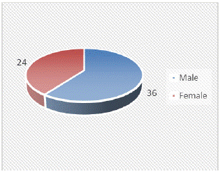
Figure 1: Gender distribution.
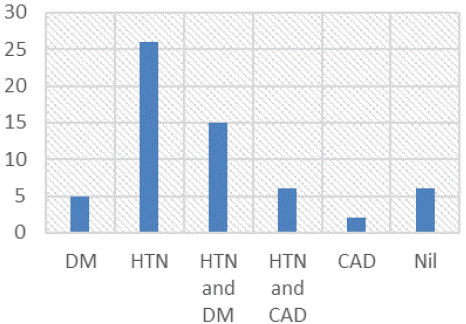
Figure 2: Co-morbid conditions.
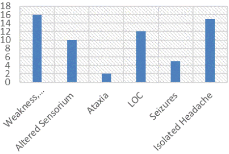
Figure 3: Clinical presentation.
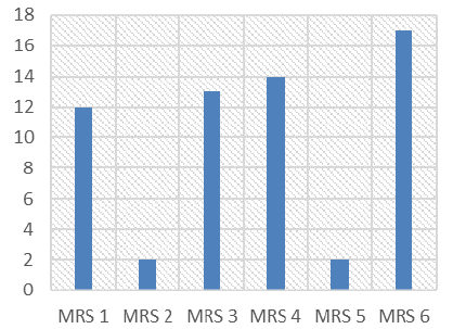
Figure 4: Outcome MRS at Discharge.
Table 2 depicts the co-morbid conditions among the study participants. It was observed in our study that 78.33% had systemic hypertension alone or with associated co morbidities, 25% had both HTN and DM, 10% had both HTN and CAD, 3.33% had CAD only and 10% did not have any co morbidities or identifiable causative factor. It was observed in our study that 16.67% patients had an alcoholic binge within 48 hours of the event. Further, 55% patients had MAP 110-130 mm Hg. 18.33% had 131-150 mm Hg, 5% had MAP >150 mm Hg.
![]()
    Co-Morbid
    Frequency
    Percentage


    DM
    5
    8.33


    HTN
    26
    43.33


    HTN and DM
    15
    25.00


    HTN and CAD
    6
    10.00


    CAD
    2
    3.33


    Nil
    6
    10.00


    Total
    60
    100
Table 2: Co-morbid conditions.
Table 3 depicts the clinical presentation among study participants. It was observed that 26.67% presented with motor weakness in the form of hemiplegia. 20% presented with LOC. 3.33% presented with ataxia. 16.67% had alteration in sensorium with loss of consciousness. 8.33% had seizures. 25% presented with isolated headache as the isolated manifestation.
![]()
    Clinical    Presentation
    Frequency
    Percentage


    Weakness, Hemiplegia
    16
    26.67


    Altered Sensorium
    10
    16.67


    Ataxia
    2
    3.33


    LOC
    12
    20.00


    Seizures
    5
    8.33


    Isolated Headache
    15
    25.00


    Total
    60
    100
Table 3: Clinical presentation.
At the time of admission, patients were assessed on the basis of Glasgow Comma Scale (GCS). It was observed in our study that 31.67% had admission Glasgow Comma Scale (GCS) of 15 without any worsening during hospital stay. 21.67% had admission GCS 11- 14. 28.33% had admission GCS 5-10. 20% had admission GCS <5.
The patients in our study were also evaluated on the basis of National Institute of Health Stroke Scale (NIHSS). It was observed that 38.3% had NIHSS < 5, 36.7% had NIHSS 6-15, 5% had NIHSS 16-25, and 20% had NIHSS >25.
At the time of discharge patients in our study were assessed on the basis of Glasgow Outcome Scale (GOS) and Modified Rankin Scale (MRS).
Table 4 depicts the ICH location in our study. On brain imaging it was found in our study that 50% patients were capsuloganglionic and 18.3% was of lobar location, brain stem in 10.0%, cerebellum in 13.3%, subarachnoid hemorrhage in 3.3% and mixed in 5.0% patients.
![]()
    ICH    Location
    Frequency
    Percentage


    Capsulo ganglionic
    30
    50.0


    Lobar
    11
    18.3


    Brain stem
    6
    10.0


    Cerebellum
    8
    13.3


    Subarachnoid hemorrhage
    2
    3.3


    Mixed
    3
    5.0


    Total
    60
    100
Table 4: ICH Location.
It was observed in our study that 28.3% had lethal outcome with GOS-1, 3.3% had GOS-2 and 23.3 % had GOS-3 had poor outcome, 21.7% had GOS-4 and 23.3 % had GOS-5 had better outcome. Further, 28.33% had lethal outcome with MRS 6. 3.33% had lethal outcome with MRS 5. 23.33% had lethal outcome with MRS 4. 21.67% had lethal outcome with MRS 3. 3.33% had lethal outcome with MRS 2. 20% had lethal outcome with MRS 1.
Discussion
In this observational study we describe the various risk factors, clinical parameters and final outcome of patients with intra cerebral hemorrhage.
Among patients with lethal outcome, 29.4 % between (51-60), 47.1% between (61-70), 5.9 % >70 years. 53% were in the above 60 years 17.6 % were < 50 yrs. This implies that more mortality is observed with increasing age. This observation is consistent with the previous studies in the literature like Sunil et al., Maya et al and Nilsson OG et al [10-12].
Among patients who had lethal outcome GOS 1- about 27.8% were males and 29.2% were females. Among patients with poor outcome - GOS 2.8% were males 4.2% were females, GOS 3- 30.6% were males and 12.5% were females. Among patients with better outcome GOS 4- 19.4% were males 25% were females, GOS 5- 19.4% were males and 29.2% were females. The lethal and poorer outcome observed had been more in males when compared to females in this study. Among better outcome group females had better outcome than males. These were not statistically significant the p value being 0.576 These observations were similar with the previous studies like study by Sunil et al who have observed that among patients who had died 72% were males and 28% were females in their study. But Maya et al., in their study had stated that male patients had better out-come than female [10,11].
Among patients with lethal outcome of GOS 1- 23.5% had GCS <5, 17.6% had GCS [5-10], 17.6 % >GCS [11-14] and 41.2% had GCS 15. Among patients with lethal outcome of GOS-2 0% GCS <5, 0% had GCS [5-10], 50% had GCS [11-14] and 50% had GCS 15. Among patients with lethal outcome of GOS-3 14.3% had GCS <5, 21.4% had GCS [5-10], 21.4% >GCS [11-14] and 42.9 had GCS 15. Among patients with lethal outcome of GOS-4 30.8% had GCS <5, 30.8 % had GCS [5-10], 38.5% >GCS [11- 14] and 0% had GCS 15. Among patients with lethal outcome of GOS-5, 7.1% had GCS <5, 64.3% had GCS [5-10] 7.1% had GCS [11-14] and 21.4 had GCS 15. Patients had lethal outcome if GCS < 5, a poorer outcome for GCS [5-10] and a better outcome if GCS>10.
Among patients with lethal outcome of GOS-1, 41.2% had NIHSS =5, 35.3% had NIHSS 6-15, 0% had NIHSS 16-25 and 23.5% had NIHSS >25. Among patients with lethal outcome of GOS-2, 50% had NIHSS =5, 50% had NIHSS 6-15, 0% had NIHSS-16-25 and 0% had NIHSS >25. Among patients with lethal outcome of GOS-3 50 %had NIHSS =5, 21.4% had NIHSS 6-15, 14.3% had NIHSS 16-25 and 14.3% had NIHSS >25. Among patients with lethal outcome of GOS- 4 15.4% had NIHSS =5, 46.2% had NIHSS 6-15, 7.7% had NIHSS 16- 25 and 30.8% had NIHSS >25. Among patients with lethal outcome of GOS-5 21.4% had NIHSs =5, 57.1% had NIHSS 6-15, 7.1% had NIHSS 16-25 and 14.3% had NIHSS >25, Patients who presented with NIHSS >25, 82.9% had died and only 17.1 had survived. This again emphasis clearly that like Glasgow coma scale that the outcome as measured by the Glasgow outcome scale has inverse correlation with NIH stroke scale. The correlation is statistically significant (p- 0.000) Hence Glasgow coma scale and NIH stroke scale at the time of admission are most helpful in prognosticating a patient with intra cerebral bleed.
Among patients with blood pressure with mean arterial pressure more than 150 mmHg, 33.3% had died, only 33.3% had survived with better outcome GOS 4 and GOS 5. Among patients with blood pressure with mean arterial pressure between 130-150 mmHg about 36.4% had died and 28.6% had survived with a better outcome. Among patients with blood pressure with mean arterial pressure between 110-130 mmHg only 21.2% had died and 35.7% had survived with a better outcome. Among patients with blood pressure with mean arterial pressure between less than 110 mmHg, only 38.5% had died, 30.8% had survived with a better outcome and 46.2% had survived with a better outcome (GOS 4 and 5). The correlation is statistically significant (p 0.028 *).
Among the patients who had lethal outcome GOS 1 about 5.9% were diabetic patients, 41.2% had isolated or hypertension associated diseases, and in about 23.5% of patients the etiology was unidentifiable. The reason for this observation should have been multifactorial. The correlation was not statistically significant. But these observations differed from the observations in the study by Maya et al. [11].
The lethal and poorer outcome is more with increased age. The lethal and poorer outcome was more in males compared to females. The lethal outcome is also influenced by the associated co morbid conditions. Among patients with lethal outcome significant proportion had associated diabetic status. The Glasgow Outcome Score (GOS) has inverse correlation with NIHSS score and a direct correlation with Glasgow Coma Scale (GCS). The presence of Intra ventricular extension has poorer or lethal outcome, whereas absence of Intra ventricular extension had better outcome. The lethal outcome was more with infra tentorial and better outcome was more with supra tentorial. The volume of ICH is direct correlation with the outcome GOS at discharge. The outcome Modified Rankin Score had a direct correlation with the volume of intracerebral bleed. The more the Mean Arterial Pressure (MAP), lethal and poorer was the outcome. Lesser MAP was associated with a better the outcome. More the volume of the bleed more the lethal and poor outcome with supra tentorial location. But with infra tentorial even small volume bleed had significant lethal outcome. The capsulo ganglionic and lobar location had better outcome compared to the brainstem location.
Conclusion
ICH remains a disease with high morbidity and mortality. The study concluded that poor outcome of ICH patients was significantly associated with location of Hematomas Arterial Blood Pressure, NIHSS at baseline, age and comorbid conditions.
References
- Ziai WC, Carhuapoma JR. Intracerebral Hemorrhage. Continuum (Minneap Minn). 2018; 24: 1603-1622.
- Qureshi AI, Mendelow AD, Hanley DF. Intracerebral haemorrhage. Lancet. 2009; 373: 1632–1644.
- Qureshi AI, Tuhrim S, Broderick JP, Batjer HH, Hondo H, Hanley DF. Spontaneous intracerebral hemorrhage. N Engl J Med. 2001; 344: 1450– 1460.
- Garcia JH, Ho KL. Pathology of hypertensive arteriopathy. Neurosurg Clin N Am. 1992; 3: 497–507.
- Broderick J, Connolly S, Feldmann E, et al. Guidelines for the management of spontaneous intracerebral hemorrhage in adults: 2007 update: a guideline from the American heart association/American stroke association stroke council, High Blood Pressure Research Council, and the quality of care and outcomes in research interdisciplinary working group. Circulation. 2007; 116: 391–413.
- Cadilhac DA, Dewey HM, Vos T, Carter R, Thrift AG. The health loss from ischemic stroke and intracerebral haemorrhage: evidence from the North East Melbourne Stroke Incidence Study (NEMESIS) Health and Quality of Life Outcomes. 2010; 14: 49.
- Lee HY, Hwang JS, Jeng JS, Wang JD. Quality-adjusted life expectancy (QALE) and Loss of QALE for Patients with Ischemic Stroke and Intracerebral Hemorrhage. Stroke. 2010; 41: 739–744.
- van Asch CJ, Luitse MJ, Rinkel GJ, van der Tweel I, Algra A, Klijn CJM. Incidence, case fatality, and functional outcome of intracerebral haemorrhage over time, according to age, sex, and ethnic origin: a systematic review and meta-analysis. The Lancet Neurology. 2010; 9: 167–176.
- Kulshrestha M, Vidyanand. An Analysis of the Risk Factors and the Outcomes of Cerebrovascular Diseases in Northern India. J Clin Diagn Res JCDR. 2013; 7: 127–31.
- Sunil K Narayan, P Sivaprasad I, Sharma Sushma, Ratnakar K Sahool, Tarun Kumar Dutta. Etiology and outcome determinants of intracerebral hemorrhage in a south Indian population, A hospital-based study. Ann Indian Acad Neurol. 2012; 15: 263-6.
- Maya P Danovska, Margarita L Alexandrova, Nachko I Totsev, Irena I Gencheva, Plamen G. Clinical and neuroimaging studies in patients with acute spontaneous intracerebral hemorrhage. Stoev 1 Journal of IMAB – Annual Proceeding (Scientific Papers). 2014; 20: 489-494.
- Nilsson OG, Lindgren A, Stahl N, Brandt L, Saveland H. Incidence of intracerebral and subarachnoid hemorrhage in southern Sweden. J Neurol Neurosurg Psychiatry. 2000; 69: 601–7.