Clinical Image
Austin Neurosurg Open Access. 2014;1(1): 1005.
Aneurysm-Induced Oculomotor Palsy in Neurofibromatosis Type 1
Kimihiro Nagatani*, Hideo Osada, Fumihiro Sakakibara, Satoru Takeuchi, Naoki Otani, Kojiro Wada and Kentaro Mori
Department of Neurosurgery, National Defense Medical College, Japan
*Corresponding author: Kimihiro Nagatani, Department of Neurosurgery, National Defense Medical College, 3-2 Namiki, Tokorozawa, Saitama 359-8513, Japan
Received: April 25, 2014; Accepted: April 28, 2014; Published: April 30, 2014
Keywords
Oculomotor palsy; Intracranial aneurysm; Neurofibromatosis type 1; Von Recklinghausen's disease; Clipping; Surgery.
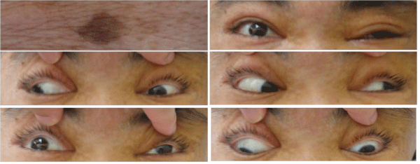
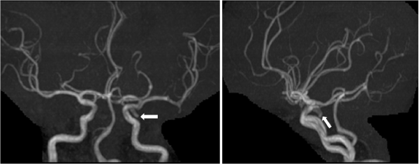
A 44–year–old woman with café au lait spots on her skin (Figure 1; upper left), who had a history of neurofibromatosis type 1 (NF1) and no history of intracranial lesions, presented with retrobulbar headache associated with left oculomotor palsy (Figure 1), which began 3 days before admission. Magnetic resonance imaging revealed no subarachnoid hemorrhage; however, magnetic resonance angiography revealed a left saccular aneurysm (diameter, 8 mm) projecting inferolaterally, at the junction of the internal carotid artery and posterior communicating artery (Figure 2). Because her painful oculomotor palsy indicated impending aneurysmal rupture, she underwent emergency surgery. The left oculomotor nerve was compressed by the aneurysm dome, and the aneurysm neck was successfully clipped. Her oculomotor palsy disappeared after the surgery.
Figure 1: Retrobulbar headache associated with left oculomotor palsy.
Figure 2: Projecting inferolaterally, at the junction of the internal carotid artery and posterior communicating artery.
Besides intracranial tumors such as optic gliomas, an association between NF1 and intracranial aneurysms has recently been suggested [1]. Screening NF1 patients for intracranial aneurysms might be warranted.
References

