Mini Review
Austin Neurosurg Open Access. 2014;1(4): 1016.
Sacral Dimple–the Role and Yield of Imaging
Sy C1, Nyame V1 and Haridas A1,2*
1Department of Neurological Surgery, Wayne State University School of Medicine, USA
2Department of Pediatric Neurosurgery, Children's Hospital of Michigan, USA
*Corresponding author: Haridas A, Department of Pediatric Neurosurgery, Children's Hospital of Michigan, 3901 Beaubien Street, Detroit, MI 48201, USA
Received: May 20, 2014; Accepted: June 05, 2014; Published: June 09, 2014
Abstract
The finding of sacral dimples in newborns has been considered as a cutaneous sign for underlying Occult Spinal Dysraphism (OSD). As such, even isolated findings are worked up with a screening ultrasound and often a Magnetic Resonance Image (MRI) of the lumbar spine. This is an effort to avoid missing a detrimental malformation and to allow for early treatment. These modalities are not without risks, especially as newborns must often undergo anesthesia for MRI procedures. When sacral dimples are found without other cutaneous signs/lesions the probability that an OSD exists is low and falsepositive ultrasound findings can lead to unneeded health risks. A screening ultrasound should only be performed when sacral dimples are found with other cutaneous signs, especially those that have demonstrated higher associations with underlying OSD.
Keywords: Sacral dimple; Sacral pit; Ultrasound; Magnetic resonance imaging; Occult spinal dysraphism; Newborn screening
Abbreviations
Occult Spinal Dysraphism (OSD); Magnetic Resonance Imaging (MRI)
Introduction
Sacral dimples are one of the commonest spinal cutaneous abnormalities seen in the neonatal period. These dimples or "pits" result from incomplete closure of the neural tube during embryogenesis. The relationship of these findings with defects that can cause subsequent neurological sequelae and disability has created an atmosphere of defensive practitioners. Of the different cutaneous signs that correlate with underlying OSD, a recent study involving 1000 newborns found that sacral dimples were the most common finding at 12.8% (more common than myelomeningoceles: 0.5%, acrochordons: 0.1%, and dermoid cysts: 0.1%) [1].
Having a low threshold for further neurological workup is not without reason. A recent report demonstrates that issues can occur and manifest even in adulthood-including motor weakness, incontinence, and chronic pain [2]. As such, early imaging has extended beyond the traditional ultrasound in an effort to avoid overlooking an underlying dysraphism. This surge in pursuing additional imaging is emboldened by associated findings which include hair tufts, family history, neurological signs, skin discoloration/depigmentation, skin folds, deviated gluteal clefts, and soft tissue masses to name a few. Ultrasound screening has proven to be cheap, non-invasive and portable. It is the case, however, that false positive ultrasound findings may then subject infants to MRIs-a modality that is time-consuming,expensive, and includes anesthesia risks (most troubling being hypoxemia) [3]. In this paper, we wish to highlight the relatively low diagnostic yield of imaging in regards to sacral dimples and encourage vigilant clinical decision-making instead of further tests that are likely to incur unneeded costs as well as avoidable patient risks.
Low-Yield of Imaging with Isolated Sacral Dimples
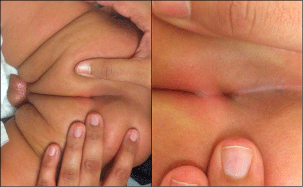
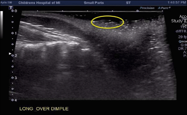
In one study of 943 patients referred for cutaneous stigmata, 68% (638 patients) had a sacral dimple. Of these 638 patients, the resultant ultrasound was normal in 600 patients (where one patient had fatty filum on MRI requiring surgery), and 38 patients exhibited abnormal ultrasounds (with 4 undergoing subsequent surgical repair) [4]. Based on this study by Chern et al., approximately 5% of patientswith cutaneous stigmata will have abnormal lumbar ultrasonography and surgery was only required in less than 1% of infants. In another study of 216 patients who were subjected to ultrasound imaging, the authors found that having multiple indications, as opposed to findings of isolated sacral dimples were, only at that point, six times more likely to yield the diagnosis of spinal dysraphism [5]. Other signs, in contrast, may warrant further investigation to avoid detrimental outcomes as recently outlined in a recent case of midline hypertrichosis in a newborn [6]. These findings are supported by other studies that propose further imaging only when two or more cutaneous lesions are found [7,8]. This questions the value of imaging in detecting closed defects such as spinal lipoma, cord tethering, or fatty filum in the presence of only a sacral dimple without any other cutaneous stigmata. Clinical (Figure 1), as well as ultrasound (Figure 2) findings from a typical patient are provided.
Figure 1: Sacral dimple in a 4 month old (image is magnified on the right).
Figure 2: Sagittal ultrasound showing normal conus level and no underlying spinal dysraphism (yellow circle is approximate area of cutaneous sacral dimple).
When to get an Ultrasound or MRI?
As a rule, lumbar MRI should always be preceded by a screening ultrasound given the disparity in absolute risk between the two modalities. With this in mind, the biggest concern then turns towards the sensitivity of ultrasound as a screening tool. A study by Sasani et al. demonstrates an overall discordance of 16.58% between ultrasound and MRI findings [9]. Meanwhile, a recent comparative study using MRI as the gold standard showed that while ultrasound can be very specific for underlying occult spinal dysraphism (67- 100%) it is not nearly as sensitive (27-86%) [10]. Although definitive data is still lacking, as aforementioned, the presence of at least one additional cutaneous finding increases this sensitivity exponentially [5]. In fact, several other cutaneous findings have been found to warrant prompt investigation regardless of their association withsacral dimples or not, including spinal lipomas and hairy patches as amongst the most prominent [11,12]. The level of urgency associated with such findings should not be extended to that of isolated sacral dimples until further data concludes otherwise. A recent prospective observational study showed that ultrasound had 96% sensitivity, specificity and positive predictive value when compared with MRI in low risk skin dimples and deviated gluteal clefts. In fact, the authors feel that simple dimples and deviated gluteal clefts do not require any imaging whatsoever [13].
Conclusion
Sacral dimples are benign lesions, overall, especially as a solitary finding with no associated skin stigmata, and as such do not require further imaging. Current data shows that a screening ultrasound is appropriate only when other signs/lesions are found along with a sacral dimple. Further studies including MRI are required to further stratify other cutaneous findings and their association with underlying occult spinal dysraphism. Ultimately, we hope that clinical acumen prevails and unnecessary imaging is avoided, especially in low risk lesions such as sacral dimples.
References
- Haveri FT, Inamadar AC2. A cross-sectional prospective study of cutaneous lesions in newborn. ISRN Dermatol. 2014; 2014: 360590.
- Mete M, Umur AS, Duransoy YK, Barutcuo X011f Lu M, Umur N, Gurgen SG, et al. Congenital Dermal Sinus Tract of the Spine: Experience of 16 Patients. J Child Neurol. 2014.
- Malviya S, Voepel-Lewis T, Eldevik OP, Rockwell DT, Wong JH, Tait AR. Sedation and general anaesthesia in children undergoing MRI and CT: adverse events and outcomes. Br J Anaesth. 2000; 84: 743-748.
- Chern JJ, Kirkman JL, Shannon CN, Tubbs RS, Stone JD, Royal SA, et al. Use of lumbar ultrasonography to detect occult spinal dysraphism. J Neurosurg Pediatr. 2012; 9: 274-279.
- McGovern M, Mulligan S, Carney O, Wall D, Moylett E. Ultrasound investigation of sacral dimples and other stigmata of spinal dysraphism. Arch Dis Child. 2013; 98: 784-786.
- Chiu HY, Liao YH. Images in clinical medicine. Occult spinal dysraphism. N Engl J Med. 2014; 370: 466.
- Guggisberg D, Hadj-Rabia S, Viney C, Bodemer C, Brunelle F, Zerah M, et al. Skin markers of occult spinal dysraphism in children: a review of 54 cases. Arch Dermatol. 2004; 140: 1109-1115.
- Weprin BE, Oakes WJ. Coccygeal pits. Pediatrics. 2000; 105: 69.
- Sasani M, Asghari B, Asghari Y, Afsharian R, Ozer AF. Correlation of cutaneous lesions with clinical radiological and urodynamic findings in the prognosis of underlying spinal dysraphism disorders. Pediatr Neurosurg. 2008; 44: 360-370.
- Chern JJ, Aksut B, Kirkman JL, Shoja MM, Tubbs RS, Royal SA, et al. The accuracy of abnormal lumbar sonography findings in detecting occult spinal dysraphism: a comparison with magnetic resonance imaging. J Neurosurg Pediatr. 2012; 10: 150-153.
- Schropp C, Sörensen N, Collmann H, Krauss J. Cutaneous lesions in occult spinal dysraphism--correlation with intraspinal findings. Childs Nerv Syst. 2006; 22: 125-131.
- Williams H. Spinal sinuses, dimples, pits and patches: what lies beneath? Arch Dis Child Educ Pract Ed. BMJ Publishing Group Ltd and Royal College of Paediatrics and Child Health. 2006; 91: 75.
- Ben-Sira L, Ponger P, Miller E, Beni-Adani L, Constantini S. Low-risk lumbar skin stigmata in infants: the role of ultrasound screening. J Pediatr. 2009; 155: 864-869.

