Case Report
Austin Neurosurg Open Access. 2014;1(4): 1019.
Cavernous Angioma of the Dural Convexity Mimicking a Meningioma
Kashlan ON1, Sack JA2 and Ramnath S1*
1Department of Neurosurgery, University of Michigan, USA
2Department of Neurosurgery, University of California-San Diego, USA
*Corresponding author: Ramnath S, Department of Neurosurgery, University of Michigan, 1500 E. Medical Center Dr., Room 3552 TC, Ann Arbor, MI 48109-5338, USA
Received: May 29, 2014; Accepted: August 08, 2014; Published: August 12, 2014
Abstract
Background: Dural-based cavernous angiomas represent a small proportion of all intracranial cavernous angiomas. Moreover even though these lesions have a similar histological appearance as their intra-axial counterparts, they have different imaging characteristics. This fact leads to frequent misdiagnosis via imaging. We present a case of a dural-based occipital convexity cavernous angioma thought to be a meningioma preoperatively followed by a review of the recent literature.
Case Presentation: A 56-year-old man with a known left occipital mass presented with a recent history of visual scintillations. Imaging showed an enhancing mass felt to represent a meningioma. At the time of his operation, a red to purple appearing extra cerebral mass was removed, and its histology was consistent with cavernous angioma.
Conclusions: It is difficult to distinguish an isolated dural based convexity cavernous angioma from a meningioma based on radiology. This case report highlights the importance of keeping both etiologies in the differential diagnosis of a dural-based lesion.
Keywords: Cavernous angioma; Cavernoma; Cavernous malformation; Meningioma mimicker
Abbreviations
CT: Computed Tomography; MRI: Magnetic Resonance Imaging
Case Presentation
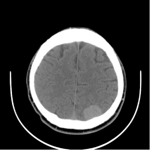
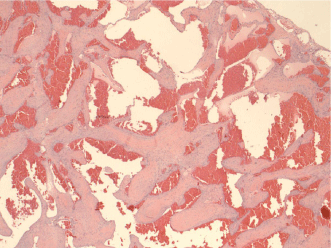
A 56-year-old man presented with a 6-month history of episodic visual scintillations of bright lights in the right visual field. His neurologic findings were normal, with no visual field deficits. A left occipital mass had been found incidentally seven years earlier on a CT scan performed at an outside hospital following a closed head injury. Repeat CT and MRI scans showed an enhancing mass in the left occipital convexity measuring 2.5 cm X 3.2 cm X 3cm as shown in Figures 1 & 2. His non-contrast CT scan report from seven years earlier described a 1.8cm X 2cm mass. Preoperatively, this mass was thought to be an enlarging meningioma. At craniotomy, it was noted that there was no erosion or hyperostosis of the overlying skull. After incising the dura, a dense, red mass was easily dissected off the cortical surface with no invasion of the brain. The mass histologically showed numerous vascular channels divided by various thicknesses of connective tissue septae typical of cavernous angiomas as shown in Figure 3. The patient's postoperative course was uneventful and his imaging demonstrated a gross total resection. At his 1 year postoperative visit, the patient had no evidence of recurrence or development of other lesions.
Figure 1: Non-contrast head CT demonstrating left parieto-occipital lesion.
Figure 2: Pre-operative brain MRI with (a) pre-contrast T1, (b) post-contrast T1, (c) FLAIR, and (d) diffusion-weighted axial images.
Figure 3: Pathology of lesion demonstrating a findings characteristic of a cavernous angioma.
Discussion/Conclusion
Cavernous angiomas are vascular malformations composed of enlarged sinusoidal vessels arranged in clusters, enclosed by a thin endothelial wall without interposed tissue within. They lack smooth muscle, an elastic lamina, and are sometimes calcified or ossified. The lumina can be thrombosed and show attempts at re-canalization. The term "cavernous angioma" has been used interchangeably with "cavernous hemangioma," "cavernous malformation," or "cavernoma". These lesions are accepted as vascular abnormalities rather than as neoplastic processes [1]. Their origin remains obscure [2].
Cavernous angiomas are most commonly seen within the brain parenchyma. However, they are also found intraspinally, or arising from the dura as illustrated by this case report. Extra-axial dural-based cavernous angiomas are extremely rare when compared to their intra-axial counterparts [3]. Published data from Lewis et al. identified two types of dural cavernous angiomas [2]. The first, which is more common, includes ones that present in the dura of the middle fossa usually in vicinity of the cavernous sinus. The second type consists of dural-based lesions elsewhere such as the convexity, cerebral and cerebellar falx, the tentorium, posterior fossa, and the floor of the anterior fossa [2,4]. The separation of the two types is important due to the differences in the patient population affected and the more aggressive clinical course of dural-based cavernous angiomas in the middle fossa [2].
To better understand these rare lesions, a literature review was performed searching for case reports published after 1992 dealing with dural-based cavernous angiomas occurring outside the middle cranial fossa [1,4,6-9,11-25]. These results were compared to published data from the Lewis et al manuscript, which included a review of the literature published prior to 1992 [2]. A summary of the cases found is shown in Table I. As was noted by Lewis et al, headache was the most common chief complaint in our review, occurring in 12 of the 22 patients; however, this prevalence is lower than the 75% (9 of 12 patients) seen in their paper [2]. Moreover, the cerebral convexities continue to be the most common area affected by dural-based cavernous angiomas outside the middle cranial fossa occurring in 10 out of 22 patients (45.5%). Even though noted in the literature to have no gender preference, our review indicated that there may be a male predominance with 16 of the 22 cases affecting males; this is in contrast to Lewis et al where only 10 out of 18 of patients affected were male [2]. If the data are combined, 26 out of the 40 total cases evaluated (65%) occurred in males, which strengthen the idea of a possible male predominance in patients affected with dural-based cavernous angiomas occurring outside the middle fossa. With regards to prognosis, these lesions are amenable to gross total resection and do not have a propensity to recur [4].
Case
Chief Complaint
Age
Sex
Location
MRI T1 Appearance
MRI T2 Appearance
MRI Enhancement
CT Appearance
CT Enhancement
Angiogram
Tsutsumi S, et al.4
Headache
43
M
Cerebellar
Hypointense
Mixed
Yes
Isodense
-----
-----
Zeng, X et al.13
Seizures
37
F
Falx
-----
Hyperintense
Yes
-----
-----
-----
Episode of confusion
58
M
Cerebellar falx
Isointense
Hyperintense
Yes
Hyperdense
-----
-----
Sakakibara Y et al.15
Left face and upper extremity numbness
59
M
Convexity
Isointense
-----
Yes
-----
-----
Pooling of medium in late venous phase
Gutiérrez-González R et al.16
Anosmia
47
F
Anterior cranial fossa floor
Hypointense
Hyperintense
Yes
-----
-----
-----
Incidental finding
47
M
Cerebellar falx
Isointense
Hyperintense
Yes
Hyperdense
-----
-----
Headache
15
M
Convexity
Hypointense
Mixed
Yes
Hyperdense
Yes
-----
Occipital headache, intermittent left field scintillations
15
M
Tentorium
Mixed
Mixed
Yes
-----
-----
Hypervascular with pooling of contrast medium in late venous phase
Hwang SW et al.9
Vertigo, headaches
61
M
Convexity
Isointense
Hyperintense
Yes
-----
-----
-----
Boockvar JA et al.19
Headaches, decreasing visual acuity
31
M
Superior sagittal sinus (superior to torcula)
-----
-----
Yes
-----
-----
Avascular mass
Ophthalmic migraine
63
F
Falx
Hypointense
Hyperintense
Yes
-----
Yes
-----
Headaches, facial pain
18
F
Convexity
Isointense
Hyperintense
Yes
-----
-----
Tumor blush
Hyodo A et al.8
Altered mental status
77
M
Convexity
Hyperintense
Hypointense
Minimal
-----
-----
-----
Rushton AW et al.6
Headaches, vomiting, blurred vision
5
M
Cerebellar
-----
-----
-----
Mixed
-----
-----
Blurred vision
53
M
Tentorium
Mixed
-----
Yes
-----
-----
Hypervascular
Headaches, vomiting
5
M
Cerebellar
-----
-----
-----
Hyperdense
Yes
-----
Headaches, vomiting, loss of consciousness
78
F
Convexity
-----
-----
-----
Subdural hematoma
-----
-----
Headaches, seizures, left visual blurring
35
M
Convexity
Mixed
Mixed
Yes
-----
Minimal
Negative
Revuelta R et al.23
Headaches
66
M
Convexity
Isointense
Hyperintense
-----
-----
Yes
-----
Episodic ataxia of left limbs
60
M
Cerebellar
-----
-----
-----
Hyperdense
Yes
Vascular lesion fed by occipital artery
Seizures
77
F
Convexity
-----
-----
-----
-----
Yes
-----
Right visual scintillations
56
M
Convexity
Hypointense
Hyperintense
Yes
Hyperdense
-----
-----
Table 1: Summary of 22 Cases of Dural-Based Cavernous Angiomas outside the Middle Fossa since 1992.
Even though histologically similar to intraparenchymal cavernous angiomas, dural-based cavernous angiomas have a completely different appearance on CT, MRI and angiography [5]. This fact makes accurate pre-operative diagnosis very difficult, as imaging findings can be varied and resemble meningiomas. On CT, dural-based cavernous angiomas outside the middle fossa are mostly hyperdense, but could be isodense. At times, they may be calcified [6-8]. They also may cause either bony erosion or hyperostosis of the overlying skull [6,9]. They almost always enhance on CT. On MRI, these lesions are usually either isointense or hypointense on T1 sequences. Much like on CT, they also usually enhance on MRI. Based on our review, dural-based cavernous angiomas outside the middle fossa are hyperintense on T2 sequences 64.3% of the time (9 out of 14 patients) and were not seen to be isointense in any of the cases. This is different from meningiomas, which are isointense almost half the time [10]. However, meningiomas can be hyperintense on T2 sequences as well so having a T2 hyperintense lesion is far from specific for dural-based cavernous angioma outside the middle fossa. Interestingly, these lesions can have dural tails like meningiomas, and may also cause significant perilesional edema [11,12]. Angiography can show a tumor blush, hypervascularity, an avascular mass, can be completely negative, or demonstrate pooling of medium during the late venous phase.
As is apparent, there is no definitive differentiation that can be made between dural cavernous angiomas and meningiomas with regards to imaging. Therefore, it is important for the neurosurgeon to keep this entity among the differentials when planning resection of a lesion that seems to be a meningioma.
References
- Biondi A, Clemenceau S, Dormont D, Deladoeuille M, Ricciardi GK, Mokhtari K, et al. Intracranial extra-axial cavernous (HEM) angiomas: tumors or vascular malformations? See comment in PubMed Commons below J Neuroradiol. 2002; 29: 91-104.
- Lewis AI, Tew JM Jr, Payner TD, Yeh HS. Dural cavernous angiomas outside the middle cranial fossa: a report of two cases. See comment in PubMed Commons below Neurosurgery. 1994; 35: 498-504.
- Riant F, Bergametti F, Fournier HD, Chapon F, Michalak-Provost S, Cecillon M, et al. CCM3 Mutations Are Associated with Early-Onset Cerebral Hemorrhage and Multiple Meningiomas. See comment in PubMed Commons below Mol Syndromol. 2013; 4: 165-172.
- Tsutsumi S, Yasumoto Y, Saeki H, Ito M. Cranial dural cavernous angioma. See comment in PubMed Commons below Clin Neuroradiol. 2014; 24: 155-159.
- Rosso D, Lee DH, Ferguson GG, Tailor C, Iskander S, Hammond RR, et al. Dural cavernous angioma: a preoperative diagnostic challenge. See comment in PubMed Commons below Can J Neurol Sci. 2003; 30: 272-277.
- Rushton AW, Ng HK, Metreweli C. Dural cavernous haemangioma with bony infiltration. See comment in PubMed Commons below Clin Radiol. 1999; 54: 406-408.
- Vogler R, Castillo M. Dural cavernous angioma: MR features. See comment in PubMed Commons below AJNR Am J Neuroradiol. 1995; 16: 773-775.
- Hyodo A, Yanaka K, Higuchi O, Tomono Y, Nose T. Giant interdural cavernous hemangioma at the convexity. Case illustration. See comment in PubMed Commons below J Neurosurg. 2000; 92: 503.
- Hwang SW, Pfannl RM, Wu JK. Convexity dural cavernous malformation with intradural and extradural extension mimicking a meningioma: a case report. See comment in PubMed Commons below Acta Neurochir (Wien). 2009; 151: 79-83.
- Elster AD, Challa VR, Gilbert TH, Richardson DN, Contento JC. Meningiomas: MR and histopathologic features. See comment in PubMed Commons below Radiology. 1989; 170: 857-862.
- Joshi V, Muzumdar D, Dange N, Goel A. Supratentorial convexity dural-based cavernous hemangioma mimicking a meningioma in a child. See comment in PubMed Commons below Pediatr Neurosurg. 2009; 45: 141-145.
- Shen WC, Chenn CA, Hsue CT, Lin TY. Dural cavernous angioma mimicking a meningioma and causing facial pain. See comment in PubMed Commons below J Neuroimaging. 2000; 10: 183-185.
- Zeng X, Mahta A, Kim RY, Saad AG, Kesari S. Refractory seizures due to a dural-based cavernoma masquerading as a meningioma. See comment in PubMed Commons below Neurol Sci. 2012; 33: 441-443.
- Melone AG, Delfinis CP, Passacantilli E, Lenzi J, Santoro A. Intracranial extra-axial cavernous angioma of the cerebellar falx. See comment in PubMed Commons below World Neurosurg. 2010; 74: 501-504.
- Sakakibara Y, Matsumori T, Taguchi Y, Koizumi H. Supratentorial high convexity intradural extramedullary cavernous angioma: case report. See comment in PubMed Commons below Neurol Med Chir (Tokyo). 2010; 50: 328-329.
- Gutiérrez-González R, Casanova-Pe&nTilde;o I, Porta-Etessam J, Martínez A, Boto GR. Dural cavernous haemangioma of the anterior cranial fossa. See comment in PubMed Commons below J Clin Neurosci. 2010; 17: 936-938.
- Ito M, Kamiyama H, Nakamura T, Nakajima H, Tokugawa J. Dural cavernous hemangioma of the cerebellar falx. See comment in PubMed Commons below Neurol Med Chir (Tokyo). 2009; 49: 410-412.
- Mori H, Koike T, Endo S, Takii Y, Uzuka T, Takahashi H, et al. Tentorial cavernous angioma with profuse bleeding. Case report. See comment in PubMed Commons below J Neurosurg Pediatr. 2009; 3: 37-40.
- Boockvar JA, Stiefel M, Malhotra N, Dolinskas C, Dwyer-Joyce C, LeRoux PD, et al. Dural cavernous angioma of the posterior sagittal sinus: case report. See comment in PubMed Commons below Surg Neurol. 2005; 63: 178-181.
- Lee AG, Parrish RG, Goodman JC. Homonymous hemianopsia due to a dural cavernous hemangioma. See comment in PubMed Commons below J Neuroophthalmol. 1998; 18: 250-254.
- Hsiang JN, Ng HK, Tsang RK, Poon WS. Dural cavernous angiomas in a child. See comment in PubMed Commons below Pediatr Neurosurg. 1996; 25: 105-108.
- Suzuki K, Kamezaki T, Tsuboi K, Kobayashi E. Dural cavernous angioma causing acute subdural hemorrhage--case report. See comment in PubMed Commons below Neurol Med Chir (Tokyo). 1996; 36: 580-582.
- Revuelta R, Teixeira F, Rojas R, Juambelz P, Romero V, Valdes J, et al. Cavernous hemangiomas of the dura mater at the convexity. Report of a case and therapeutical considerations. See comment in PubMed Commons below Neurosurg Rev. 1994; 17: 309-311.
- Goel A, Achwal S, Nagpal RD. Dural cavernous haemangioma of posterior cranial fossa. See comment in PubMed Commons below J Postgrad Med. 1993; 39: 222-223.
- Perry JR, Tucker WS, Chui M, Bilbao JM. Dural cavernous hemangioma: an under-recognized lesion mimicking meningioma. See comment in PubMed Commons below Can J Neurol Sci. 1993; 20: 230-233.


