Case Report
Austin Neurosurg Open Access. 2025; 11(1): 1076.
Myeloproliferative Neoplasm Presenting as Spinal Myelopathy: Early Recognition and Management
Ndaro Daniel1,2* and Hugh P. Sims-Williams1,2
1Tenwek Hospital, Bomet, Kenya
2Loma Linda University, Carlifornia, USA
*Corresponding author: Ndaro Daniel, Tenwek Hospital, Bomet, Kenya; Loma Linda University, Carlifornia, USA Email: ndarodaniel@gmail.com
Received: April 13, 2025 Accepted: April 24, 2025 Published: April 28, 2025
Abstract
Myeloproliferative neoplasms (MPNs) are a group of hematological disorders marked by excessive blood cell production, with spinal involvement being an uncommon presentation, particularly in young adults. This case report describes a 24-year-old male who presented with progressive lower limb weakness and back pain, ultimately diagnosed with chronic myeloid leukemia (CML) in the blast phase, manifesting as spinal myelopathy due to extramedullary hematopoiesis. MRI revealed extradural masses at L5-S1 and T3-T9 levels, causing spinal cord compression. Hematological investigations showed marked leukocytosis and anemia; bone marrow biopsy confirmed an MPN, and immunohistochemistry suggested B-lymphoid blast phase CML. Despite limitations in confirming the BCR-ABL mutation, clinical and pathological findings supported the diagnosis.
Management focused on supportive therapy, corticosteroids, and multidisciplinary planning. The patient showed significant neurological improvement with corticosteroids, and surgery was deferred in favor of initiating systemic chemotherapy. This case underscores the diagnostic and therapeutic challenges of MPN-related spinal myelopathy, emphasizing the need for early recognition, appropriate imaging, and coordinated care. It also highlights the importance of considering hematologic malignancies in the differential diagnosis of unexplained myelopathy, especially in resource-limited settings. Timely medical management, even without immediate surgical intervention, can result in favorable neurological outcomes. This report adds to the limited literature on spinal manifestations of MPNs and advocates for heightened clinical suspicion and multidisciplinary collaboration in managing such rare presentations.
Abbreviations
MPN: Myeloproliferative Neoplasm; CML: Chronic Myeloid Leukemia; GCS: Glasgow Coma Scale; CT: Computed Tomography; MRI: Magnetic Resonance Imaging; CD: Cluster of Differentiation; TdT: Terminal deoxynucleotidyl Transferase; BCR-ABL: Breakpoint Cluster Region-Abelson.
Introduction
Myeloproliferative neoplasms (MPNs) encompass a spectrum of hematological disorders characterized by excessive production of blood cells. The major subtypes include chronic myeloid leukemia (CML), polycythemia vera, essential thrombocythemia, and primary myelofibrosis. While these conditions can lead to systemic complications, spinal involvement remains an infrequent manifestation [1]. When present, spinal myelopathy typically results from extramedullary hematopoiesis or direct spinal cord compression. The occurrence of such a presentation in young adults is exceedingly rare, making the case under discussion particularly significant.
Case Presentation
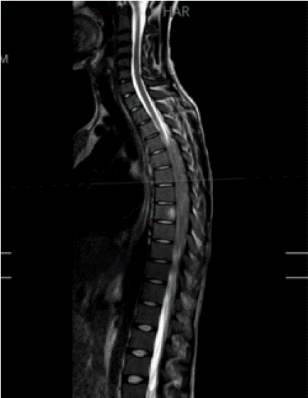
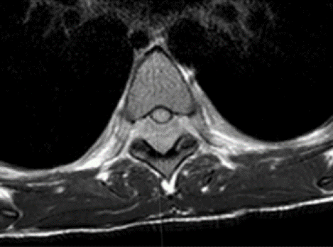
A 24-year-old male with no prior medical history presented with a one-month history of bilateral lower limb weakness, initially preceded by persistent, non-radiating lower back pain. Over time, the weakness progressed asymmetrically, with the right lower limb being more affected than the left, ultimately necessitating the use of a walking aid. The patient denied any history of trauma, weight loss, fever, or previous infections. On examination, he was hemodynamically stable with a Glasgow Coma Scale (GCS) score of 15. Neurological evaluation revealed normal upper limb motor strength (5/5), while the lower limbs exhibited profound weakness-graded 2/5 on the right and 0/5 on the left. Sensory function remained intact, reflexes were normal, and no pathological reflexes were elicited. Bowel and bladder functions were preserved. Imaging studies revealed a large extradural mass at the right L5-S1 level, causing significant displacement of the thecal sac, with a secondary mass extending from T3 to T9, resulting in spinal canal narrowing. Additionally, CT imaging demonstrated massive splenomegaly. MRI confirmed the presence of extradural masses with mixed signal intensities, accompanied by mild to moderate cord compression at the T4-T10 levels (Figure 1,2).

Figure 1: T2 Sagittal MRI of the spine showing the thoracic epidural mass.

    Figure 2: T2 axial MRI at the T6 vertebra showing the epidural mass with
cord compression.
Hematological investigations showed marked leukocytosis (386,000/μL) and normocytic normochromic anemia. Bone marrow biopsy confirmed hypercellular marrow consistent with an MPN. Immunohistochemical analysis revealed blast cells expressing CD34, Pax5, and TdT, supporting a diagnosis of CML in the blast phase (B-lymphoid lineage). Although the Philadelphia chromosome/BCRABL mutation could not be confirmed due to technical limitations, the overall findings were strongly indicative of CML. The patient was diagnosed with a myeloproliferative neoplasm presenting as spinal myelopathy due to extramedullary hematopoiesis. Differential diagnoses included spinal tumors (primary or metastatic), inflammatory conditions (e.g., multiple sclerosis), spinal infections (e.g., tuberculosis, abscesses), and trauma-related myelopathy.
The patient was initially managed with supportive care, including corticosteroids to reduce inflammation, anticoagulation to mitigate thrombotic risk, and analgesia for pain control. A multidisciplinary team comprising a hematologist, neurosurgeon, and orthopedic surgeons was involved in treatment planning.
The patient showed significant neurological improvement following corticosteroid administration. Given the nature of the disease, which is systemic and requires chemotherapy, and the observed response to steroids, decompressive surgery was deferred to avoid delaying chemotherapy. Instead, close monitoring and followup imaging were prioritized. There are plans to initiate targeted therapy pending confirmation of the BCR-ABL mutation.
Following corticosteroid therapy, the patient experienced substantial neurological improvement, regaining the ability to walk short distances unaided. Repeat imaging demonstrated partial regression of the extradural masses [3]. The patient continues to be monitored on an outpatient basis, with plans for further testing to confirm the BCR-ABL mutation and determine eligibility for targeted chemotherapy. Long-term surveillance remains necessary to assess for disease progression or recurrence.
Discussion
The presentation of MPNs with spinal myelopathy is exceptionally rare [1], particularly in young patients. Diagnosis is often delayed due to the rarity of such cases and the broad differential diagnoses, which include spinal tumors, infections, and inflammatory conditions. This case underscores the importance of maintaining a high index of suspicion for hematological disorders in patients presenting with unexplained neurological deficits.
Managing spinal myelopathy secondary to MPNs poses significant challenges, requiring a delicate balance between spinal cord decompression and systemic hematological treatment. In cases of extramedullary hematopoiesis, a multidisciplinary approach is essential to optimize outcomes [2]. Timely interventions, including corticosteroids and appropriate chemotherapy, can enhance neurological function and improve quality of life.
While reports of spinal myelopathy secondary to MPNs are scarce, early recognition and intervention-including decompressive surgery, when necessary, followed by targeted hematological management-are critical in achieving favorable neurological recovery [1].
Conclusion
This case highlights the rare and complex nature of MPNs presenting with spinal myelopathy, particularly in young patients. Early recognition of hematological abnormalities, prompt imaging, and a collaborative management approach are vital for optimizing patient outcomes. This report reinforces the need for heightened clinical awareness and a multidisciplinary strategy in addressing such rare presentations in low- and middle-income countries.
References
- Horwood E, Dowson H, Gupta R, Kaczmarski R, Williamson M. Myelofibrosis presenting as spinal cord compression. J Clin Pathol. 2003; 56: 154-156.
- Hawkins AN, Breton JM, Bhatnagar A, Paudel N, Voyadzis JM. Spinal extramedullary hematopoiesis mimicking an epidural tumor in a patient with high-risk polycythemia vera: illustrative case. J Neurosurg Case Lessons. 2024; 8: CASE23659.
- Subahi EA, Abdelrazek M, Yassin MA. Spinal cord compression due to extramedullary hematopoiesis in patient with Beta thalassemia major. Clin Case Rep. 2020; 9: 405-409.