
Mini Review
Austin Neurosurg Open Access. 2015; 2(4): 1040.
Evaluation of the Immediate and Early Role of Decompressive Craniectomy in the Treatment of Refractory ICP in Cases of STBI in Egypt: A Clue for Responders
Mohamed El-Fiki*
Professor of Neurosurgery, University Of Alexandria, Egypt
*Corresponding author: Mohamed El-Fiki, Professor of Neurosurgery, University Of Alexandria, 63 Sidi Gaber Street, Cleopatra, Alexandria, Egypt
Received: August 21, 2015; Accepted: September 23, 2015; Published: September 28, 2015
Abstract
Introduction: Decompressive craniotomy may be performed in several indications including severe traumatic brain injuries. To date, there is no specific drug treatment and many promising agents in pre-clinical animal models have failed in clinical trials. Evidence-based guidelines for traumatic brain injury management have not made a major impact on recovery. Decompressive craniectomy procedure was successful in decreasing intracranial pressure in most cases but has failed to change prognosis. Selecting patients who may improve using new selection criteria will increase the procedure acceptance and define criteria for success.
Patients and Methods: Alexandria study recruited 80 patients with severe traumatic brain injuries & increased intracranial pressure above 20 mm H2O who presented with Glasgow Coma Score mean of 5.83. Surgical decompression was performed via wide uni- or bi-lateral frontotemporo-parietal hemicraniectomy and augmentation duraplasty.
Results: All patients showed decreased intracranial pressure postoperatively. This was not reflected as improved outcome except in those who sustained decreased ICP for one week. Eight patients showed good recovery (10%), 8 others were moderately disabled (10%), 5 patients were severely disabled (6%), while 16 remained vegetative (20%). Mortality was encountered in 43 cases (54%).
Conclusion: Decompressive craniectomy decreased high intracranial pressure in patients with severe traumatic brain injuries with high morbidity and mortality. Only patients who maintained a lowered intracranial pressure below 20 mm H2O showed clinical recovery. Patients who reached good functional outcome were those in whom lowered intracranial pressure was maintained after decompressive craniectomy for more than one week. Patients who showed a later secondary increased intracranial pressure are candidates for further studies & treatment alternatives and may be spared decompressive craniectomy.
Keywords: Craniectomy; Severe Traumatic Brain Injuries; Decompressive Craniectomy; Neurotrauma
Abbreviations
WHO: World Health Organization; DC: Decompressive Craniectomy; RTA: Road Traffic Accidents; STBI: Severe Traumatic Brain Injuries; ICP: Intracranial Pressure; ICU: Intensive Care Unit; CPP: Cerebral Perfusion Pressure; CSF: Cerebrospinal Fluid; PRx: Pressure Reactivity Index; mABP: mean Arterial Blood Pressure; SSS: Superior Sagittal Sinus; pO2: Partial Pressure of Oxygen
Introduction
The WHO report on Road Traffic Accidents (RTA) in March 2013 included 99% of world population. Egypt RTA rate is one of the highest where 12 fatal accidents occurred daily with 12300 death & 154000 injuries in 2009. Cyclists & pedestrians death accounts for 27% world wide and 45% in Eastern Mediterranean. Increased Intracranial Pressure (ICP) due to Severe Traumatic Brain Injuries (STBI) is the main etiologic factor for mortality. To date, there is no specific drug treatment for acute brain injury, and many pre-clinical animal drug trials have failed clinically. Evidence-based guidelines for TBI management in several countries have not made a major impact on recovery.
According to the Monroe Kellie Doctrine; in STBI, when the compensatory mechanisms start to fail, smaller increases in cerebral swelling/edema or minimal enlargement of a mass lesion will initiate incremental increases in decompensated ICP to a hazardous level that might be fatal. DC is performed to reverse ICP to compensated levels.
In order to alleviate the dire consequences of increased ICP Decompressive Craniectomy (DC) may be performed for indications such as STBI, extensive hemispherical infarctions, intracranial hemorrhages, or edema of infections [1-4]. Conflicting results of DC are published using different techniques and endpoints as well as study designs. Numerous complications may arise in a sequential fashion and at specific time points following DC [5]. However the procedure proved successful in decreasing ICP in most cases although it has failed to change prognosis. STBI management guidelines consider DC as the last -tier option. Given the favorable results of some prospective studies, DC procedure achieved popularity as a theoretical favorable remedy for patients with STBI and intractable ICP [6]. Selecting patients who may improve will increase the procedure acceptance and define criteria for success.
University of Alexandria, Egypt trial Objective was to evaluate the early role (first week) of DC in treatment of traumatic brain injury in terms of lowering increased ICP, 1-week outcome, rate of complications and ICP after one week of DC.
Patients and Methods
From Jan 2009-July 2014, the study recruited 80 patients with STBI (GCS < 9) & persistent increased ICP > 20 mm H2O for more than 12 hours who were admitted to the Neuro-ICU at the departments of neurosurgery, University of Alexandria, Egypt. They were screened with CT scans that were reported according to Marshall Scale [7]. ICP monitoring was implemented. Refractory cases of raised ICP who showed failure of best conservative measures to lower high ICP value were subjected to DC. Forty-three patients were randomized to primary DC, while another 37 patients crossed-over to DC from best medical treatment when their ICP persistently increased above 20 cm H2O for at least 12 hours in spite of occasional barbiturate administration. The two groups thus became a single cohort of 80 patients. DC was performed via wide uni- or bi-lateral frontotemporoparietal hemicraniectomy not less than 12 cm in longest dimension. An island of bone was left to cover the SSS. Supplementary subtemporal decompression was performed if impending or starting uncal herniation were suspected (Figure 1). Augmentation duraplasty was performed in all patients. Outcome of DC was evaluated as regards ICP value, procedure related complications and factors affecting outcome. Patient’s ICP was compared to their preoperative value in outcome analysis (patients were used as their own controls). Statistical analysis was performed (Student’s t-test for continuous variables and the Χ² test for categorical variables). A value of p < 0.05 was considered significant. SPSS software version 19.0 was used. The ethical committee of the University of Alexandria, Faculty of Medicine, approved the study. Informed consent was obtained from next of kin when available. Some patients were brought to hospital as unrecognized comatose patients.
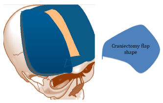
Figure 1: Extent of DC in Alexandria study (blue). SSS flap (clay colored).
Results
The current study encompasses 80 adult patients who were subjected to DC after STBI at Alexandria Main University Hospitals. DC was performed in 43 of them at initial randomization to surgery because of persistently high ICP above 20 mm HO2 and GCS less than 9. Another 37 patients were also operated when they failed standard best medical treatment including barbiturate administration and their ICP surpassed 20 mm HO2 (Figure 2). Most patients were in their 4th or 6th decade of age (Figure 3). Patients were in very bad clinical state with GCS 4-6 in most cases and generally less than 9 with a mean of 5.83 (Figure4). Reflecting the severity of the causative trauma, 53 patients (66%) have extra-cranial thoracic or abdominopelvic injuries as well as fractures of long bones associated with their head trauma (Figure 5). Associated injuries were accountable for hypotensive episodes during the first 24 hours of ICU confinement where the systolic BP dropped to less than 90 mm Hg in 37 patients (Figure 6). Hypoxic episodes to pO2 below 90% were noted in 44 patients during that same period (Figure 7). CT scan was performed for all patients on admission and repeated 6 hours later or whenever deemed necessary. Marshal CT classification was used to standardize reporting [7]. DC was performed through a wide craniectomy not less than 12 cm in longest dimension on one or both sides according to the clinical status of the patients. An island of bone was left over the SSS in bilateral cases to protect venous drainage (Figure 1). Fortyfour patients sustained a hypoxic episode during ICU confinement after STBI 73% of them died and 27% survived after DC. In the mean time 72% of the 36 patients who did not suffer hypoxic episodes survived DC and only 10 patients of them died in spite of DC. On the other hand hypotension also had a deleterious effect on outcome in spite of DC. Out of 37 hypotensive patients with STBI who were managed with DC only 5 patients (13%) survived, while 32 patients (87%) succumbed to their STBI. At the same time out of 43 patients who did not suffer hypotensive episodes 32 patients (74%) survived and only 11 patients (26%) passed away after their severe head injury & DC (Figure 7).
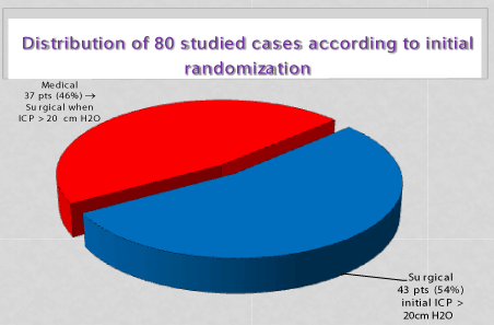
Figure 2: Distribution and randomization of cases.
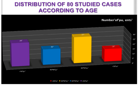
Figure 3: Age distribution.
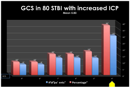
Figure 4: GCS distribution.
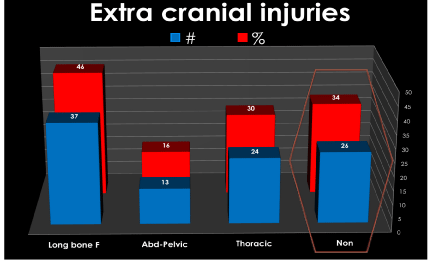
Figure 5: Sites of extracranial injuries.
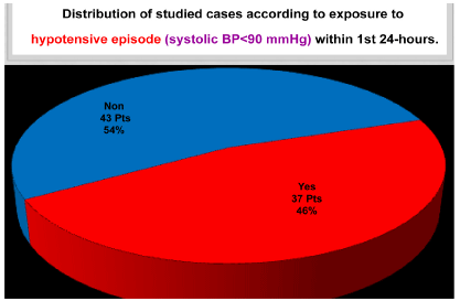
Figure 6: Hypotensive Episodes during the first 24 hours.
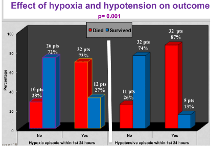
Figure 7: Effect of Hypoxia and Hypotension of Outcome of DC in STBI.
Complications were frequently encountered in more than half of the patients (52%) mostly as CT hypo dense hypo-perfused suspected hemispherical, lobar or scattered infarctions or pathological edema. Pneumonias prevailed in 10% of patients. Hydrocephalus was confronted in later scans in 8% of cases (Figure 8).
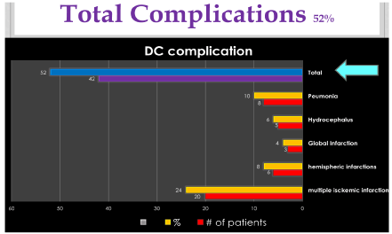
Figure 8: Total complications.
DC succeeded in decreasing ICP to lower than 20 mm HO2 in all patients (Figure 9A and 9B). One week after DC, only patients whose ICP remained below 20 mm HO2 enjoyed good recovery, mild impairment or severe disability. Eight patients (10%) showed good recovery, 8 others were moderately disabled, 5 patients (6%) were severely disabled while 16 patients (20%). remained vegetative. Mortality was encountered in 43 cases (54%) in spite of DC (Figure 10).
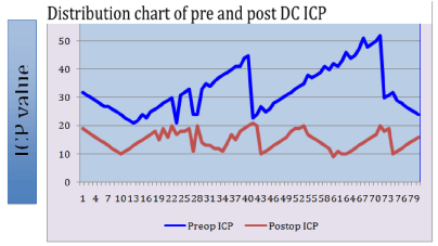
    Figure 9A:  Distribution chart of individual ICP measurements pre- and post-
DC.
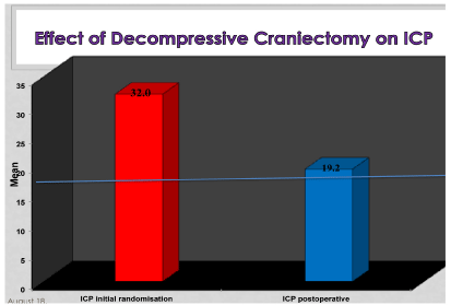
Figure 9B: Effect of Decompressive Craniectomy on ICP.
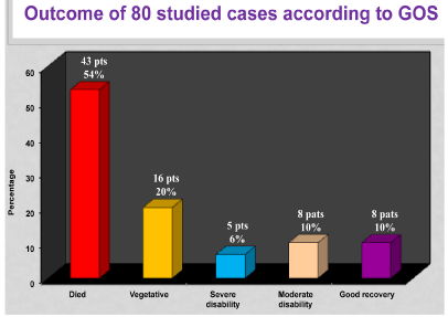
Figure 10: Outcome of DC according to GOS.
Associated extra cranial injuries were encountered in 53 patients (66%) reflecting the severity of causative trauma and were significantly associated with one-week mortality (p= 0.019). More than 70% of patients who have associated injuries (37 patients) died in spite of DC and best medical treatment; while 82% of the 27 patients without associated injuries survived (Figure 11).
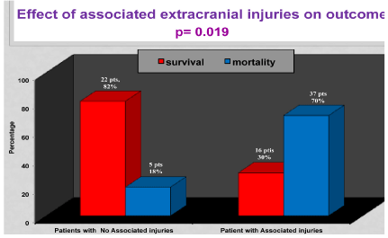
Figure 11: Effect of Extracranial Injuries on Outcome of DC in STBI.
Patients in whom DC succeeded to lower ICP but failed to maintain the lowered ICP value below 20 mm H2O for one week became vegetative or passed away (Figure 12). In that figure the light green horizontal arrow defines the targeted 20 mm HO2 ICP level of the surgical DC procedure. Those patients with moderate or severe disability as well as patients who enjoyed good recovery continued to maintain an average ICP value below the threshold of 20 mm H2O one week after DC. In spite of the fact that vegetative patients or those who perished achieved an immediate postoperative decline of ICP to the desired level, they failed to maintain the lowered ICP during the next (vertical purple arrow).
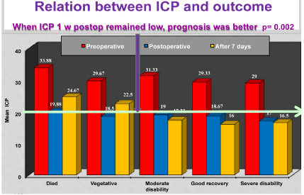
Figure 12: Relation between ICP and Outcome of DC in STBI.
Discussion
Managing STBI in specialized neuro ICU, safe transfer, better triage with GCS, and admission CT scans improved outcome compared to victims managed in general ICU. Guidelines for management of STBI when applied resulted in minor improvement of mortality. When CSF was drained, mortality dropped from 43% to 21 % in a meta-analysis [8] suggesting an impact of monitoring and reducing ICP in Neuro ICU. Maturity and refining ICP management of patients as well as rational perfections of DC patient selection are worth investigation. Because of individual time variations of STBI patient’s response to optimum treatment, a trial of ICP optimization in DC is indispensible.
International prospective randomized controlled trials with different selection criteria and different ICP threshold were conducted [9,10]. The cohort profiles and criteria for entry and randomization between the DECRA [9] and RESCUEicp [10] were very different. Hence the results from the DECRA study did not deter recruitment into RESCUEicp that is now complete. The results of DECRA study are discouraging, while those of the RESCUEicp are not available yet. In 17 African patients, outcome was good in all those with mild head injury, in 66.7% of the moderate, and 56% of STBI [11]. ICP monitoring together with brain tissue oxygenation and consequent CT or MRI examinations are valuable tools in management of STBI [12].
In the current study in Alexandria DC was performed in 80 patients with STBI, 43 of them because of persistently high ICP above 20 mm HO2 for more than 12 hours after initiating ICP monitoring because of GCS less than 9. Another 37 patients were also operated later when they failed standard best medical treatment including barbiturate administration and their ICP surpassed 20 mm HO2 after initial response (Figure 2). The threshold of ICP is variable in most published series [8,9,12,13]. The study excluded children since their response to trauma might be different than adults [14,15]. Most patients were in their 4th or 6th decade of age (Figure 3) comparable to other published series.
Patients were recruited to the study if GCS was less than 9 reflecting the bad clinical status. Most patients have a GCS of 4-6 with a mean of 5.83 (Figure 4). DECRA study included patients with GCS of 3- 8 or Marshal Grade III lesions with no mass lesions. The RESCUEicp study GCS was 3-8 in more slightly more than 2/3rd of cases, while in 30 % GCS was above 9 and reached 13-15 in 11% of the total. In the DECRA study ICP above 20 mmHg was an indication for DC any time after injury. Because there was criticism for the DECRA study for possible very early interference and possibly performing DC surgery in what might not be an actual persistently elevated ICP, the current study used a lower threshold of ICP for a prolonged period of time to test for the efficacy of best medical treatment of duration for more than 12 hours. In the RESCUEicp the threshold for DC was 25 mmHg for 1-12 hours within 72 hours post-injury. They accepted contusions like Alexandria study. The current study outcome was decided at one week after DC, while the outcome of both the DECRA and RESCUEicp studies was 6 months GOS with an extended outcome of two years in RESCUEicp.
Effect of associated injuries, Hypotension and Hypoxia: Reflecting the severity of the causative trauma, 53 patients (66%) have extra-cranial thoracic or abdomino-pelvic injuries as well as fractures of long bones associated with their head trauma (Figure 5). This percentage reflects the fact that many patients were pedestrians rather than vehicle occupants. Coupled with defective road safety protocols in Egyptian highways, falls represent a good percentage especially in construction workers as a result of deficient endorsement of work place safety regulations. RESCUEicp patients were hypoxic in 4% while 13% where hypotensive at initial presentation. Associated injuries in Alexandria study were accountable for hypotensive episodes during the first 24 hours of ICU confinement where the systolic BP dropped to less than 90 mm Hg in 37 patients (Figure 6). Hypoxic episodes to pO2 below 90% were noted in 44 patients during that same period (Figure 7). Hypotension and hypoxia also might be attributed to delayed evacuation and transfer to neuro ICU with inadequate resuscitation at the scene of the accident secondary to poor equipment and personnel training availability. The high percentage of hypotension and hypoxia in the current study might be accountable for a good percentage of the encountered complications (Figure 8) as well as many of the severely disabled patients in spite of maintaining a low ICP for one week. Associated extra cranial injuries were significantly associated with one-week mortality (p= 0.019). More than 70% of patients who have associated injuries (37 patients) died in spite of DC and best medical treatment; while 82% of the 27 patients without associated injuries survived (Figure 11). Improved road and work place safety and victim’s evacuation strategies including air lift helicopters to neuro ICU where management guidelines are strictly adhered to must be emphasized.
CT scan was performed for all patients on admission and repeated 6 hours later or whenever deemed necessary depending on the clinical situation. Marshal CT classification was used to standardize reporting [13]. Patients of grade VI on Marshal Scale with mass lesions were excluded from the study as in the DECRA study. The use of Marshal Scale was advantageous in many other studies in order to decrease inter-observer bias [13,16].
The extent of DC in Alexandria is generally wider than the DECRA study and closer to the RESCUEicp i.e. more than 12 cm in largest diameter. Sub temporal decompression is added if swelling is predominantly unilateral or with midline shift. Dural repair was optional in RESCUEicp but in Alexandria study we routinely performed duraplasty to buttress the decompressed brain. Tight bandage or positioning patient head on the craniectomy side was avoided. Bilateral edema cases had a bilateral boney DC with bilateral U-shaped opening of the dura, based on the superior sagittal sinus [10] as in RESCUEicp. Some surgeons prefer opening the dura in a slit like fashion [17]. We recommend a duratomy as wide as possible repaired with an expansible duraplasty to avoid possible herniations and variable ICP underlying the intact dura. A bone island is left between anterior and posterior bony cuts as in-situ flap overlying the SSS with cutting neither the sinus nor the falx to protect venous drainage (Figure 1).
Outcome Summary of DC
The DECRA study showed that DC patients enjoyed better control of ICP and required fewer medications to control it. DC patient’s mechanical ventilation was shorter and they stayed briefer time in ICU. However at 6 months the DC patients had a worse outcome and an increased morbidity with excessively more vegetative patients [9]. Criticism to this study was widespread. Alexandria study focused on one-week outcome. In this study all operated patients responded to DC with decreased ICP postoperatively as in the DECRA study (Figure 9A and 9B) however with high incidence of complications (Figure 8) as was shown in most published series [9,10,18]. The high incidence of hypoxia and hypotension may have aggravated the incidence of complications. The most important observation of Alexandria study is that it showed for the first time that “maintained lowered ICP after DC for one week is instrumental for survival” and better prognosis. The decreased ICP in this study was reflected as an improved outcome only in those who sustained a decreased ICP for one week. Patients where ICP increased one week after DC died or became vegetative (Figure 12). To the author’s knowledge this is the first time in literature that an ICP level for “Maintained low ICP below 20 mm HO2 after one week signified a better outcome of DC”.
Bad prognosis was associated with: 1. Exposure to hypotension, hypoxia, (p= 0.001), 2. Associated extra cranial injuries, (p= 0.019), 3. Very high preoperative ICP (p= 0.002) or 4. Recurrence of increased ICP within one week. Good prognosis was associated with maintained low ICP below 20 mmHO2 for one week after DC.
Individualization of CPP management target, using continuous assessment of cerebrovascular reactivity has been tried [11]. Absolute ICP thresholds or deleterious cerebral hypoperfusion values are based on statistics ignoring individual- and time-dependent variables. That is why “Optimal CPP strategy” and “optimal mean arterial pressure” should be supplemented by “maintained individualized optimal ICP”. Parameter variations followed up overtime will detect the candidate for decompression or continuous conservative treatment.
A patient specific correlation coefficient of ICP and mABP is the pressure reactivity index (PRx), which reflects the status of cerebral vascular auto regulation [12,13]. Rather than accepting a generic, absolute ICP threshold, neurosurgeons should individualize thresholds based on patient characteristics, critical parameters, and on a risk benefit ratio of treating high ICP values to define pressure reactivity-guided ICP
Wrong Assumptions: There are three wrong Assumptions that disturb our understanding of expected outcome of DC in STBI:
1. Patients with STBI are a homogenous group
2. Patients with same GCS are a homogenous group
3. All Patients with STBI in whom we succeeded to decrease ICP should show recovery.
On the contrary STBI patients are heterogeneous & respond differently to DC even if they have the same admission GCS and the same CT scan characteristics. Some patients with refractory traumatic high ICP are candidates for decompressive craniectomy with the advantage of immediate and maintained lower ICP. ICP monitoring could guide therapeutic outcome decision-making selection of DC candidates in cases of increased ICP if the findings of this study are applied.
In all patients DC succeeded to decrease ICP below 20 cm HO2 in the immediate postoperative period. This was maintained below 20 cm H2O in those who improved. An increase of ICP to above 20 cm H2O after one week demarcates those who died or became vegetative.
Conclusion
Decompressive craniectomy decreased high ICP in patients with STBI with high morbidity and mortality. Only patients who maintained a lowered ICP below 20 mm H2O showed clinical recovery. Patients who showed a later secondary increased ICP are candidates for further studies & treatment alternatives.
Recommendations and perspectives of Neurotrauma Challenge: Improved road and work place safety and victim’s evacuation strategies including air lift helicopters to neuro ICU where management guidelines are strictly adhered to must be emphasized. The future challenges of therapy-driven clinical trials in TBI & neurotrauma in general will come from different avenues and lessons learned from other specialties. This may direct our attention to investigate other etiologies and treatment modalities in our neurosurgical endeavor to beat the problem of diffuse brain edema. Multiple factors are instrumental in production of this dismal situation & management with monitoring shall be multifaceted.
References
- Takeuchi S, Wada K, Nagatani K, Otani N, Mori K. Decompressive hemicraniectomy for spontaneous intracerebral hemorrhage. Neurosurg Focus. 2013; 34: E5.
- Neugebauer H, Heuschmann PU, Jüttler E. Decompressive Surgery for the Treatment of malignant INfarction of the middle cerebral artery - Registry (DESTINY-R): design and protocols. BMC Neurol. 2012; 12: 115.
- Suyama K, Horie N, Hayashi K, Nagata I. Nationwide survey of decompressive hemicraniectomy for malignant middle cerebral artery infarction in Japan. World Neurosurg. 2014.
- Jüttler E, Unterberg A, Woitzik J, Bösel J, Amiri H, Sakowitz OW, et al. l DESTINY II Investigators: Hemicraniectomy in older patients with extensive middle-cerebral-artery stroke. N Engl J Med. 2014; 370: 1091-1100.
- Stiver SI. Complications of decompressive craniectomy for traumatic brain injury. Neurosurg Focus. 2009; 26: E7.
- Steve Chang, Timothy R Harrington, Scott R Petersen. Craniectomy for Traumatic Brain Injury. Barrow Quarterly. 2003; 19.
- Maas AIR, Hukkelhoven CWPM, Marshall LF, Steyerberg EW. Prediction of outcome in traumatic brain injury with computed tomographic characteristics: a comparison between the computed tomographic classification and combinations of computed tomographic predictors. Neurosurgery. 2005; 57: 1173-1182.
- Carpenter KLH, Czosnyka M, Jalloh I, Newcombe VFJ, Helmy A, Shannon RJ, et al. Systemic, local, and imaging biomarkers of brain injury: more needed, and better use of those already established? Front Neurol. 2015; 6: 26.
- Cooper DJ, Rosenfeld JV, Murray L, Arabi YM, Davies AR, D'Urso P, et al. Decompressive Craniectomy in Diffuse Traumatic Brain Injury. DECRA Trial Investigators; Australian and New Zealand Intensive Care Society Clinical Trials Group. N Engl J Med. 2011; 364: 1493-1502.
- Hutchinson PJ, Kolias AG, Timofeev I, Corteen E, Czosnyka M, Menon MD, et al. Update on the RESCUEicp decompressive craniectomy trial. Crit Care. 2011; 15: P312.
- Amos Olufemi Adeleye. Decompressive craniectomy for traumatic brain injury in a developing country: An initial observational study. Indian Journal of Neurotrauma (IJNT). 2010; 7: 41-46.
- Honeybul S. Decompressive Craniectomy for Severe Traumatic Brain Injury: A Review of its Current Status. J Neurol Neurophysiol. 2012; S9.
- Adelson PD. Challenges in Pediatric Neurotrauma: Trials and Tribulations. AANS Neurosurgeon Quarterly. 2014; 23.
- Kochanek PM, Carney N, Adelson PD, Ashwal S, Bell MJ, Bratton S, et al. Chapter 3. Indications for intracranial pressure monitoring. Pediatric Critical care Medicine. Supplement 1_Suppl, Guidelines for the Acute Medical Management of Severe Traumatic Brain Injury in Infants, Children, and Adolescents. 2nd Edn. 2012; 13: S11-S17.
- Kochanek PM, Carney N, Adelson PD, Ashwal S, Bell MJ, Bratton S, et al. Chapter 4. Threshold for treatment of intracranial hypertension. Pediatric Critical Care Medicine. 2012; 13: S18-S23.
- Fakhry SM, Trask AL, Waller MA, Watts DD. IRTC Neurotrauma Task Force. Management of brain-injured patients by an evidence-based medicine protocol improves outcomes and decreases hospital charges. J Trauma. 2004; 56: 492-499.
- Yoo DS, Kim DS, Cho KS, Huh PW, Park CK, Kang JK. Ventricular pressure monitoring during bilateral decompression with dural expansion. J Neurosurg. 1999; 91: 953-959.
- Al-Jishi A, Saluja RS, Al-Jehani H, Lamoureux J, Maleki M, Marcoux J. Primary or secondary decompressive craniectomy: different indication and outcome. Can J Neurol Sci. 2011; 38: 612-620.