
Case Report
Austin Neurosurg Open Access. 2022; 8(1): 1073.
Massive Obstruction of the Spinal Canal and Progressive Cervical Myelopathy due to Osseous Hypertrophy of a Facet Joint: A Case Report
Bayrak C¹*, Müller S¹, Back W² and Scheller C¹
1Klinikum Bremerhaven-Reinkenheide, Department of Neurosurgery, Academic Teaching Hospital of the University of Göttingen, Germany
2PaDMoL (Institute of Pathology), Bremerhaven, Germany
*Corresponding author: Can Bayrak, 1Klinikum Bremerhaven-Reinkenheide, Department of Neurosurgery, Academic Teaching Hospital of the University of Göttingen, Germany
Received: August 30, 2022; Accepted: September 28, 2022; Published: October 05, 2022
Abstract
Introduction: Cervical spondylotic myelopathy is one of the most common causes of cervical spinal cord dysfunction.
Case Report: A 65-year-old patient was admitted with slowly-progressing myelopathic symptoms. Imaging studies indicated a massive lesion, which was thought to be a primary bone tumor. Resection at Segment C4 was performed, and histopathology showed hypertrophic facet joint.
Discussion: Age-related degenerative changes and adaptation mechanisms result in hypertrophy of the joint and synovial cyst formation. In our case, it was enormous, and could be easily mistaken for a tumorous process.
Conclusion: Such extensive hypertrophic changes are rarely addressed in the literature and should be considered as differential diagnosis.
Keywords: Spinal cord diseases; Spinal cord compression; Zygapophyseal joint; Bone neoplasms
Abbreviations
CSM: Cervical Spondylotic Myelopathy; CT: Computed Tomography; MRT: Magnetic Resonance Tomography; T8: 8th Thoracic Vertebra; C4: 4th Cervical Vertebra
Introduction
One of the most common causes of cervical spinal cord dysfunction is Cervical Spondylotic Myelopathy (CSM) [1]. The clinical course of CSM is quite variable, from an insidious and stepwise decline in neurological function to rapid deterioration. Different pathophysiological factors are involved at each step during the development of the disease. Anatomically the spinal cord can be compressed due to various reasons such as protruding vertebral discs, deformed vertebral bodies, facet joint hypertrophy, osteolytic lesions, hypertrophic ligamentum flavum, and ossified posterior longitudinal ligament [1]. Early recognition and treatment are essential for an optimal clinical outcome. Surgical intervention has been shown to be superior for moderate and severe cases of CSM [2]. During ten years from 1993 to 2002, the incidence of CSM and a subsequent number of cervical spinal surgery demonstrated a 7-fold increase in the United States [1]. Even though it is one of the most common causes ofcervical spinal cord dysfunction, there has been only one other case report addressing this issue in the literature: unilateral facet joint pathology causing rapidly progressive CSM [3].
Our case had a chronic clinical course for two years. We report a patient with exceptional facet joint hypertrophy, compressing more than 50% of the spinal canal, thus causing myelopathy. It presents a prominent facet joint hypertrophy, which was initially suspected to be a primary bone tumor.
Case Report
A 65-year-old male was admitted to the neurosurgical inpatient clinic due to progressive hypesthesia from T8 level downwards, hyperreflexia in the lower extremities, unsteady a tactic gait, and stool and urine incontinence for two years. Cervical- CT images (Figure 1) demonstrated progressive reduction of the spinal canal (more than 50%, reducing the canal diameter from 1,5x2,4 cm to 0,7x1,2 cm) and raised bony structure involving both lamina and Vertebral joint at the left side. Cervical MRI (Figure 2) showed an unclear tumorous lesion at the C4 level with absolute stenosis of the Spinal Canal and displacement of the cervical spinal cord to the right side, starting from the bone to the left facet joint at this level. MR topography showed no intradural involvement. We first suspected a space-occupying process deriving from bone on behalf of the radiological findings, and surgical intervention was planned. Resection of the suspected lesion at the C4 level was performed. Dural sac examination under a microsurgical approach showed a hard, nodular structure that was challenging to ablate with the punch tool because the process was adherent to the dura. The lesion firstly was downsized with a diamond burr to detach it from the dura and afterwards it was in toto removed. After this procedure the dural sac unfolded again, thus signalizing the release of the dural compression (Figure 1 & Figure 2). After surgery, the gait and incontinence symptoms of the patient improved clinically. The histopathological examination of the resected obstructive tissue showed degenerative changes in the facet joint and the formation of a synovial cyst without neoplastic changes or malignancy (Figure 5).
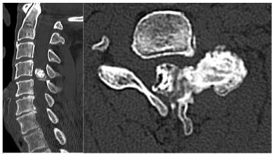
    Figure 1: Showing pre-operative CT scans, sagittal and horizontal views of

the cervical spine, progressive reduction of the spinal canal (more than 50%),

and raised bony structure involving both lamina and vertebral joint on the

left side.
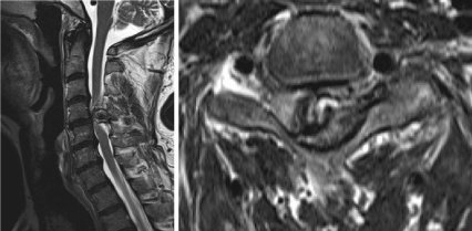
    Figure 2: Cervical MRI shows the cervical spinal cord compression due to an

unclear tumorous lesion at the C4 level and cervical spinal cord displacement

to the right side, starting from the bone to the left facet joint at this level.
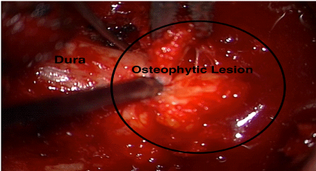
    Figure 3: Intraoperative demonstration under the microscope, in to removal

of the suspected lesion.
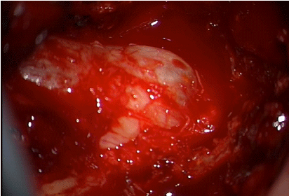
    Figure 4: After removing the structure, unfolded dural sac indicating

compression release.
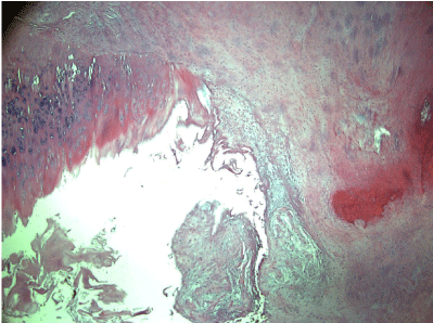
    Figure 5: Fibrotic periarticular tissue with pseudocyst formation representing

a synovial cyst (Hematoxylin and Eosin (HE), x40).
Discussion
The facet joint is a diarthrodial joint with opposing articular cartilage surfaces that reduce friction and a capsule covering the joint space [4]. Along with the disc, the facet joints transfer stress. They guide and constrain spinal motions due to their geometry and mechanical functions. It has only recently become the focus of experimental, biomechanical, and clinical studies. They prevent two adjacent vertebrae from engaging in relative motions that could overload and damage the surrounding spinal structures, mainly the disc, the nerve roots, the spinal column, and the spinal cord, resulting in various symptoms. The pathophysiology of CSM can be explained with a “static and dynamic model.” Degenerative changes result in static compression and exacerbate the compression of the spinal cord during various movements. As a result, static and dynamic compression leads to axonal stretching, spinal cord ischemia from vascular constriction, and venous congestion [5]. Degenerative changes of the fibrous tissues and the loss of the cushioning cartilage may result in ruptures of the capsule, resulting in leakage of the inflammatory cytokines into the intraspinal space, irritating nerve roots and triggering pain signaling and triggering synovial cyst formation. At the molecular level, neural cells undergo apoptosis instead of necrosis with CSM [6]. The case report highlights the significance of recognizing differences in the individual anatomy. It presents a rare case with prominent facet joint hypertrophy that could have been easily mistaken as a tumorous process. This highlights the importance of degenerative changes, dynamic and static adaptative factors, which result in massive hypertrophy with synovial cysts of the facet joint. Only one case in the literature reported a unilateral facet joint hypertrophy resulting in rapidly progressing CSM [3]. In this case, the rapid progression and the demonstration of the “facet joint gap” sign on the cervical radiogram was acute. Our case illustrates a unilateral facet joint pathology that constricted the cervical spinal canal to a much larger degree, resulting in more pronounced clinical symptoms. In this case, the chronic nature of the symptoms indicated a slowly growing and, therefore, possibly benign timorous process compressing the spinal cord. However, such extensive hypertrophic adaptative tissue reactions should be considered in the differential diagnosis.
References
- Bakhsheshian J, Mehta VA, Liu JC. Current Diagnosis and Management of Cervical Spondylotic Myelopathy. Glob Spine J. 2017; 7: 572-586.
- Kadanka Z, Bednařík J, Novotný O, Urbánek I, Dušek L. Cervical spondylotic myelopathy: conservative versus surgical treatment after 10 years. Eur Spine J. 2011; 20: 1533-1538.
- Takeshima Y, Okamoto A, Yokoyama S, Nakagawa I, Nakase H. Unilateral Degenerative Facet Joint Pathology Eliciting Rapidly Progressive Cervical Spondylotic Myelopathy. Cureus. 2021; 13: e14238.
- Jaumard NV, Welch WC, Winkelstein BA. Spinal Facet Joint Biomechanics and Mechanotransduction in Normal, Injury and Degenerative Conditions. J Biomech Eng. 2011; 133: 71010-NaN.
- Nouri A, Tetreault L, Singh A, Karadimas SK, Fehlings MG. Degenerative Cervical Myelopathy: Epidemiology, Genetics, and Pathogenesis. Spine. 2015; 40: E675-693.
- Karadimas SK, Gatzounis G, Fehlings MG. Pathobiology of cervical spondylotic myelopathy. Eur Spine J Off Publ Eur Spine Soc Eur Spinal Deform Soc Eur Sect Cerv Spine Res Soc. 2015; 24: 132-138.