
Case Report
Austin Neurosurg Open Access. 2023; 9(1): 1074.
Giant Cell Tumor of the Sphenoid with Clivus Extension in a Pediatric Patient
Garrido Ruiz PA¹*; Román Garrido M²
1Department of Neurosurgery, Hospital Universitario de Salamanca, Salamanca, Spain
2Department of Primary Care, Hospital Universitario de Salamanca, Salamanca, Spain
*Corresponding author: Patricia Alejandra Garrido Department of Neurosurgery, Hospital Universitario de Salamanca, Neurosurgery Service, 4th Floor Block D, P.o de San Vicente, 182, 37007 Salamanca, Spain. Tel: 616792305 Email: 02717445H@saludcastillayleon.es; agrjandri_92@hotmail.com
Received: March 14, 2023 Accepted: April 21, 2023Published: April 28, 2023
Abstract
We present a case of giant cell tumor of the clivus in a pediatric patient. The case is very interesting because this benign tumor is generally located in the epiphysis of long bones, and only 1% is found in the cranial vault and the base of skull. The sphenoid bone and the petrous portion of the temporal bone are the most commonly affected cranial locations, probably due to their endochondral ossification.
Keywords: Giant cell tumor; Clivus; Pediatric; Endoscopic
Case Presentation
The patient is a 13-year-old girl with a personal history of migraine and tinnitus, with a normal MRI conducted the year before. She presented oppressive temporal-parietal headache and complete ophthalmoplegia of the left eye, with ipsilateral blindness and no light perception. A CT scan reveals an expansile soft tissue mass centered in the clivus that expands anteriorly to the sphenoid bone and the posterior ethmoidal cells, with associated remodeling and patchy lytic areas in adjacent bone structures. A brain MRI shows a 4.4x2.4x2.3cm mass that suggests fibrous dysplasia or plasmacytoma.
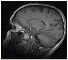
Figure 1: Sagittal brain MRI without contrast (hyperintense lesion).
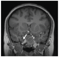
Figure 2: Coronal brain MRI without contrast (hyperintense lesion).
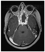
Figure 3: Axial brain MRI with contrast (hypercaptant lesion).
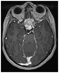
Figure 4: Axial brain MRI with contrast (hypercaptant lesion).
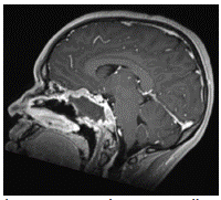
Figure 5: Sagittal brain MRI with contrast (hypercaptant lesion)
Discussion
A transnasal biopsy was performed, with a histological diagnosis of giant cell tumor of bone. The 18F-FDG PET-CT and brain CT for neuronavigation that were conducted 10 days later revealed a growing mass in its anteroposterior diameter. he lesion was excised with endoscopic transnasal surgery.
Conclusion
Giant cell tumors present slow growth and local aggressiveness and can cause compression symptoms in adjacent structures. They can extend to the lungs, which mean that it is important to start treatment as soon as possible with complete surgical excision for curation and in order to prevent recurrence. One potential complementary treatment is denosumab, which is still in trial stage. Currently, the use of bisphosphonates and radiotherapy is the complementary treatment. This last technique is still controversial because it can cause sarcomatous transformation and is reserved for cases of recurrence.
References
- Agrawal A, Gali R, Shanthi V, Ramakrishna BA, Mohan KV. Giant cell tumor of the clivus with presence of epithelioid histiocytes. Asian J Neurosurg. 2014; 9: 48–9.
- Yamamoto M, Fukushima T, Sakamoto S, Tomonaga M. Giant cell tumor of the sphenoid bone: Long-term follow-up of two cases after chemotherapy. Surg Neurol. 1998; 49: 547–52
- Zorlu F, Selek U, Soylemezoglu F, Oge K. Malignant giant cell tumor of the skull base originating from clivus and sphenoid bone. J Neurooncol. 2006; 76: 149–52.
- Chawla S, Henshaw R, Seeger L, Choy E, Blay JY, et al. Safety and efficacy of denosumab for adults and skeletally mature adolescents with giant cell tumour of bone: Interim analysis of an open-label, parallel-group, phase 2 study. Lancet Oncol. 2013; 14: 901–8.
- Iacoangeli M, Di Rienzo A, Re M, Alvaro L, Nocchi N, et al. Endoscopic endonasal approach for the treatment of a large clival giant cell tumor complicated by an intraoperative internal carotid artery rupture. Cancer Manag Res. 2013; 5: 21–4.
- Wolfe JT, 3rd, Scheithauer BW, Dahlin DC. Giant-cell tumor of the sphenoid bone. Review of 10 cases. J Neurosurg. 1983; 59: 322–7.
- Epstein N, Whelan M, Reed D, Aleksic S. Giant cell tumor of the skull: A report of two cases. Neurosurgery. 1982; 11: 263–7.
- AL Rhoton. Rhoton's Cranial Anatomy and Surgical Approaches. Oxford University Press, 2019. Congress of Neurological Surgeons (CNS).
- M Greenberg. Handbook of Neurosurgery, Thieme, 2019. 9th Edition.
- JM Gonzalez Darder. Abordajes Neuroquirurgicos de la Patologia Craneal y Cerebral. Elsevier Espan~a. 2015.