Letter to the Editor
Austin J Nucl Med Radiother. 2016; 3(1): 1018.
Giant Hydrocele in a Decompansated Cirrhotic Patient: Not Always Up Sometimes Down
Ergenc H, Eminler AT*, Ilce HT, Koksal AS and Parlak E
Faculty of Medicine, Department of Internal Medicine, Gastroenterology and Nuclear Medicine, Sakarya University, Turkey
*Corresponding author: Ahmet Tarik Eminler, Faculty of Medicine, Department of Gastroenterology, Sakarya University, Korucuk, Sakarya, Turkey
Received: June 21, 2016; Accepted: June 22, 2016; Published: June 24, 2016
Abstract
Hydrocele denotes a pathological accumulation of serous fluid in a body cavity. Ascites is the most common complication of cirrhosis and it causes an increase in the intra abdominal pressure. Here in we report an alcoholic cirrhotic patient with giant hydrocele that is communicating with ascites. We showed the peritoneo-scrotal communication by intraperitoneal injection of Tc-99m macro aggregated albumin. Communicating shunt between peritoneal cavity and scrotum should be considered in the decompensated cirrhotic patient with resistant scrotal enlargement.
Keywords: Hydrocele, Ascites, Cirrhosis
Letter to the Editor
Hydrocele denotes a pathological accumulation of serous fluid in a body cavity. Also the term is used usually to describe “hydrocele testis” which is the accumulation of fluids around a testicle. It is often caused by fluid secreted from a remnant piece of peritoneum wrapped around the testicle, called the tunica vaginalis [1]. Most pediatric hydroceles are congenital and adult hydroceles are usually secondary. The latter can present acutely from local injury, infection, radiotherapy, or increased intra abdominal pressure [2]. Ascites is the most common complication of cirrhosis and it causes an increase in the intra abdominal pressure. Here in we report an alcoholic cirrhotic patient with giant hydrocele that is communicating with ascites.
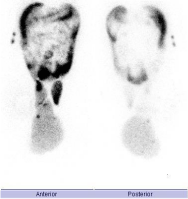
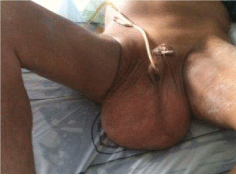
60 years old male patient with a history of decompensate alcoholic cirrhosis was admitted to the hospital with the complaints of progressive symptomatic scrotal enlargement over the last few months (Figure 1). Abdominal ascites was not clinically evident, but ultrasound examination showed minimally ascites between the intestinal loops. The scrotal Doppler ultrasonography performed for existing scrotal swelling and detected an advanced increase in the amount of bilateral scrotal fluid and has been interpreted as hydrocele. The swollen hemiscrotum was painless and transilluminated, providing evidence of a hydrocele. To detect the presence of a communicating hydrocele, peritoneal Scintigraphy was performed by intraperitoneal injection of 1 mCi Tc-99m macro aggregated albumin was injected with in the minimal ascites detected by ultrasonography. The images showed radio tracer accumulation within the peritoneal cavity and bilateral scrotum that more prominent on the right side (Figure 2). A hydrocele resulting from a communicating shunt between the peritoneal cavity and scrotum was confirmed.

    Figure 1:  The images taken in the twentieth minute show a heterogeneous
manner accumulate of the radioactive substance (Tc-99m macro aggregated
albumin) bilateral scrotal region image of sintigraphi.

    Figure 2:  Bilateral scrotal enlargement that is more prominent at the right
side (hydrocele) photos of scrotum.
The increased intra abdominal pressure caused by massive ascites could give rise to a patent shunt that permits flow of peritoneal fluid in to the scrotum. Intraperitoneal injection of a radiotracer for visualization of a peritoneo-scrotal communication has been used for many years [3]. Recently, Tc-99m macro aggregated albumin is used for this reason [4]. Adult hydroceles are usually secondary and the incidence of adult hydroceles is rising with increasing use of the peritoneal cavity for peritoneal dialysis, ventriculoperitoneal shunts, and renal transplants. Ascites existing in decompensated cirrhotic patient stand to leak up to the pleural cavity and causes hepatic hydrotorax. However, ascites in the peritoneal cavity may communicate with the tunica vaginalis and causes hydrocele in some rare cases. Diuretic therapy is ineffective for these patients although some surgical procedures or transplantation may be treatment options [2,4].
In summary, communicating shunt between peritoneal cavity and scrotum should be considered in the decompansated cirrhotic patient with resistant scrotal enlargement.
References
- Abdel-Rheem MS. Cause of primary vaginal hydrocele and ascites in advanced liver cirrhosis. Am J Surg. 1983; 146: 647-651.
- Hsu CC, Chen YW, Chuang YW, Huang YF. Ascites with a communicating hydrocele detected by peritoneal Scintigraphy. Clin Nucl Med. 2004; 29: 326.
- Ducassou D, Vuillemin L, Wone C, Ragnaud JM, Brendel AJ. Intraperitoneal injection of technetium-99m sulfur colloid in visualization of a peritoneovaginalis connection. J Nucl Med. 1984; 25: 68-69.
- De Gottardi A, Abraldes JG. An uncommon cause of massive scrotal enlargement. Liver Int. 2010; 30: 995.