Case Report
Austin J Nucl Med Radiother. 2021; 6(1): 1026.
Role of 18F-FDG PET-CT in CNS Lymphoma-A Case Report
Zohra FT¹*, Sarker AK², Hosen J¹, Hasnat MA¹, Hosen J¹, Sharmin RA¹ and Ahasan MM¹
1Institute of Nuclear Medical Physics, AERE, Savar, Dhaka, Bangladesh
2Institute of Nuclear Medicine and Allied Science, Mitford, Dhaka, Bangladesh
*Corresponding author: Fatema Tuz Zohra, Institute of Nuclear Medical Physics, AERE, Savar, Dhaka, Bangladesh
Received: April 15, 2021; Accepted: April 30, 2021; Published: April 06, 2021
Abstract
The actual role of 18F-FDG PET/CT in evaluating primary brain lymphoma is still an open issue. Brain lymphoma usually show elevated 18F-FDG uptake, often higher than other brain tumors or inflammatory processes, but the metabolic behavior of this lymphoma is not still understood. Central nervous system lymphoma is a rare non-Hodgkin lymphoma in which malignant (cancer) cells from lymph tissue form in the brain and/or spinal cord (primary CNS) or spread from other parts of the body to the brain and/or spinal cord (secondary CNS).A 55 year-old man presented with headache. Magnetic Resonance Imaging (MRI) revealed a well-enhanced mass lesion in the left frontal lobe. A surgical specimen obtained through left orbito-pterional craniotomy revealed a Diffuse Large B-Cell Lymphoma (DLBCL). 18F FDG PET-CT scan showed multiple hypodensehypermetabolic lesions in brain. Multiple hypodense focal hypermetabolic areas were seen in right frontal lobe, left frontal lobe and left temporal lobe. There was also a subcentimetrichypermetabolic sub-carinal lymph node. The activity was diminished on follow-up PET-CT after 8 courses of chemotherapy. This case indicates that FDG PET-CT scan can aid identify the atypical primary CNS lymphoma for staging workup and can be a useful tool to see treatment response.
Keywords: Primary central nervous system lymphoma; FDG PET-CT; Treatment response
Introduction
Central nervous system lymphoma is a rare. Primary Non- Hodgkin’s Lymphoma (NHL) of the Central Nervous System (CNS) is uncommon and generally affects the brain. CNSL accounts for 3-4 % of all primary brain tumors and 4-6 % of extranodal lymphomas; diffuse large B-cell lymphoma (DLBCL) is the most common histological type [1-4]. Diffuse large B-cell lymphoma (DLBCL) is the most common form of CNS NHL. The most frequent involved site of disease in CNSL is the brain, followed by eyes, spinal cord, nerves and leptomeninges [4]. Bailey first described CNSL as “perithelial sarcoma” of the CNS and Henry in 1974 recognized its lymphoid origin [5]. The majority of these tumors (95%) are considered Diffuse Large B-Cell Lymphomas (DLBCLs) [6,7]. Brain lymphoma is commonly related to immunodeficiency, especially with Acquired Immunodeficiency Syndrome (AIDS), but can develop also in immunocompetent population. Over the past three decades the incidence of PCNSL has increased especially in the immunocompetent population [8,9]. Contrast-enhanced Magnetic Resonance Imaging (MRI) of the brain and/or spine is the standard diagnostic modality when CNSL is suspected. It often shows common morphological features such single or multiple uniformly well enhancing lesions located in the peri-ventricular areas and basal ganglia, associated with moderate edema and absence of necrosis, but MRI may have several limitations. Although CT and MR imaging are still the most important modalities in the diagnosis of CNSL, modern metabolic imaging modalities other than conventional morphological imaging are increasingly used to improve accurate diagnosis of CNSL. We present a case of CNS NHL developing NL subsequently during the initial courses of chemotherapy following subtotal surgical resection of the brain lymphoma. As far as we know, this is the first reported case in which primary brain NHL with NL, without other visceral lesions, manifested subsequently during the initial chemotherapy [10].
18F FDG PET/CT imaging and interpretation
The Patients underwent 18F-FDG PET/CT before any treatment (local surgery, chemotherapy, radiotherapy and/or combination); PET/CT was performed after at least 6 h fasting. An activity of 3.5- 4.5 MBq/kg of 18F-FDG was administered intravenously; images were acquired 60 min after injection from the vertex to the mid-thigh on a Discovery 690 tomograph or Discovery ST PET/CT tomograph (General Electric Company-GE®-Milwaukee, WI, USA) with standard parameters (CT: 80 mA, 120 Kv without contrast; 2.5-4 min per bed- PET-step of 15 cm) and the reconstruction was performed in a 128×128 or 256×256 matrix and 60 cm field of view. Patient were instructed to void before imaging scan, no oral or intravenous contrast agents were administrated or bowel preparation used for any patient. PET images were analyzed both visually and semiquantitatively. Readers had knowledge of clinical history and every focal tracer uptake deviating from physiological distribution and background was regarded as suggestive of lymphoma; it was defined as intense 18F-FDG activity higher than the surrounding tissue on visual analysis.
Case Study
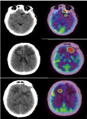
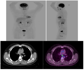
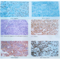
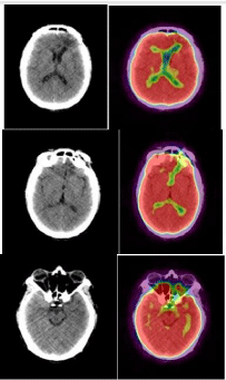
A 55 year-old man presented with headache. Clinical complaining of nausea vertigo, headache and weight loss and due to repetitive vomiting. Histological diagnosis was Diffuse Large B-Cell Lymphoma (DLBCL). Magnetic Resonance Imaging (MRI) revealed a wellenhanced mass lesion in the left frontal lobe. A surgical specimen obtained through left orbito-pterional craniotomy revealed a diffuse large B-cell lymphoma (DLBCL). 18F FDG PET-CT scan showed multiple hypodensehypermetabolic lesions in brain. (Figure 1) Shows hypodense focal hypermetabolic areas in right rontal lobe (SUVmax 29.0), left frontal lobe (SUVmax 27.6) and left temporal lobe (SUVmax 32.3). (Figure 2) shows Maximum Intensity Projection (MIP) view shows focal hypermetabolic area in the brain. There is a subcentimetrichypermetabolic (SUVmax 4.3) sub-carinal lymph node. Multiple hypodense focal hypermetabolic areas were seen in right frontal lobe, left frontal lobe and left temporal lobe. There was also a subcentimetrichypermetabolic sub-carinal lymph node. (Figure 3) Shows Histopathology and Immunohistochemistry of primary CNS Non-Hodgkins lymphoma. The activity was diminished on follow-up PET-CT after 8 courses of chemotherapy (Figure 4). This case indicates that FDG PET-CT scan can aid identify the atypical primary CNS lymphoma for staging workup and can be a useful tool to see treatment response.

    Figure 1: Shows hypodense focal hypermetabolicareas in right rontal lobe
(SUVmax 29.0), left frontal lobe (SUVmax 27.6) and left temporal lobe
(SUVmax 32.3).

    Figure 2: Maximum Intensity Projection (MIP) view shows focal
hypermetabolic area in the brain. There is a subcentimetrichypermetabolic
(SUVmax 4.3) sub-carinal lymph node.

    Figure 3: Shows Histopathology and Immunohistochemistry of primary CNS
Non-Hodgkins lymphoma.

    Figure 4: Shows complete remission of intracranial lesions after 8 courses
of chemotherapy.
Discussion
Direct involvement of the CNS in Hodgkin’s lymphoma is rare, with an incidence of about 0.02%, and to our knowledge has never been described in NLPHL [11]. However, various paraneoplastic events in the nervous system, in particular the CNS, have been described in Hodgkin’s lymphoma patients and are summarized [12]. Diffuse Large B Cell Lymphoma (DLBCL) is the largest subtype of Non-Hodgkin’s Lymphomas (NHLs) and is characterized by relatively frequent extranodal presentation. In these cases, the most common extranodal localizations are stomach, CNS, bone, testis and liver [13]. We presented 18F-FDG PET/CT images in case of neurolymphomatosis from non-Hodgkin lymphoma. The combination of clinical history, physical examination, PET/CT, MRI and follow-up were used to confirm final diagnosis [14]. Furthermore, an early FDG-PET assessment of response to chemotherapy is becoming a routine part of management in HL and histologically aggressive NHL [11]. Therapy of choice is steroids and immediate lymphoma-directed chemotherapy [15]. Many of the reported cases of PCNSBL used HD-MTX +/-, some variation of cyclophosphamide, doxorubicin, vincristine, and prednisone (CHOP) therapy [16]. It is an inflammatory syndrome of the Central Nervous System (CNS), prominently involving the brainstem and pons. The clinical manifestations of CLIPPERS are characterized by gait ataxia, dysarthria, diplopia, and altered facial sensation. The Magnetic Resonance Imaging (MRI) shows a punctate and nodular pattern of gadolinium enhancement “peppering” in the pons, brainstem, white matter, and other adjacent structures. Perivascular lymphohistiocytic and predominant T cells infiltration in brain biopsy specimen is another striking characteristic [17]. In our case, the patient was finally diagnosed as PTCL-NOS by the pulmonary nodular and the skin biopsies. She was timely treated with chemotherapy and obtained complete remission. For example, hypermetablic lesions in PET-CT may help locate the biopsy parts. The patient in our study found hypermetablic lesions in bilateral lungs and skin after PET-CT and the following biopsy confirmed lymphoma. The present case proposes that biopsies in extracerebral lesions under the assisting examination of PET-CT can be helpful in further identification. Further studies are required to determine the relationship between lymphoma and CLIPPERS and the potential value of systemic imaging in discriminating lymphoma from CLIPPERS during the early stages of the disease and during the follow-up period [17].
Conclusion
In conclusion, 18F-FDG pathological uptake in primary brain DLBCL lymphoma occurred in most of the population evaluated, being independently associated with diameter maximum of the lesion and morphological appearance. Brain lesions with typical MRI appearance and large diameter are likely to have 18F-FDG uptake. A case of primary central nervous system lymphoma presenting with peripheral nerve involvement was described. MRI appears to be the method of choice for detecting brain disease in patients with primary brain lymphoma, whereas 18F-FDG PET/CT seems to play a relevant role in the assessment of extra-cerebral disease.
References
- Miller DC, Hochberg FH, Harris NL, Gruber ML, Louis DN, Cohen H. Pathology with clinical correlation of primary central nervous system non- Hodgkin’s lymphoma. Cancer. 1994; 74: 1383-1397.
- Behin A, Hoang-Xuan K, Carpentier AF, Delattre JY. Primary brain tumours in adults. Lancet. 2003; 361: 323-331.
- Ricard D, Idbaih A, Ducray F, Lahutte M, Hoang-Xuan K, Delattre JY. Primary brain tumours in adults. Lancet. 2012; 379: 1984-1996.
- Hochberg FH, Miller DC. Primary central nervous system lymphoma. J Neurosurg. 1988; 68: 835-853.
- Ney DE, Deangelis LM. Management of central nervous system lymphoma. In: Non- Hodgkin Lymphomas. Philadelphia: Lippincott Willilams & Wilkins. 2010; 527-539.
- Shenkier TN, Blay JY, O’Neill BP, Poortmans P, Thiel E, Jahnke K, et al. Primary CNS lymphoma of T-cell origin: a descriptive analysis from the international primary CNS lymphoma collaborative group. J ClinOncol. 2005; 23: 2233-2239.
- Louis DN, Ohgaki H, Wiestler OD, Cavenee WK, Burger PC, Jouvet A, et al. The 2007 WHO classification of tumours of the central nervous system. ActaNeuropathol. 2007; 114: 97-109.
- Van der Sanden GA, Schouten LJ, van Dijck JA, van Andel JP, van der Maazen RW, Coebergh JW. Primary central nervous system lymphomas: incidence and survival in the Southern and Eastern Netherlands. Cancer. 2002; 94: 1548-1556.
- Olson JE, Janney CA, Rao RD, Cerhan JR, Kurtin PJ, Schiff D, et al. The continuing increase in the incidence of primary central nervous system non- Hodgkin lymphoma: a surveillance, epidemiology, and end results analysis. Cancer. 2002; 95: 1504-1510.
- Mori Y, Yamamoto K, Ohno A, Fukunaga M, Nishikawa A. Primary Central Nervous System Lymphoma with Peripheral Nerve Involvement: Case Report. Cureus. 2019; 11 e5675.
- Hong CM, Lee SW, Lee HJ, Song BI, Kim HW, Kang S, et al. Neurolymphomatosis on F-18 FDG PET/CT and MRI findings: a case report. Nuclear medicine and molecular imaging. 2011; 45: 76-78.
- Vetter M, Tzankov A, Engert A, Mehling M, Herrmann R, Rochlitz C. Hodgkin’s lymphoma and paraneoplastic phenomena in the central nervous system: a case report and review of the literature. Case Reports in Oncology. 2011; 4: 106-114.
- Airaghi L, Greco I, Carrabba M, Barcella M, Baldini IM, Bonara P, et al. Unusual presentation of large B cell lymphoma: a case report and review of literature. Clinical & Laboratory Haematology. 2006; 28: 338-342.
- Canh NX, Van Tan N, Tung TT, Son NT, Maurea S. 18F-FDG PET/CT in Neurolymphomatosis: Report of 3 Cases. Asia Oceania Journal of Nuclear Medicine and Biology. 2014; 2: 57.
- Vetter M, Tzankov A, Engert A, Mehling M, Herrmann R, Rochlitz C. Hodgkin’s lymphoma and paraneoplastic phenomena in the central nervous system: a case report and review of the literature. Case Reports in Oncology. 2011; 4: 106-114.
- Bower K, Shah N. Primary CNS Burkitt lymphoma: a case report of a 55-Yearold cerebral palsy patient. Case reports in oncological medicine. 2018; 2018: 5869135.
- Liu XH, Jin F, Zhang M, Liu MX, Wang T, Pan BJ, et al. Peripheral T cell lymphoma after Chronic Lymphocytic Inflammation with Pontine Perivascular Enhancement Responsive to Steroids (CLIPPERS): a case report. BMC neurology. 2019; 19: 266.