
Case Report
Ann Obes Disord. 2016; 1(2): 1009.
Internal Hernia and Biliary Tract Lithiasis after Rouxen- Y Gastric Bypass: the Diagnostic Dilemma and its Therapeutic Approach
Chiappetta S*, Khan MS and Stier C
Department of Obesity and Metabolic Surgery, Sana Klinikum Offenbach, Germany
*Corresponding author: Sonja Chiappetta, Department of Obesity and Metabolic Surgery, Sana Klinikum Offenbach, Starkenburgring 66, 63069 Offenbach am Main, Germany
Received: March 25, 2016; Accepted: July 01, 2016; Published: July 04, 2016
Abstract
Two of the most common surgical long-term complications after Laparoscopic Roux-en-Y Gastric Bypass (LRYGB) are Small Bowel Obstruction (SBO) due to Internal Hernia (IH) and Biliary Tract Lithiasis (BTL). Due to elevated cholestatic and pancreatic enzymes in IH and BTL differential diagnoses can be difficult and IH has to be suspected in every acute or chronical abdominal pain after LRYGB.
We report a clinical case of IH mimicking BTL in a patient after LRYGB. In this clinical case exact diagnosis and treatment was performed using a standardized diagnostic flow chart. During diagnostic laparoscopy internal herniation of the entire common channel was seen through Peterson space. A chyloperitoneum is a typical sign for chronic herniation and can confirm IH.
Keywords: Roux-en-Y gastric bypass; Long term complications; Internal hernia; Biliary tract lithiasis; Chyloperitoneum
Abbreviations
LRYGB: Laparoscopic Roux-En-Y Gastric Bypass; SBO: Small Bowel Obstruction; BTL: Biliary Tract Lithiasis; IH: Internal Hernia; UDCA: Ursodeoxycholic Acid; MRCP: Magnetic Resonance Cholangiopancreatography
Introduction
Since its inception half a century ago, bariatric surgery has become the most rapidly increasing area of surgery almost worldwide and especially in the western countries. The most commonly performed procedures worldwide are Laparoscopic Roux-en-Y Gastric Bypass (LRYGB) and sleeve gastrectomy.
In LRYGB stomach is reduced to a small gastric pouch with a capacity of 30 ml. Approximately 50 cm distal to ligament of Treitz, the jejunum is divided, and the bottom end of jejunum is anastomosed to this newly created gastric pouch (roux limb). The top end of divided jejunum (biliopancreatic limb) is anastomosed to jejunum at 150 cm distal to gastrojejunal anastomosis (Figure 1). LRYGB works by two principal mechanisms: restriction and malabsorption. Restriction is achieved by reducing the size of stomach and malabsorption results from bypassing part of jejunum.
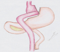
Figure 1: Schematic Figure of LRYGB.
The two most common surgical long-term complications after LRYGB are Small Bowel Obstruction (SBO) and Biliary Tract Lithiasis (BTL).
Internal Hernia (IH) is recognized the most frequent cause of SBO (> 50%) and the incidence of IH is in the range of 1-5% [1,2]. Mean time to intervention for internal hernia repair is reported to be after 413 +/- 46 days and occurs after an average excess body weight loss of 59% +/- 3.3 [2].
The three classic locations for IH after antecolic LRYGB are Peterson Space (between Roux limb´s mesentery and transverse mesocolon, (Figure 2)), mesenteric defect and Brolin space (at the jejuno-jejunostomy). Diagnosis is based on clinical presentation, plain abdominal X-ray and upper gastrointestinal studies. While patients with LRYGB may be unable to vomit, liquid accumulation within the closed loop often determines elevated cholestatic or pancreatic enzymes [3], imitating a cholecystolithiasis [4], choledocholithiasis or pancreatits. Severe gastric dilatation can further cause gastric wall necrosis with clinical signs of an acute abdomen. Multislice CT spiral scan can fail to disclose internal hernia in up to two-third of cases [5,6]. Only the diagnosis of transmesenteric internal hernias can often be made because of presence of the clustered loops with a mesenteric swirl [7].
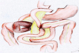
Figure 2: Internal Hernia in Peterson Space after LRYGB.
Due to the decreased awareness of some physicians bowel gangrene from strangulated internal hernia is still a clinical problem [8,9]. The necessity of early emergency surgery with explorative laparoscopy to avoid severe intestinal complications and related mortality in SBO is well known [10].
Thus, IH has to be suspected in every acute or chronic abdominal pain after LRYGB. If there is suspicion of IH then diagnostic laparoscopy is mandatory.
The incidence of cholelithiasis following obesity surgery ranges from 6.7% to 52.8%, increasing further if either weight loss rate exceeds 1.5 kg/week or when Excess Weight Loss of more than 24% [10,11]. A meta-analysis confirmed the effectiveness of Ursodeoxycholic Acid (UDCA) prophylaxis with 8.8% of those taking UDCA developing gallstones compared to 27.7% for placebo [12]. Gallstone-related complications are common and prophylactic cholecystectomy is still a controversial discussion [13]. Because of the lack of endoscopic access to the duodenum using endoscopic retrograde Cholangiopancreatography (ERCP), choledocholithiasis in a post-gastric bypass patient can be hard to treat [14].
Due to elevated cholestatic and pancreatic enzymes in IH and BTL differential diagnoses can be difficult to some extent. In this clinical case report, we describe a clinical case of IH mimicking BTL in a patient after LRYGB. In this case exact diagnosis and effective management was done using a standardized diagnostic flow chart. Two flow chart diagrams are mentioned here (Figure 3,4). Figure 3 shows a management plan in patients with acute abdominal pain. Acute abdominal pain is defined as severe abdominal pain of unclear etiology that is less than 24 hours in duration [15]. Figure 4 shows a management plan in patients with intermittent or chronic abdominal pain that has been present for at least two months [15].
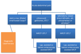
Figure 3: Diagnostic algorithm in acute abdominal pain after LRYGB.
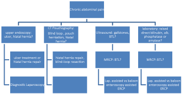
Figure 4: Diagnostic algorithm in chronic abdominal pain after LRYGB.
If patient presents with acute or chronic abdominal pain, labs (pancreatic enzymes, alkaline phosphatase and total-direct bilirubin), ultrasound and plain x-ray of the abdomen should be carried out. If labs are deranged and ultrasound reveals gall stones with dilated common biliary duct, magnetic resonance Cholangiopancreatography (MRCP) should be done as a next investigation. On the basis of MRCP findings of BTL, laparoscopic assisted or baloon enteroscopy assisted ERCP should be performed.
If plain x-ray of the abdomen shows dilated loops with air fluid levels then diagnostic laparoscopy is indicated. In case of nonpathological plain x-ray diagnostic laparoscopy is even mandatory.
In case of chronic abdominal pain, we also perform CT Pouchography to evaluate the size of gastric pouch, the presence of a blind loop and hiatal hernia with pouch herniation because they are considered as differentials for chronic abdominal pain in post LRYGB patients.
It is worth mentioning here that the mortality rate of LRYGB is less than 1% [16] but post LRYGB, the internal hernias have rapid progression to bowel ischemia and a bad outcome. The mortality rate of IH can be as high as 43.8% [17]. Furthermore, one meta-analysis reported mortality rate of concomitant gallstones and BTL from 0.33% to 14.67% [18].
Clinical Case
A 25-year old male patient with a BMI of 26.6 kg/m² (height 1.87 m, weight 93 kg) presented to our Emergency Room in November 2013, with abdominal pain for one day associated with nausea. Abdominal pain was colicky in nature and 8 out of 10 in severity. The pain was localized to paraumbilical area with some radiation to upper abdomen. The patient had never experienced such symptoms before and after LRYGB. There was no history of absolute constipation and patient had his last normal bowel movements on the morning of presentation. The patient’s bariatric history started in November 2011, when he underwent LRYGB at an initial weight of 149 kg (BMI 42.6 kg/ m²). Intra- and postoperative course was uneventful and the patient had regular follow ups in our clinic.
During his follow-up visits, at 6 weeks (weight 126.2 kg) we detected sludge in the gallbladder on abdominal ultrasound and therapy with UDCA was started for 6 months. At 6 months followup (weight 106 kg), gallstones were detected on ultrasound of the abdomen and laparoscopic cholecystectomy was recommended.
At 2 years follow-up the patient had a weight loss of 56 kg (weight 93 kg, BMI 26.6 kg/m²) and a good quality of life. We again recommended laparoscopic cholecystectomy but due to self-reported organizational issues cholecystectomy was not planned.
On examination in Emergency Room, his vital signs were stable. Abdomen was tender in paraumbilical area, with no guarding or rigidity. There was no abdominal distension and bowel sounds were normal. Ultrasound of the abdomen detected multiple small gallstones with normal wall thickness of the gallbladder. Laboratory findings showed an elevated bilirubin of 1.88 mg/dl (direct 0.63 mg/ dl, indirect 1,3 mg/dl) and raised serum amylase level of 210 U /l (normal range 0-137). On repeated labs in the next morning, there was further increasing bilirubin level of 2.53 mg/dl. Choledocholithiasis was suspected due to the colicky abdominal pain and also deranged bilirubin levels with slightly raised pancreatic enzyme levels. We performed MRCP and no stones were seen within the non-dilated bile duct. Thus, diagnostic laparoscopy was planned to rule out internal herniation after LRYGB. Intraoperative chyloperitoneum was detected (Figure 5). Internal herniation of the entire common channel was seen through the Peterson space. Common channel herniation was completely reduced. No indication was seen for small bowel resection. Peterson Space was closed with a non-absorbable suture. Simultaneous cholecystectomy was performed. Examination of chyloperitoneum showed elevated triglycerides of 326 mg/dl with a blood count of 63 mg/dl. Postoperative course was uneventful and the patient was discharged on the 2nd postoperative day.
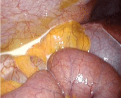
    Figure 5: Chyloperitoneum can be a typical sign of chronic internal herniation
after LRYGB.
Discussion
Due to elevated cholestatic and pancreatic enzymes in both IH and BTL, differential diagnoses can be difficult. A standardized diagnostic algorithm is mandatory to rule out patients with IH and to avoid severe intestinal complications.
During primary surgery of LRYGB we support closure of the mesenteric defect with non-absorbable suture. Closure of mesenteric defect is associated with significantly lower incidence of internal hernia rate. However, closure of the mesenteric defects might be associated with increased risk of early small bowel obstruction caused by kinking of the jejunojejunostomy [19].
We also recommend that all patients with either acute abdominal pain or chronic abdominal pain after LRYGB should undergo the diagnostic alogrithms (Figure 2,3) with laboratory blood tests, ultrasound of the abdomen, plain x-ray, upper GI endoscopy and early laparoscopy to detect internal hernia and to avoid strangulation with intestinal gangrene [15]. In case of elevated cholestatic and pancreatic enzymes a choledocholithiasis and pancreatitis should be excluded via MRCP.
In Patients with chronic abdominal pain after LRYGB, CT Pouchography should also be done. If it reveals hiatal hernia with a concomitant gastroesophageal reflux disease in the upper GI endoscopy, then hiatal hernia can be repaired laparoscopically. If any blind loop at gastojejunostomy is appreciated on CT, blindloop syndrome could be a cause for postprandial chronic pain. If CT Pouchography is unremarkable, diagnostic laparoscopy is mandatory (Figure 3).
During laparoscopy a chyloperitoneum can confirm internal hernia after LRYGB [20,21]. A true chylous effusion is defined as the presence of ascitic fluid with high fat (triglyceride) content, usually higher than 110 mg/dl [20]. In case of suspected IH the three classic locations have to been inspected: Peterson Space, the mesenteric defect and Brolin space. During laparoscopic surgery characterization of the alimentary limb, the biliopancreatic limb and the common channel have to be performed and bowel lengths have to be measured. Also marker sutures at time of primary surgery can help to identify the limbs. If no identification is possible, laparoscopic control of the anatomy can be achieved by examining small intestine in reverse direction starting from the ileocecal valve [22].
Conclusion
Chyloperitoneum can confirm internal hernia after LRYGB and the three aforementioned classic locations for internal hernia should be examined if chyloperitoneum is detected.
To improve diagnosis of IH and BTL after LRYGB we strongly recommend that every patient with acute or chronic abdominal pain after LRYGB should undergo our proposed management algorithm. BTL has always to be ruled out by MRCP, if elevated cholestatic or pancreatic enzymes are present.
References
- Smith SC, Edwards CB, Goodman GN, Halversen RC, Simper SC. Open vs laparoscopic Roux-en-Y gastric bypass: comparison of operative morbidity and mortality. Obes Surg. 2004; 14: 73-76.
- Ahmed AR, Rickards G, Husain S, Johnson J, Boss T, O'Malley W. Trends in Internal Hernia Incidence after laparoscopic Roux-en-Y Gastric Bypass. Obesity Surgery. 2007; 17: 1563-1566.
- Spector D, Perry Z, Shah S, Kim JJ, Tarnoff ME, Shikora SA. Roux-en-Y gastric bypass: hyperamylasemia is associated with small bowel obstruction. Surg Obes Relat Dis. 2015; 11: 38-43.
- Kolk A, Sportel IG, te Riele WW, Hazebroek EJ, van Ramshorst B, Wiezer MJ. Internal hernia following laparoscopic gastric bypass. Ned Tijdschr Geneeskd. 2012; 156: A4393.
- Gunabushanam G, Shankar S, Czerniach DR, Kelly JJ, Perugini RA. Small-bowel obstruction after laparoscopic Roux-en-Y gastric bypass surgery. J Comput Assist Tomogr. 2009; 33: 369-375.
- Patel RY, Baer JW, Texeira J, Frager D, Cooke K. Internal Hernia complications of gastric bypass surgery in the acute setting: spectrum of imaging findings. Emerg Radiol. 2009; 16: 283-289.
- Goudsmedt F, Deylgat B, Coenegrachts K, Van De Moortele K, Dillemans B. Internal Hernia After Laparoscopic Roux-en-Y Gastric Bypass: a Correlation Between Radiological and Operative Findings. Obes Surg. 2015; 25: 622-627.
- Reiss JE, Garg VK. Bowel gangrene from strangulated Petersen's space hernia after gastric bypass. J Emerg Med. 2014; 46: e31-34.
- de Bakker JK, van Namen YW, Bruin SC, de Brauw LM. Gastric bypass and abdominal pain: think of Petersen hernia. JSLS. 2012; 16: 311-313.
- Campanile FC, Boru CE, Rizzello M, Puzziello A, Copaescu C, Cavallaro G, et al. Acute complications after laparoscopic bariatric procedures: update for the general surgeon. Langenbecks Arch Surg. 2013; 398: 669-686.
- Shiffman ML, Sugerman HJ, Kellum JM, Brewer WH, Moore EW. Gallstone formation after rapid weight loss: a prospective study in patients undergoing gastric bypass surgery for the treatment of morbid obesity. Am J Gastroenterol. 1991; 86: 1000-1005.
- Uy MC, Talingdan-Te MC, Espinosa WZ, Daez ML, Ong JP. Ursodeoxycholic acid in the prevention of gallstone formation after bariatric surgery: a meta-analysis. Obes Surg. 2008; 18: 1532-1538.
- Amstutz S, Michel JM, Kopp S, Egger B. Potential Benefits of Prophylactic Cholecystectomy in Patients Undergoing Bariatric Bypass Surgery. Obes Surg. 2015; 25: 2054-2060.
- Schreiner MA, Chang L, Gluck M, Irani S, Gan SI, Brandabur JJ, et al. Laparoscopy-assisted versus balloon enteroscopy-assisted ERCP in bariatric post-Roux-en-Y gastric bypass patients. Gastrointest Endosc. 2012; 75: 748-756.
- Rasquin A, Di Lorenzo C, Forbes D, Guiraldes E, Hyams JS, Staiano A, et al. Childhood functional gastrointestinal disorders: child/adolescent. Gastroenterology. 2006; 130: 1527-1537.
- Benotti P, Wood GC, Winegar DA, Petrick AT, Still CD, Argyropoulos G, et al. Risk factors associated with mortality after Roux-en-Y gastric bypass surgery. Ann Surg. 2014; 259: 123-130.
- Akyildiz H, Artis T, Sozuer E, Akcan A, Kucuk C, Sensoy E, et al. Internal hernia: complex diagnostic and therapeutic problem. Int J Surg. 2009; 7: 334-337.
- Lu J, Cheng Y, Xiong XZ, Lin YX, Wu SJ, Cheng NS. Two-stage vs single-stage management for concomitant gallstones and common bile duct stones. World J Gastroenterol. 2012; 18: 3156-3166.
- Stenberg E, Szabo E, Ågren G, Ottosson J, Marsk R, Lönroth H, et al. Closure of mesenteric defects in laparoscopic gastric bypass: a multicentre, randomised, parallel, open-label trial. Lancet. 2016; 387: 1397-1404.
- Hidalgo JE, Ramirez A, Patel S, Acholonu E, Eckstein J, Abu-Jaish W, et al. Chyloperitoneum after laparoscopic Roux-en-Y gastric bypass (LRYGB). Obes Surg. 2010; 20: 257-260.
- Hanson M, Chao J, Lim RB. Chylous ascites mimicking peritonitis after laparoscopic Roux-en-Y gastric bypass for morbid obesity. Surg Obes Relat Dis. 2012; 8: e1-2.
- Martin MJ, Beekley AC, Sebesta JA. Bowel obstruction in bariatric and nonbariatric patients: major differences in management strategies and outcome. Surg Obes Relat Dis. 2011; 7: 263-269.