Clinical Image
Austin J Obstet Gynecol. 2014;1(2): 1.
Dicephalic Parapagus Conjoined Twins
Rodych Jennifer, Young Carmen and Jain Venu*
Department of Obstetrics and Gynecology, University of Alberta, Canada
*Corresponding author: Jain Venu, Department of Obstetrics and Gynecology, Maternal Fetal Medicine, University of Alberta, 5S131 Lois Hole Hospital, Royal Alexandra Hospital, 10240 Kingsway Avenue, Edmonton, Alberta, T5H 3V9, Canada
Received: July 02, 2014; Accepted: July 28, 2014; Published: July 30, 2014
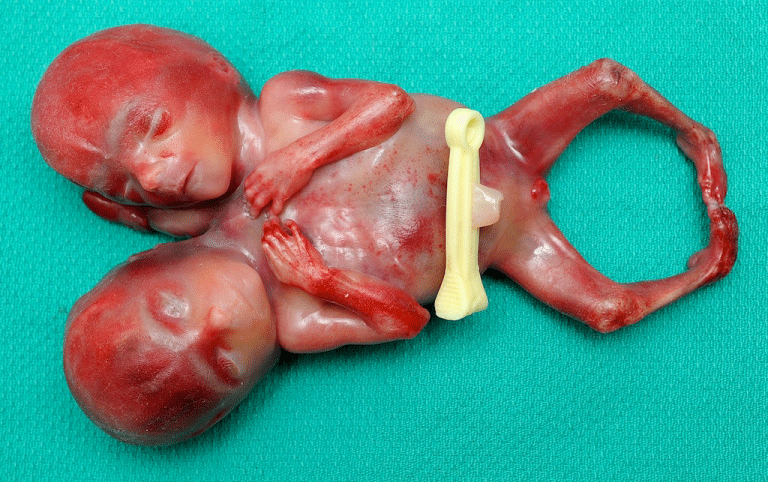
A 28-year-old G2P1 was referred for conjoined twins diagnosed during a routine 18-week ultrasound. Ultrasounds at 5 and 8 weeks reported a possible dichorionic-diamniotic gestation, and a singleton with an adjacent cyst, respectively. Ultrasound revealed dicephalic parapagus tribrachius bipus conjoined twins [1]. The fetuses were joined at the thorax with two hearts fused medially (Figure 1). A single stomach, pelvis and bladder, with two kidneys, three arms and two legs with bilateral clubbed feet were visualized. After counseling, the patient chose to terminate pregnancy by induction of labor (Figure 2). Conjoined twins occur in the order of 1/50000-1/100000 gestations, with 28% as parapagus unions [2]. Only 33% of conjoined twins are diagnosed by ultrasound before 10 weeks [3]. With advancing gestation, inseparable fetal movements and conjoined anatomy become more apparent. Conjoined twins should be considered in all cases of monochorionic twinning. Late diagnoses present more difficult options for parents and physicians.
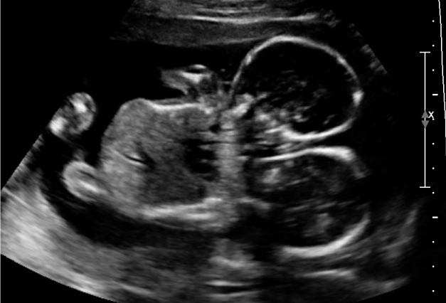
Figure 1: Ultrasound image.