Case Report
Austin J Obstet Gynecol. 2015;2(1): 1035.
Uterine Rupture at Term Following a Prior Wedge Resection for Interstitial Pregnancy
Gonzales SK1*, Adair CD2 and Gist WE1
1Department of Obstetrics and Gynecology, University of Tennessee College of Medicine, USA
2Department of Maternal Fetal Medicine, University of Tennessee College of Medicine, USA
*Corresponding author: Kyle S. Gonzales, Department of Obstetrics and Gynecology, University of Tennessee – Chattanooga, 979 East Third Street, Suite C-720, Chattanooga, TN, USA
Received: February 10, 2015; Accepted: March 25, 2015; Published: April 02, 2015
Abstract
Background: Uterine rupture following fundal surgery for interstitial pregnancy is a rare event. Literature on wedge resection for treatment of an interstitial ectopic discuss success rates in removal of the ectopic pregnancy – but a paucity of data exists on subsequent deliveries, rates of rupture, delivery modes, and timing. The timing of delivery for a mother with a prior interstitial pregnancy involving a wedge resection is controversial, and the current literature does not adequately assess the risks of continuing the pregnancy beyond 36 weeks to the patient or the fetus.
Case: Our case represents a near catastrophic result after complete uterine rupture of a term fetus with a history of prior wedge resection for an interstitial pregnancy. The G2P0010 patient presented at 37 1/7 weeks gestation with complaints of severe acute abdominal pain. Upon delivery the fundus of the uterus had ruptured at the area of the prior surgical site.
Conclusion: Early delivery at 36 weeks without amniocentesis for lung maturity, following the guidelines of a mother with a prior classical cesarean, will establish a safe delivery window to maximize benefit to both mother and the fetus.
Keywords: Uterine rupture; Interstitial; Wedge resection; Delivery timing
Case Presentation
A 32 year-old, Gravida 2, Para 0010 woman presented to our obstetrical triage with complaints of acute onset of severe abdominal pain at 37 1/7 weeks gestation. Her prenatal care had been uncomplicated. Her history was significant for an exploratory laparotomy with a left interstitial ectopic pregnancy 6 years previously. A cornual wedge resection was performed and products of conception were noted at the cornua. The tubal lumen was cauterized. The cornual defect was closed in two layers of running locked sutures, using monofilament suture. Planned management for this pregnancy was delivery via cesarean section at 38 weeks gestation.
She was extremely uncomfortable and in moderate distress. Maternal blood pressure was 135/90, pulse was 115 beats per minute, and she was a febrile. External fetal monitoring revealed a baseline of 90 beats per minute. Abdominal ultrasound confirmed a live intrauterine pregnancy with fetal heart tones of approximately 90, in the presence of a rigid abdomen.
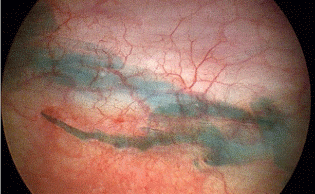
Given the clinical findings and history, a diagnosis of uterine rupture was made. Immediate cesarean section under general anesthesia resulted in the delivery of a live born male weighing 2616 grams, with APGAR scores of 0 at 1 minute, 4 at 5 minutes, and 7 at 10 minutes. Upon surgical exploration, the placenta was noted to be extravasated into the abdomen with complete placental detachment. The fundus of the uterus had a large defect reaching from the left adnexa to the right adnexa at the prior surgical site (Figure 1). The rupture site was noted to be hemostatic. The hysterotomy site was closed first in one running locked suture. The fundal rupture was then repaired in two layers of running locked sutures of multifilament suture. Both uterine sites were noted to be hemostatic. The patient’s initial hemoglobin level was 13.6g/dL, and her post-operative level was 10.9 g/dL. The infant was admitted to NICU. Cord gas values were arterial pH of 6.68, and a base deficit of 7mmol/L. After 12 months of follow up, the infant has no abnormal neurologic findings.

Figure 1: Uterine rupture extending the width of the uterus.
Discussion
Interstitial pregnancy is a rare event with a reported incidence of 1 in 2500 to 5000 live pregnancies [1]. An interstitial pregnancy can be defined as one which implants in the proximal tubal segment and lies within the muscular uterine wall [2], and is found lateral to the round ligament [2]. Prior to transvaginal ultrasound and Betahuman chorionic gonadotropin assay, rupture of an interstitial ectopic occurred between 8-16 weeks due to the distensibility of the myometrium covering the interstitial segment of the tube [2,3] leading to a mortality rate as high as 2.5% [4]. Earlier detection rates have led to more conservative treatment options. However, the complications of future pregnancies from these treatments are still unknown or speculative at best [2].
There are several case reports of vaginal deliveries following uterine surgery for interstitial pregnancies without rupture or adverse events [3,5,6]. However, a uterine rupture was reported after a forceps assisted vaginal delivery [7], and Su et al. report a uterine rupture and placental extravasations after vaginal delivery of a term infant [8]. Uterine ruptures have occurred as early as the 2nd and early 3rd trimesters as well [9-11]. Rupture may also differ by the type of treatment performed. Newer developments in laparoscopic and hysteroscopy have allowed for less invasive surgery, perhaps not involving the full thickness of the uterus, or using electrocautery loops [12,13].
Uterine rupture during subsequent pregnancies following an operation that involves the myometrium is a known complication [8,10,14]. Uterine rupture rates after myomectomy procedures range from 0.49-1.7% in subsequent pregnancies depending on laparoscopy vs. laparotomy [15]. The surgical steps of removal of an interstitial pregnancy are similar to that of a myomectomy. However, due to the low incidence of interstitial pregnancies, the rate of rupture is unknown and difficult to assess.
Uterine rupture can be a devastating event for both the mother and the fetus [15]. The fetus is at risk for hypoxia, acidosis or other sequela or even death [15]. Uterine rupture can occur prior to the onset of labor. Because of this early delivery is recommended [15]. Spong et al. recommend delivery between 37-38 weeks for prior myomectomy, and 36-37 weeks for a prior classical cesarean surgery [15, 16]. There is not a specific recommendation for women with a prior wedge resection for interstitial pregnancies.
Conclusion
The risk of rupture after an interstitial pregnancy is low, but as seen in our case, without access to immediate obstetrical intervention, results have the potential to be catastrophic. The delivery mode and timing remains somewhat nebulous. There are multiple case reports of successful term vaginal deliveries following wedge resections for interstitial pregnancies. However, there are also reports of attempted vaginal deliveries with dire results. A cesarean section is most often cited as the preferred mode of delivery, but timing is often an issue. According to the most recent guidelines put forth by Spong et al. the timing of cesarean section should be set at 37-38 weeks with a history of a myomectomy [15]. Our patient may not have benefited from these recommendations, being she at 37 weeks and 1 day. Other literature recommends cesarean delivery prior to the onset of labor, but as presented in this case report, the time from onset of regular uterine contractions to time of rupture can be minimal. We suggest a more stringent regimen of delivery plan for patients with a prior wedge resection for interstitial pregnancy, following the recommendation by Spong et al. for prior classical cesarean deliveries – delivery at 36- 37 weeks without amniocentesis for lung maturity [15,16]. The overall risk of rupture is unknown and difficult to assess due to the multitude of treatment modalities, as well as different definitions and locations of interstitial ectopic pregnancies. This unknown risk however, must not be assumed benign.
References
- Damario MA, Rock JA. Ectopic Pregnancy. Rock JA, Jones III HW, editors. In: TeLinde’s Operative Gynecology. Philadelphia: Lippincott Williams & Wilkins; 2008; 816.
- Hoffman BL, Schorge JO, Schaffer JI, Halvorson LM, Bradshaw KD, Cunningham FG, et al. Ectopic Pregnancy. Williams Gynecology. Dallas: McGraw-Hill. 2012: 212.
- Ng S, Hamontri S, Chua I, Chern B, Siow A. Laparoscopic management of 53 cases of cornual ectopic pregnancy. FertilSteril. 2009; 92: 448-452.
- Moawad NS, Mahajan ST, Moniz MH, Taylor SE, Hurd WW. Current diagnosis and treatment of interstitial pregnancy. Am J Obstet Gynecol. 2010; 202: 15-29.
- Anderson BB. Successful intrauterine term pregnancy after resection of corneal pregnancy. Eur J ObstetGynecolReprod Biol. 1999; 84: 99-100.
- Sagiv R, Golan A, Arbel-Alon S, Glezerman M. Three conservative approaches to treatment of interstitial pregnancy. J Am Assoc GynecolLaparosc. 2001; 8:154-158.
- Auamkul S. Rupture of gravid uterus after cornual resection. J Reprod Med. 1970; 5: 218-220.
- Su CF, Tsai HJ, Chen GD, shih YT, Lee MS. Uterine rupture at scar of prior laparoscopic cornuostomy after vaginal delivery of a full-term healthy infant. J ObstetGynaecol Res. 2008; 34: 688-691.
- Downey GP, Tuck SM. Spontaneous uterine rupture during subsequent pregnancy following non-excision of an interstitial ectopic gestation. BJOG. 1994; 10: 162-163.
- Weissman A, Fishman. Uterine following conservative surgery for interstitial pregnancy. Eur J ObstetGynecolReprod Biol. 1992; 44: 237-239.
- Loret de Mola JR, Austin CM, Judge NE, Assel BG, Peskin B, Goldfarb JM. Cornual heterotopic pregnancy and cornual resection after in vitro fertilization/embryo transfer: a report of two cases. J Reprod Med. 1995; 40: 606-610.
- Moon HS, Choi YJ, Park YH, Kim SG. New simple endoscopic operations for interstitial pregnancies. Am J Obstet Gynecol. 2000; 182: 114-121.
- Tulandi T, Vilos G, Gomel V. Laparoscopic treatment of interstitial pregnancy. Obstet Gynecol. 1995; 85: 465-467.
- Pelosi MA III, Pelosi MA. Spontaneous uterine rupture at thirty-three weeks subsequent to previous superficial laparoscopic myomectomy. Am J Obstet Gynecol. 1997; 177: 1547-1549.
- Spong CY, Mercer BM, D’Alton M, Kilpatrick S, Blackwell S, Saade G. Timing of Indicated Late-Preterm and Early-Term Birth. Obstet Gynecol. 2011; 118: 323-33.
- Stotland NF, Lipschitz, Caughey AB. Delivery strategies for women with a previous classical cesarean delivery: a decision analysis. Am J of Obstet Gynecol. 2002; 187: 1203-1238.