
Case Report
Austin J Obstet Gynecol. 2015; 2(4): 1048.
Early Ultrasound Diagnosis of Placenta Accreta: A Case Report
Bortoletto P¹*, Shulman L², Confino E³, Cohen L4, Fritsch M5 and Pavone ME³
¹Northwestern University, Feinberg School of Medicine, USA
²Division of Reproductive Genetics, Feinberg School of Medicine, USA
²Division of Reproductive Endocrinology and Infertility, Feinberg School of Medicine, USA
4Division of Obstetric and Gynecologic Ultrasound, Feinberg School of Medicine, USA
5Department of Pathology, Feinberg School of Medicine, USA
*Corresponding author: Bortoletto P, Northwestern University, Feinberg School of Medicine, St. Clair Street, Suite 14-200, Chicago, IL, 60611, USA
Received: August 17, 2015; Accepted: September 22, 2015; Published: September 25, 2015
Abstract
Background: With the rising rate of cesarean section, there has been an increase in the incidence of abnormal placentation in subsequent pregnancies, leading to the clinical complications of placenta accreta (PA) and cesarean scar ectopic pregnancies. The majority of cases of PA are unexpected and initially identified intraoperatively or during third trimester ultrasound.
Case: A 38-year-old G3P1011 with a previous low transverse lowersegment cesarean delivery who conceived using letrozole/IUI had an initial viability ultrasound at 5 weeks 5 days which was suspicious for an early placenta accreta versus cesarean scar pregnancy. Subsequent serial ultrasounds revealed placental lakes, a moth eaten appearance, and increased vascularity, concerning for placenta accreta.
Results: She underwent a repeat cesarean section with a high vertical incision during which placenta was found to be adherent to the uterus and the decision was made to proceed with hysterectomy. Placental pathology showed a mature placenta with extensive placenta increta and focal placenta percreta involving the right lateral uterus in addition to a complete placenta previa. She experienced a 7L blood loss requiring 6 units of pRBC and was managed in the ICU for 4 days until progressing to discharge along with a healthy 3.2kg male infant.
Conclusion: Our case represents a unique case in which a placenta accreta was detected by transvaginal ultrasound in a woman at 5 weeks and 5 days gestation who conceived with the assistance of intrauterine insemination. To our knowledge, this is the earliest case of suspected placenta accreta by ultrasonography.
Keywords: Placenta accreta, Assisted reproductive technology and Cesarean scar pregnancy
Introduction
Placenta Accreta (PA) may be defined as an abnormal adherence, in whole or in part, of the placenta to the uterine wall with subsequent failure to separate after delivery. The pathologic basis is a deficiency in the decidua basalis, which allows for chorionic tissue, including placental villi, to rest directly on myometrium, or penetrate into the muscle without any interposed decidua, obscuring a normal cleavage plane for placental separation. Given the absence of this protective layer, villi may also enter directly into the veins of the myometrium, explaining the hemorrhage encountered in attempted forced removal of the placenta. For this reason PA usually remains undetected until a catastrophic hemorrhage develops during delivery. There has been no shortage of case reports on the topic, with the first historical account of this condition dating back to Plater in the seventeenth century [1]. PA is not an uncommon phenomenon, with the prevalence estimated to be around 1 in 2500 pregnancies in 1985 [2]. The average reported incidence has increased ten-fold in the last 50 years, from 0.03 to 0.3%, with the highest incidence, 0.9%, reported in a study based on clinical diagnostic criteria [3]. The increase in PA in recent years is attributed to the increase in the prevalence of known risk factors. The most thoroughly studied of these is a previous cesarean section, with the risk increasing progressively with the number of repeated sections [4]. Maternal age and coexistent placenta previa are also known independent risk factors for development of PA [5].
As a result of this increasing incidence, patients and providers are more frequently confronted with difficult decisions such as whether to undergo a planned cesarean hysterectomy or to transfer care to a high-risk specialist. The prenatal diagnosis of PA, which has been well reported in several publications, has allowed both obstetricians and anesthesiologists to collaborate in decreasing maternal morbidity and mortality [6]. The diagnosis of PA can be made with the use of ultrasonography. Gray scale ultrasonography, color Doppler and three dimensional color Doppler imaging have all been described with varying specificity and sensitivity [7]. The accuracy of ultrasound for the prediction of placenta accreta is generally reported to be good, although not as high as previously thought, with sensitivities ranging from 77-97% [8]. We herein describe a case in which abnormal findings suggestive of a PA were detected by ultrasound at approximately 5 weeks’ gestation in a patient who conceived with Assisted reproductive technology (ART).
Case Presentation
A 38-year-old G3P1011 with one spontaneous abortion and one low transverse lower-segment cesarean delivery who conceived using letrozole/IUI had an initial viability ultrasound at 5 weeks 5 days which was suspicious for an early placenta accreta. Her history is notable for morbid obesity (BMI 54), PCOS, and type 2 DM. The patient received her first ultrasound at 5 weeks and 5 days after presenting with a bleeding episode. This initial ultrasound (Figure 1) raised concern for a low-lying accreta versus cesarean scar pregnancy due the location of the gestational sac just above the cesarean scar, increase in color Doppler, and evidence of deep trophoblastic invasion. Subsequent placental lakes were noted 7 days later (Figure 2) with scans at 11, 12 and 16 weeks showing signs of multiple lacunae, a moth eaten appearance, and increased vascularity consistent with placenta accreta (Figure 3). The development of a complete anterior placenta previa was also noted. Given the evidence of both placenta previa and accreta on serial ultrasounds, in addition to the standard obstetrics practice at our institution, it was recommended that she undergo a planned cesarean section at 34 weeks with plan for postpartum hysterectomy. Given the scheduled pre-term cesarean section, the patient received 2 doses of 12.5mg Betamethasone for fetal lung maturity and had 10u of packed Red Blood Cells (pRBC) and 4u of FFP stored in the event of hemorrhage during the procedure.
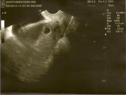
    Figure 1:  Transvaginal fetal ultrasound at 5 weeks 5 days gestation. A sagittal
section shows a suspected low-lying placenta accreta with trophoblastic
invasion suggestive of placenta accreta versus cesarean section scar
implantation. * Deep trophoblastic invasion.
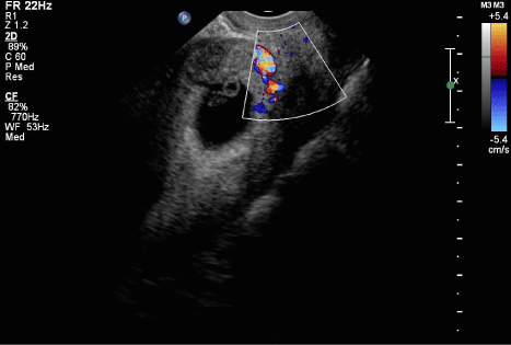
    Figure 2:  Transvaginal Doppler ultrasound at 6 weeks 6 days gestation. A
sagittal section shows a suspected low-lying placenta accreta. The blood
vessels shown are the site of implantation and trophoblastic invasion.
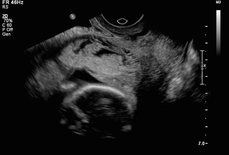
    Figure 3:  Transvaginal fetal ultrasound at 16 weeks 3 days gestation. A
sagital section shows signs of multiple lacunae, a moth eaten appearance,
and increased vascularity consistent with placenta accrete.
Results
She underwent a cesarean section with a high vertical incision and the fetus was delivered with a footling breech. The placenta was found to be adherent to the uterus and the decision was made to proceed with hysterectomy (Figure 4). Due to difficulty in transecting the uterine arteries for hemostatic control and ongoing blood loss, the gynecologic oncologist was called to the OR per the hospitals blood loss protocol. Upon achieving successful hemostasis with the aid of Floseal and a single piece of Gelfoam, the uterus with attached placenta was removed and sent for pathologic examination. Placental pathology showed a mature placenta with extensive placenta increta and focal placenta percreta involving the right lateral uterus in addition to a complete placenta previa (Figure 5). The patient experienced a 7L blood loss requiring 5 units of pRBC and 4 units of FFP intra-operatively. She was intubated for < 24hrs and managed conservatively for 3 more days in the intensive care unit where she received 1 additional unit of pRBC on post-op day 1 for hypotension and tachycardia. She was discharged home on post-operative day 3 with an asymptomatic hemoglobin. The neonate was a 3210-g boy with Apgar scores of 2 and 7 at 1 and 5 minutes, respectively. After initial examination, the newborn was transferred to the neonatal intensive care unit. His course was complicated by mild jaundice, not requiring treatment, and poor oral intake. He was discharged to home on day 4 of life with his mother.
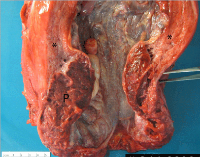
    Figure 4:  Anterior view of retained placenta (P) with deep invasion of placental
tissue throughout the wall of the lower uterine segment with extension to the
serosal surface in many areas consistent with increta, myometrium.
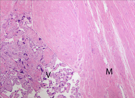
    Figure 5:  Classic histopathologic image of focal placenta accreta. Chorionic
villi (V) are in direct contact with myometrium (M) with no intervening decidua,
or other chorionic layers in this case. 100X magnification.
Conclusion
Our case represents a unique case in which a placenta accreta was suspected in a woman at 5 weeks and 5 days gestation who conceived with the assistance of intrauterine insemination. To our knowledge, this is the earliest suspected diagnosed case of placenta accreta, with earlier cases reported at 9 and 18 weeks, respectively [9,10]. The patient in question received standard serial ultrasounds as part of her assisted reproductive care, which incidentally allowed us to track the sonographic progression from questionable PA to definitive PA with eventual histopathologic corroboration. One of the potential confounders in diagnosing an early PA is its sonographic similarity to a cesarean section scar ectopic pregnancy. Although in the latter scenario the gestational sac is usually not contained within the uterine cavity, absolute distinctions are likely artificial, and these two entities more likely form a continuum with overlap in outcome and management [11]. In addition to overlap in imaging, Timor- Trisch reported that early PA and placental implantation in cesarean scar pregnancy are histopathologically indistinguishable [12]. It is becoming more apparent that perhaps these two entities more likely form a disease continuum with overlap in outcome and clinical management.
When considering patients receiving ART, Jackson, et al. demonstrated in a meta-analysis that singleton IVF pregnancies are associated with numerous adverse perinatal outcomes, including perinatal mortality, preterm delivery, low birth weight, and small for gestational age, even after controlling for maternal age and parity [13]. Additionally, Sun et al. noted a significantly increased risk in preeclampsia with IUI compared to IVF [14]. Although no association between pregnancies achieved by ART and PA has been previously described in the literature, Esh-Broder et al. reported that the odds of developing PA are significantly higher in IVF pregnancies than in spontaneous pregnancies [15]. In addition to PA, Romundstad et al. found that placenta previa occurred six times more often in singleton pregnancies after assisted reproduction compared with naturally conceived pregnancies, although they could not distinguish if this was due to maternal factors or factors related to ART [16]. To explain these differences, it is believed that perhaps a relationship exists between aberrant placental morphologic features and superficial implantation and/or inadequate orientation of the blastocyst after IVF and IUI [17]. Knowing this, obstetricians should be aware of the association between defects in placental implantation and ART. Early ultrasound evaluation is recommended for this patient population, as early diagnosis is possible, as evidenced by our case report.
References
- Sedlis A, Finn Jw, Loughran Ch. Placenta accreta in cesarean scar; report of a case. Obstet Gynecol. 1957; 9: 575-579.
- Clark SL, Koonings PP, Phelan JP. Placenta previa/accreta and prior cesarean section. Obstet Gynecol. 1985; 66: 89-92.
- Gielchinsky Y, Rojansky N, Fasouliotis SJ, Ezra Y. Placenta accreta--summary of 10 years: a survey of 310 cases. Placenta. 2002; 23: 210-214.
- Silver RM, Landon MB, Rouse DJ, Leveno KJ, Spong CY, Thom EA, et al. Maternal morbidity associated with multiple repeat cesarean deliveries. Obstet Gynecol. 2006; 107: 1226-1232.
- Miller DA, Chollet JA, Goodwin TM. Clinical risk factors for placenta previa-placenta accreta. Am J Obstet Gynecol. 1997; 177: 210-214.
- Japaraj RP, Mimin TS, Mukudan K. Antenatal diagnosis of placenta previa accreta in patients with previous cesarean scar. J Obstet Gynaecol Res. 2007; 33: 431-437.
- Shih JC, Jaraquemada JMP, Su YN, Shyu MK, Lin CH, Lin SY, et al. Role of three-dimensional power Doppler in the antenatal diagnosis of placenta accreta: comparison with gray-scale and color Doppler techniques. Ultrasound in Obstetrics and Gynecology. 2009; 33:193-203.
- Bowman ZS, Eller AG, Kennedy AM, Richards DS, Winter TC, Woodward PJ2, et al. Accuracy of ultrasound for the prediction of placenta accreta. Am J Obstet Gynecol. 2014; 211: 177.
- Wheeler TC, Anderson TL, Kelly J, Boehm FH. Placenta previa increta diagnosed at 18 weeks' gestation. Report of a case with sonographic and pathologic correlation. J Reprod Med. 1996; 41: 198-200.
- Chen YJ, Wang PH, Liu WM, Lai CR, Shu LP, Hung JH. Placenta accreta diagnosed at 9 weeks' gestation. Ultrasound Obstet Gynecol. 2002; 19: 620-622.
- Rosen T. Placenta accreta and cesarean scar pregnancy: overlooked costs of the rising cesarean section rate. Clin Perinatol. 2008; 35: 519-529, x.
- Timor-Tritsch IE, Monteagudo A, Cali G, Palacios-Jaraquemada JM, Maymon R, Arslan AA, et al. Cesarean scar pregnancy and early placenta accreta share common histology. Ultrasound in Obstetrics & Gynecology. 2014; 43: 383-395.
- Jackson RA, Gibson KA, Wu YW, Croughan MS. Perinatal outcomes in singletons following in vitro fertilization: a meta-analysis. Obstet Gynecol. 2004; 103: 551-563.
- Sun LM, Walker M, Cao HL, Yang Q, Duan T, Kingdom J. Assisted Reproductive Technology and Placenta-Mediated Adverse Pregnancy Outcomes. Obstetrics & Gynecology. 2009; 114: 818-824.
- Esh-Broder E, Ariel I, Abas-Bashir N, Bdolah Y, Celnikier DH. Placenta accreta is associated with IVF pregnancies: a retrospective chart review. BJOG. 2011; 118: 1084-1089.
- Romundstad LB, Romundstad PR, Sunde A, von Düring V, Skjaerven R, Vatten LJ. Increased risk of placenta previa in pregnancies following IVF/ICSI; a comparison of ART and non-ART pregnancies in the same mother. Hum Reprod. 2006; 21: 2353-2358.
- Jauniaux E, Englert Y, Vanesse M, Hiden M, Wilkin P. Pathologic features of placentas from singleton pregnancies obtained by in vitro fertilization and embryo transfer. Obstetrics & Gynecology. 1990; 76: 61-64.