Clinical Image
Austin J Obstet Gynecol. 2017; 4(1): 1068.
Placenta Previa Percreta: An Essential Diagnosis not to Miss!
Hurni Y¹*, Lipp von Wattenwyl B¹, Bonollo M¹, Ferrero L¹, Wyttenbach R² and Canonica C¹
¹Department of Obstetric and Gynecology, Ospedale Regionale Bellinzona e Valli, Switzerland
²Department of Radiology, Ospedale Regionale Bellinzona e Valli, Switzerland
*Corresponding author: Hurni Y, Department of Obstetrics and Gynecology, Ospedale Regionale Bellinzona e Valli - Bellinzona, Switzerland
Received: March 28, 2017; Accepted: April 04, 2017; Published: April 07, 2017
Clinical Image
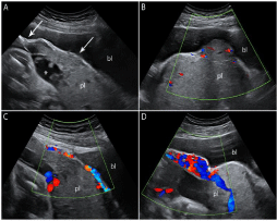
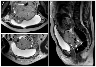
A 34-year-old woman, II-gravida I-para, presents at 12 weeks of gestation for painless vaginal bleeding with a transvaginal ultrasound showing a low-lying placenta. The patient had a history of a prior caesarean section two years prior due to breech presentation. The combination of low-lying placenta and a prior caesarean section remarkably increases the risk of an abnormally invasive placenta. A high index of suspicion is therefore required when evaluating such a pregnant woman. Careful examination revealed characteristic findings, suggesting placenta praevia percreta with bladder invasion on both greyscale and colour Doppler ultrasound (Figure 1) [1,2]. The diagnosis was supported by additional features observed on MRI (Figure 2) [2,3]. Placenta praevia percreta is among the greatest diagnostic and treatment challenges in current obstetrics. Its incidence is rising in association with the rising rate of caesarean sections. A multidisciplinary team approach is essential in managing this potentially catastrophic condition. But first, do not miss the diagnosis!

    Figure 1: Transabdominal ultrasound at 30 weeks of gestation, A: Loss of
hypoechoic plane in myometrium underneath the placental bed, reduced
myometrial thickness, interruption of the bladder wall (arrows), abnormally
large and irregular placental lacunae (asterisk), and B: Placental tissue bulging
into the bladder. Colour Doppler reveals, C: Bridging vessels extending
from placenta into bladder wall and (D) uterovescical hypervascularity. pl =
placenta; bl = bladder.

    Figure 2: A, B: Axial and C: Sagittal T2-weighted magnetic resonance images
obtained at 30 weeks of gestation, showing a placenta with heterogeneous
signal, lumpy contour, rounded edges, dark intraplacental bands (arrows A, B
and C), hypointense line disruption at the myometrial interface, and placental
tissue bulging into the bladder (asterisk, A).
References
- Collins SL, Ashcroft A, Braun T, Calda P, Langhoff-Roos J, Morel O, et al. Proposal for standardized ultrasound descriptors of abnormally invasive placenta (AIP). Ultrasound Obstet Gynecol. 2016; 47: 271-275.
- Allahdin S, Voigt S, Htwe TT. Management of placenta praevia and accreta. J Obstet Gynaecol. 2011; 31: 1-6.
- Kilcoyne A, Shenoy-Bhangle AS, Roberts DJ, Sisodia RC, Gervais DA, Lee SI. MRI of Placenta Accreta, Placenta Increta, and Placenta Percreta: Pearls and Pitfalls. American Journal of Roentgenology. 2016; 208: 214-221.